

Relation of synovitis to knee pain using contrast-enhanced MRIs
- PMID: 20472593
- PMCID: PMC3885343
- DOI: 10.1136/ard.2009.121426
Relation of synovitis to knee pain using contrast-enhanced MRIs
Abstract
Background: It has been suggested that synovitis causes joint pain. On non-contrast-enhanced MRIs synovial thickening cannot be assessed and on these images synovitis has been inconsistently associated with pain.
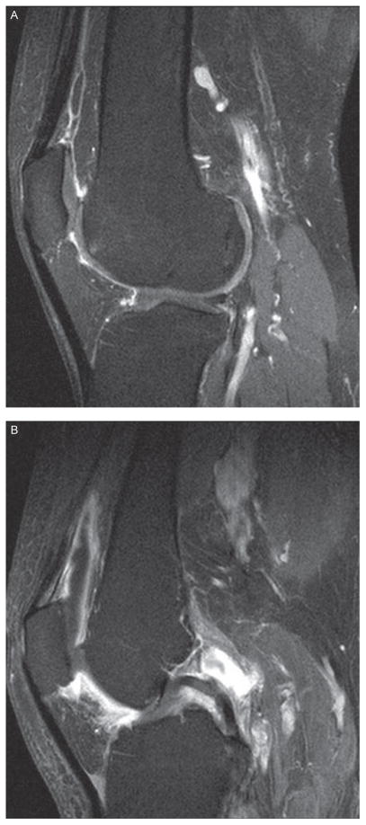
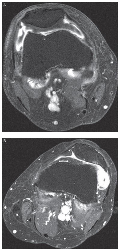
Objective: To assess synovial thickening in relation to knee pain severity among subjects in the Multicenter Osteoarthritis Study (MOST) using contrast-enhanced (CE) MRI.
Methods: MOST is a cohort study of people who have, or are at high risk of, knee osteoarthritis (OA). An unselected subset of 535 participants who volunteered underwent CE 1.5 T MRI of one knee. Synovitis was scored in six compartments and a summary score was created. Knee pain severity was assessed using the maximum item score on the Western Ontario and McMaster Osteoarthritis Index (WOMAC) pain scale. The association between synovitis and pain severity was examined using a logistic regression model adjusting for age, sex, body mass index (BMI), MRI bone marrow lesions and effusions in the whole sample and in a subgroup without radiographic OA.
Results: 454 of the 535 subjects undergoing CE MRI had complete data on synovitis and WOMAC pain. Mean age was 59 years, mean BMI 30 and 48% were women. In knees with moderate pain, 80% had synovitis. For knee pain, synovitis conferred a 9.2-fold increased odds compared with those without synovitis. In knees without radiographic OA (n=329), there was also an association of synovitis with an increased prevalence of pain.
Conclusion: Synovitis has a strong relation with knee pain severity, an association detected more clearly with CE MRI than suggested by previous studies using non-CE MRI measures of synovitis.
Conflict of interest statement
Figures
References
-
- Felson DT, Niu J, Guermazi A, et al. Correlation of the development of knee pain with enlarging bone marrow lesions on magnetic resonance imaging. Arthritis Rheum. 2007;56:2986–92. - PubMed
-
- Hill CL, Gale DG, Chaisson CE, et al. Knee effusions, popliteal cysts, and synovial thickening: association with knee pain in osteoarthritis. J Rheumatol. 2001;28:1330–7. - PubMed
-
- Torres L, Dunlop DD, Peterfy C, et al. The relationship between specific tissue lesions and pain severity in persons with knee osteoarthritis. Osteoarthr Cartil. 2006;14:1033–40. - PubMed
-
- Loeuille D, Rat AC, Goebel JC, et al. Magnetic resonance imaging in osteoarthritis: which method best reflects synovial membrane inflammation? Correlations with clinical, macroscopic and microscopic features. Osteoarthritis Cartilage. 2009;17:1186–92. - PubMed
Publication types
MeSH terms
Grants and funding
- U01 AG18820/AG/NIA NIH HHS/United States
- K23AR053855/AR/NIAMS NIH HHS/United States
- R01 NS048597/NS/NINDS NIH HHS/United States
- U01 AG18947/AG/NIA NIH HHS/United States
- U01 AG018947/AG/NIA NIH HHS/United States
- P60 AR047785/AR/NIAMS NIH HHS/United States
- K23 AR053855/AR/NIAMS NIH HHS/United States
- U01 AG18832/AG/NIA NIH HHS/United States
- U01 AG018832/AG/NIA NIH HHS/United States
- R01 AR053161/AR/NIAMS NIH HHS/United States
- U01 AG019069/AG/NIA NIH HHS/United States
- U01 AG018820/AG/NIA NIH HHS/United States
- U01 AG19069/AG/NIA NIH HHS/United States
- AR053161/AR/NIAMS NIH HHS/United States
LinkOut - more resources
Full Text Sources
Other Literature Sources
