Review
. 2010 Jul;9(7):982-90.
doi: 10.1128/EC.00030-10. Epub 2010 May 14.

Microtubule-dependent mRNA transport in fungi

Affiliations
Review

Microtubule-dependent mRNA transport in fungi

Kathi Zarnack et al. Eukaryot Cell. 2010 Jul.

Abstract

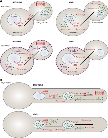
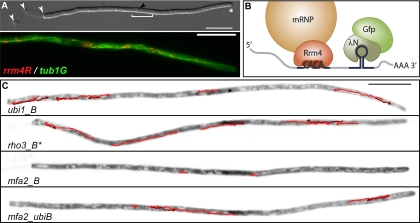
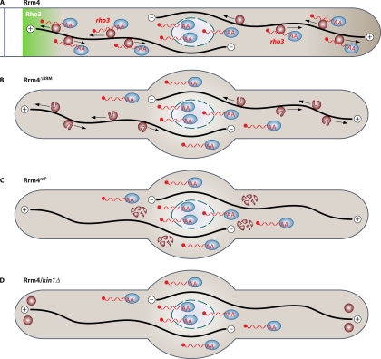
The localization and local translation of mRNAs constitute an important mechanism to promote the correct subcellular targeting of proteins. mRNA localization is mediated by the active transport of mRNPs, large assemblies consisting of mRNAs and associated factors such as RNA-binding proteins. Molecular motors move mRNPs along the actin or microtubule cytoskeleton for short-distance or long-distance trafficking, respectively. In filamentous fungi, microtubule-based long-distance transport of vesicles, which are involved in membrane and cell wall expansion, supports efficient hyphal growth. Recently, we discovered that the microtubule-mediated transport of mRNAs is essential for the fast polar growth of infectious filaments in the corn pathogen Ustilago maydis. Combining in vivo UV cross-linking and RNA live imaging revealed that the RNA-binding protein Rrm4, which constitutes an integral part of the mRNP transport machinery, mediates the transport of distinct mRNAs encoding polarity factors, protein synthesis factors, and mitochondrial proteins. Moreover, our results indicate that microtubule-dependent mRNA transport is evolutionarily conserved from fungi to higher eukaryotes. This raises the exciting possibility of U. maydis as a model system to uncover basic concepts of long-distance mRNA transport.


Figures

Fig. 1.
Fig. 1.
Actin-dependent mRNA transport in fungi. (A) Schematic representation of mRNA transport in S. cerevisiae during budding. (Top) mRNA transport results in the asymmetric distribution of the encoded protein. ASH1 mRNA (red wavy line) is transported along the actin cytoskeleton (black bar) from the mother to the daughter cell. Active transport is mediated by the SHE machinery (dark red circle), consisting of the key RNA-binding protein She2p, the potential adapter She3p, and the myosin motor Myo4p. Upon translation at the distal pole of the daughter cell, the transcription factor Ash1p predominantly enters the nucleus of the daughter cell. Genetic ablation (she2Δ) causes a mislocalization of mRNAs and translated proteins (right). (Bottom) mRNA transport is important for the symmetric distribution of the encoded protein. IST2 mRNA is transported by the same machinery to the daughter cell. The translation product localizes to the cell membrane. In the absence of mRNA transport (she2Δ), Ist2p localizes predominantly in the membrane of the mother cell (right). (B) mRNA transport in filaments of C. albicans. (Top) ASH1 mRNA is most likely transported by a related SHE machinery to the hyphal tip. Thereby, Ash1p is targeted to the first nucleus within the filament. (Bottom) In the absence of mRNA transport (she3Δ), Ash1p enters both nuclei.
Fig. 2.
Fig. 2.
Rrm4-dependent mRNA transport in hyphae of U. maydis. (A, top) Monokaryotic filament of U. maydis growing with a defined axis of polarity. The filament expands at the hyphal tip (asterisk) and inserts retraction septa at the basal pole (white arrowheads). The elongated nucleus (white bracket; the black arrowhead indicates the nucleolus) is positioned in the center of the filament. (Bottom) Rrm4-Rfp-containing mRNP particles (red) colocalize with microtubules (green, Tub1-Gfp decorated). Since colocalization is nearly complete, almost all Rrm4 particles appear orange. (B) Schematic representation of the λN-Gfp RNA live-imaging system. Rrm4 (red) target mRNAs containing CA-rich zipcodes are labeled by the recruitment of λN-Gfp (green) to its specific binding sites (box B, hairpin) in the 3′ UTR of the message. (C) Filaments of strains expressing different candidate mRNAs. ubi1_B and rho3_B are direct targets of Rrm4, whereas mfa2_B serves as a control. mfa2_ubiB is a hybrid RNA consisting of mfa2 and the CA-rich 3′ UTR of ubi1 as well as box B binding sites. Inverted frames were taken from fluorescence time-lapse movies. The movement of directed mRNA particles is tracked by red arrows (dark and light colors are used to mark overlapping particle tracks). (Reprinted from reference with permission of the publisher.)
Fig. 3.
Fig. 3.
Model depicting microtubule-dependent mRNA transport in U. maydis. (A) Rrm4-containing particles (dark red circles) transport mRNAs (red wavy lines) carrying the poly(A)-binding protein (blue ovals) bidirectionally along microtubules (black lines). The transport of rho3 mRNA might support a predominant accumulation of Rho3 at hyphal septa (45). (B) Deletion of RRM1 to RRM3 in Rrm4 causes a loss-of-function phenotype: filaments grow mostly bipolar, and target mRNAs are no longer transported along microtubules. However, the mutated Rrm4 version is still part of shuttling particles, indicating that Rrm4 is an integral part of the transport machinery and does not only hitchhike like the poly(A)-binding protein. (C) Mutations in the PABC domain of Rrm4 inhibit particle formation, thus abolishing microtubule-dependent mRNA transport. (D) Loss of the conventional kinesin Kin1 results in the accumulation of Rrm4-containing particles at both poles. Also in this case, the transport of mRNAs does not occur.

Similar articles

Cited by

References

    1. Andken B. B., Lim I., Benson G., Vincent J. J., Ferenc M. T., Heinrich B., Jarzylo L. A., Man H. Y., Deshler J. O. 2007. 3′-UTR SIRF: a database for identifying clusters of short interspersed repeats in 3′ untranslated regions. BMC Bioinformatics 8:274–287 - PMC - PubMed
    1. Aronov S., Gelin-Licht R., Zipor G., Haim L., Safran E., Gerst J. E. 2007. mRNAs encoding polarity and exocytosis factors are co-transported with cortical ER to the incipient bud in yeast. Mol. Cell. Biol. 27:3441–3455 - PMC - PubMed
    1. Aronov S., Gerst J. E. 2004. Involvement of the late secretory pathway in actin regulation and mRNA transport in yeast. J. Biol. Chem. 279:36962–36971 - PubMed
    1. Bartnicki-Garcia S. 2006. Chitosomes: past, present and future. FEMS Yeast Res. 6:957–965 - PubMed
    1. Becalska A. N., Gavis E. R. 2009. Lighting up mRNA localization in Drosophila oogenesis. Development 136:2493–2503 - PMC - PubMed

Publication types