. 2010 Aug;299(2):L192-203.
doi: 10.1152/ajplung.00426.2009. Epub 2010 May 14.

Glutaredoxin 1 regulates cigarette smoke-mediated lung inflammation through differential modulation of I{kappa}B kinases in mice: impact on histone acetylation

Affiliations

Glutaredoxin 1 regulates cigarette smoke-mediated lung inflammation through differential modulation of I{kappa}B kinases in mice: impact on histone acetylation

Sangwoon Chung et al. Am J Physiol Lung Cell Mol Physiol. 2010 Aug.

Abstract

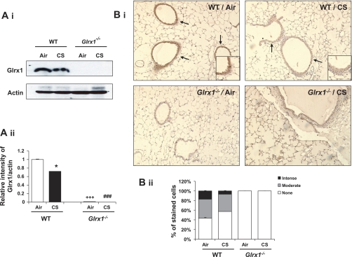
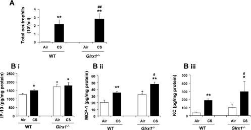
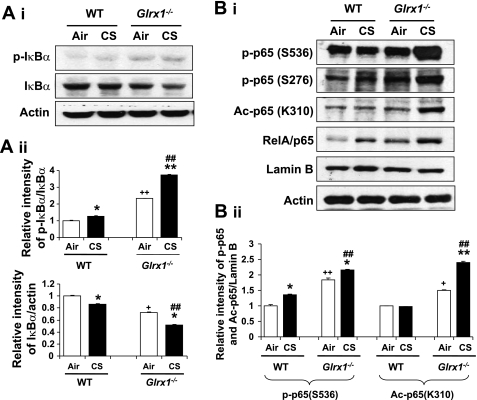
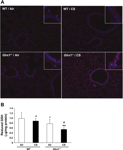
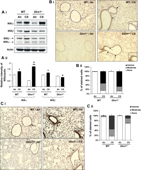
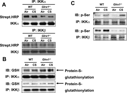
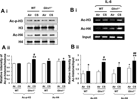
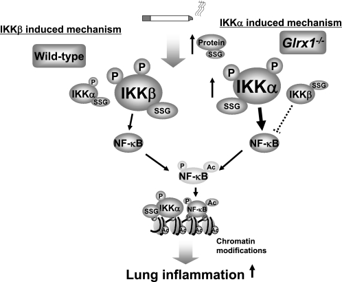
Glutaredoxin 1 (Glrx1) is a small dithiol protein that regulates the cellular redox state and redox-dependent signaling pathways via modulation of protein glutathionylation. IkappaB kinase (IKK), an essential enzyme for NF-kappaB activation, can be subjected to S-glutathionylation leading to alteration of its activity. However, the role of Glrx1 in cigarette smoke (CS)-induced lung inflammation and chromatin modifications are not known. We hypothesized that Glrx1 regulates the CS-induced lung inflammation and chromatin modifications via differential regulation of IKKs by S-glutathionylation in mouse lung. Glrx1 knockout (KO) and wild-type (WT) mice were exposed to CS for 3 days and determined the role of Glrx1 in regulation of proinflammatory response in the lung. Neutrophil influx in bronchoalveolar lavage fluid and proinflammatory cytokine release in lung were increased in Glrx1 KO mice compared with WT mice exposed to CS, which was associated with augmented nuclear translocation of RelA/p65 and its phospho-acetylation. Interestingly, phosphorylated and total levels of IKKalpha, but not total and phosphorylated IKKbeta levels, were increased in lungs of Glrx1 KO mice compared with WT mice exposed to CS. Ablation of Glrx1 leads to increased CS-induced IKKbeta glutathionylation rendering it inactive, whereas IKKalpha was activated resulting in increased phospho-acetylation of histone H3 in mouse lung. Thus, targeted disruption of Glrx1 regulates the lung proinflammatory response via histone acetylation specifically by activation of IKKalpha in response to CS exposure. Overall, our study suggests that S-glutathionylation and phosphorylation of IKKalpha plays an important role in histone acetylation on proinflammatory gene promoters and NF-kappaB-mediated abnormal and sustained lung inflammation in pathogenesis of chronic inflammatory lung diseases.


Figures

Fig. 1.
Fig. 1.
Expression of Glutaredoxin 1 (Glrx1) in lung of Glrx1 wild-type (WT) and knockout (Glrx1−/−) mice. Deficiency of Glrx1 in lungs of Glrx1−/− mice was confirmed by immunoblotting and immunohistochemistry. A: Glrx1 protein was measured in lung homogenates of WT and Glrx1−/− mice in response to air or cigarette smoke (CS) exposure. Immunoblot pictures (i) and histograms (ii) showing relative intensity of Glrx1 vs. actin bands. B: Glrx1-positive cells were identified by dark brown immunohistochemical staining (i). Insets are magnified airway regions showing decreased Glrx1 staining in response to CS compared with air exposure. ii: immunostaining scores for Glrx1 per cell in airway regions of the lung. The assessment of immunostaining intensity was performed semiquantitatively and in a blinded fashion. Solid bars, intense staining; shaded bars, moderate/weak staining; open bars, no staining. Original magnification: ×100. *P < 0.05, significant compared with respective air-exposed mice. ###P < 0.001, significant compared with CS-exposed WT mice. +++P < 0.001, significant compared with air-exposed WT mice.
Fig. 2.
Fig. 2.
Increased lung inflammatory responses in Glrx1−/− mice in response to CS exposure. A: neutrophil influx was assessed by Diff-Quik staining in cytospin slides, which were prepared using bronchoalveolar lavage fluid collected by lung lavage in air- or CS-exposed WT and Glrx1−/− mice. B: the levels of proinflammatory mediators, such as IP-10(i), MCP-1 (ii), and KC (iii), were measured by ELISA in lung homogenates of air- and CS-exposed WT and Glrx1−/− mice. Data are shown as means ± SE (n = 3–5/group). *P < 0.05, **P < 0.01, significant compared with respective air-exposed mice. #P < 0.05, ##P < 0.01, significant compared with CS-exposed WT mice. +P < 0.05, significant compared with air-exposed WT mice.
Fig. 3.
Fig. 3.
Targeted disruption of Glrx1 increased degradation of IκBα and posttranslational modifications of RelA/p65 in response to CS exposure. A: p-IκBα and IκBα levels were measured in lung cytosolic fractions of WT and Glrx1−/− mice (i). ii: histograms show relative intensity of IκBα bands vs. controls. B: posttranslational modification of RelA/p65 was assessed by measuring phosphorylation (Ser276 and Ser536) and acetylation (Ac) (Lys310) of RelA/p65 in nuclear fractions of lung by immunoblotting (i). ii: histograms show relative intensity of p65 bands vs. controls. Gel pictures shown are representative of at least 3 separate experiments. *P < 0.05, **P < 0.01, significant compared with respective air-exposed mice. ##P < 0.01, significant compared with CS-exposed WT mice. +P < 0.05, ++P < 0.01, significant compared with air-exposed WT mice.
Fig. 4.
Fig. 4.
Protein S-glutathionylation was increased in lung of Glrx1−/− mice in response to CS exposure. A: protein S-glutathionylation (PSSG) reactivity in mouse lung tissue was evaluated using Glrx1-based cysteine derivatization. Red, PSSG reactivity; blue, DNA content. Original magnification, ×100. Insets are magnified airway regions showing increased PSSG reactivity in response to CS. B: level of reduced GSH was measured in lungs of air- and CS-exposed WT and Glrx1−/− mice. Data are shown as means ± SE (n = 3–5/group). *P < 0.05, **P < 0.01, significant compared with respective air-exposed mice. #P < 0.05, significant compared with CS-exposed WT mice. +P < 0.05, significant compared with air-exposed WT mice.
Fig. 5.
Fig. 5.
Differential regulation of IKKα and IKKβ in lung of Glrx1−/− mice in response to CS exposure. A: IKKs were blotted by anti-IKKα, anti-IKKβ, and anti-IKKα/β antibody, respectively, in lung of WT and Glrx1−/− mice (i). ii: histograms showing relative intensity of IKKs vs. actin bands. Gel pictures shown are representative of at least 3 separate experiments. *P < 0.05, **P < 0.01, significant compared with respective air-exposed mice. #P < 0.05, significant compared with CS-exposed WT mice. IKKα (B) and IKKβ (C) positive cells were identified by dark brown immunohistochemical staining (i). ii: immunostaining scores for IKKs per cell in alveolar and airway regions of the lung. The assessment of immunostaining intensity was performed semiquantitatively and in a blinded fashion. Insets are magnified airway regions showing increased IKKα (B) and IKKβ (C) staining in response to CS compared with air-expressed WT and Glrx1-/- mice. Solid bars, intense staining; shaded bars, moderate/weak staining; open bars, no staining. Original magnification, ×100.
Fig. 6.
Fig. 6.
Inactivation of IKKβ and accumulation of phosphorylated IKKα in Glrx1−/− mice in response to CS exposure. A: lung homogenates were subjected to immunoprecipitation with IKKα and IKKβ antibodies, and immunoblotted with streptavidin-conjugated HRP (top) and IKKα and IKKβ (lower) antibodies. B: IKKα and IKKβ immunoprecipitates separated under nonreducing conditions, and then immunoblotted with anti-GSH (top) and IKKα and IKKβ (lower) antibodies. C: immunoprecipitated IKKα and IKKβ were analyzed for phosphorylation of serine residues using specific phospho-serine antibody, respectively. Gel pictures shown are representative of at least 3 separate experiments.
Fig. 7.
Fig. 7.
Deficiency of Glrx1 led to increased histone modification in mouse lungs in response to CS exposure. A: histone modifications were assessed by measuring phosphorylation (Ser10) and acetylation (Lys9) of histone H3, and acetylation of histone H4 (Lys12) in lung by immunoblotting using acid-extracted nuclear histone fraction (i). B: acetylated histone H3 and H4 on the IL-6 gene promoter in mouse lung were analyzed by ChIP assay. Lung homogenates were immunoprecipitated with anti-acetylated histone H3 and H4 antibodies, and chromatin modification on the promoter region of proinflammatory cytokine was detected by PCR using the primers for IL-6. Gel pictures shown are representative of at least 3 separate experiments.*P < 0.05, **P < 0.01, significant compared with respective air-exposed mice. #P < 0.05, ##P < 0.01, significant compared with CS-exposed WT mice. +P < 0.05 significant compared with air-exposed WT mice.
Fig. 8.
Fig. 8.
A schematic diagram for the role of Glrx1 in regulation of IKKα and IKKβ in response to CS. Targeted disruption of Glrx1 leads to increased lung proinflammatory response, which is associated with NF-κB activation through the differential regulation of IKKs, including glutathionylation and phosphorylation in response to CS exposure. Glrx1 deficiency leads to increased IKKα and IKKβ activation in response to CS associated with S-glutathionylation. IKKβ-S-glutathionylation leads to its inactivation and hence inhibition of NF-κB (dotted lines), whereas posttranslational modifications (S-glutathionylation and phosphorylation) on IKKα cause NF-κB activation and chromatin modifications (histone acetylation) on proinflammatory genes. IKKα and IKKβ modifications are reversible, and the inflammatory pathway is predominantly mediated by IKKβ.

Similar articles

Cited by

References

    1. Adenuga D, Yao H, March TH, Seagrave J, Rahman I. Histone deacetylase 2 is phosphorylated, ubiquitinated, and degraded by cigarette smoke. Am J Respir Cell Mol Biol 40: 464–473, 2009 - PMC - PubMed
    1. Aesif SW, Anathy V, Havermans M, Guala AS, Ckless K, Taatjes DJ, Janssen-Heininger YM. In situ analysis of protein S-glutathionylation in lung tissue using glutaredoxin-1-catalyzed cysteine derivatization. Am J Pathol 175: 36–45, 2009 - PMC - PubMed
    1. Anathy V, Aesif SW, Guala AS, Havermans M, Reynaert NL, Ho YS, Budd RC, Janssen-Heininger YM. Redox amplification of apoptosis by caspase-dependent cleavage of glutaredoxin 1 and S-glutathionylation of Fas. J Cell Biol 184: 241–252, 2009 - PMC - PubMed
    1. Biswas S, Chida AS, Rahman I. Redox modifications of protein-thiols: emerging roles in cell signaling. Biochem Pharmacol 71: 551–564, 2006 - PubMed
    1. Chariot A. The NF-kappaB-independent functions of IKK subunits in immunity and cancer. Trends Cell Biol 19: 404–413, 2009 - PubMed

Publication types