. 2010 May;47(3):434-45.
doi: 10.1177/0300985810363705.

The histopathologic and molecular basis for the diagnosis of histiocytic sarcoma and histiocyte-associated lymphoma of mice

Affiliations

The histopathologic and molecular basis for the diagnosis of histiocytic sarcoma and histiocyte-associated lymphoma of mice

X Hao et al. Vet Pathol. 2010 May.

Abstract

Histiocytic sarcoma (HS) and histiocyte-associated lymphoma (HAL) of mice are difficult to distinguish histologically. Studies of multiple cases initially diagnosed as HS or HAL allowed us to define HS as round, fusiform, or mixed cell types that were F4/80+, Mac-2+, and PAX5-; that lacked markers for other sarcomas; and that had immune receptor genes in germline configuration. Two other subsets had clonal populations of lymphocytes. The first, HAL, featured malignant lymphocytes admixed with large populations of normal-appearing histiocytes. The second appeared to be composites of lymphoma and HS. Several cases suggestive of B myeloid-lineage plasticity were also observed.


Conflict of interest statement

Declaration of Conflicting Interests

The authors declared that they had no conflicts of interest with respect to their authorship or the publication of this article.

Figures

Figure 1
Figure 1
Southern blot analyses of mouse hematopoietic neoplasms originally diagnosed as histiocytic sarcoma. Genomic organization of immunoglobulin H genes was examined by hybridization of DNA digested with EcoRI and hybridized with a JH probe. Pathology number related to the 17 DNA samples studied are available from the senior author on request. A, histiocytic sarcomas exhibit only a germline JH band. B, for HA, clonal rearrangements of JH distinct from the germline band are seen in each lane. Cases showing 2 distinct bands of equal intensity are indicative of a single clone with both IgH alleles rearranged. Cases in lanes 2, 5, and 11 with 3 or more bands are judged to be oligoclonal. The arrow associated with lane 9 points to a rearrangement on a large EcoRI fragment.
Figures 2–7
Figures 2–7
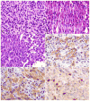
Figure 2. HS, round–cell type. Note the round nuclei, prominent nucleoli, and abundant eosinophilic cytoplasm. Figure 3. HS, spindle–cell type. Note the elongated and convoluted nuclei. Figure 4. HS, round- and spindle–cell type. Note round- (right side) and spindle-cell components (left side). Serial sections of HS stained with antibodies to the following: Figure 5. F4/80 (immunoreactive cell membrane). Figure 6. Mac–2 (immunoreactive cytoplasm and nucleus). Figure 7. PAX5 (note PAX5– HS tumor cells admixed with normal PAX5+ B cells).
Figures 8–13
Figures 8–13
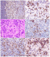
Figure 8. Histiocytic sarcoma in the liver stained with antibody to F4/80 showing high-level expression by normal Kupffer cells (right side), lower expression by the round–cell component of the histiocytic sarcoma (middle portion), and the lowest levels of expression by the spindle–cell component (left side). Figure 9. PAX5 staining of a spindle–type histiocytic sarcoma occupying the red pulp and surrounding a follicle with normal B cells. Note nuclear PAX5 staining of some spindle cells as well as normal B cells (upper left). Histopathologic and immunohistochemical features of histiocyte-associated lymphoma. Serial sections of a histiocyte–associated lymphoma stained with HE showing the following: Figure 10. B-cell lymphoma and eosinophilic histiocytes. Figure 11. Antibodies to F4/80. Figure 12. Mac–2. Figure 13. PAX5. Note the more extensive staining of the histiocyte population by Mac–2 than F4/80 as they weave among the clusters of PAX5+ cells.
Figures 14–19
Figures 14–19
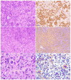
Histopathologic and immunohistochemical features of a HAL with foci of HS. Serial sections stained of a histiocytic sarcoma stained with HE (Figure 14), and antibodies to PAX5 (Figure 15). A focus of histiocytic sarcoma in the histiocyte-associated lymphoma, (Figure 16) stained for Mac-2 showing moderate histiocytic expression (Figure 17). Mitotic figures were seen in the focal case of histiocytic sarcoma (Figure 18) with a neoplastic mitotic histiocyte expressing Mac-2 (Figure 19).

Similar articles

Cited by

References

    1. Achten R, Verhoef G, Vanuytsel L, De Wolf Peeters C. Histiocyte rich, T cell rich B cell lymphoma: a distinct diffuse large B cell lymphoma subtype showing characteristic morphologic and immunophenotypic features. Histopathology. 2002;40:31–45. - PubMed
    1. Akashi K, Traver D, Miyamoto T, Weissman IL. A clonogenic common myeloid progenitor that gives rise to all myeloid lineages. Nature. 2000;404:193–197. - PubMed
    1. Arber DA, Strickler JG, Chen YY, Weiss LM. Splenic vascular tumors: a histologic, immunophenotypic, and virologic study. Am J Surg Pathol. 1997;21:827–835. - PubMed
    1. Bauer SR, Holmes KL, Morse HC, III, Potter M. Clonal relationship of the lymphoblastic cell line P388 to the macrophage cell line P388D1 as evidenced by immunoglobulin gene rearrangements and expression of cell surface antigens. J Immunol. 1986;136:4695–4699. - PubMed
    1. Carrasco DR, Fenton T, Sukhdeo K, Protopopova M, Enos M, You MJ, Di Vizio D, Nogueira C, Stommel J, Pinkus GS, Fletcher C, Hornick JL, Cavenee WK, Furnari FB, Depinho RA. The PTEN and INK4A/ARF tumor suppressors maintain myelolymphoid homeostasis and cooperate to constrain histiocytic sarcoma development in humans. Cancer Cell. 2006;9:379–390. - PubMed