. 2010 Jul;177(1):280-90.
doi: 10.2353/ajpath.2010.090592. Epub 2010 May 14.

Bisphosphonates cause osteonecrosis of the jaw-like disease in mice

Affiliations

Bisphosphonates cause osteonecrosis of the jaw-like disease in mice

Yanming Bi et al. Am J Pathol. 2010 Jul.

Abstract

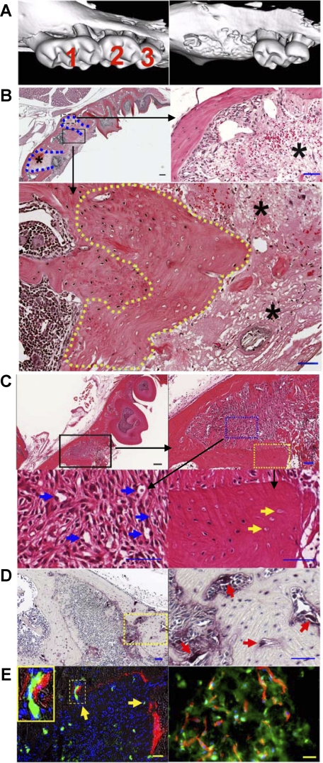
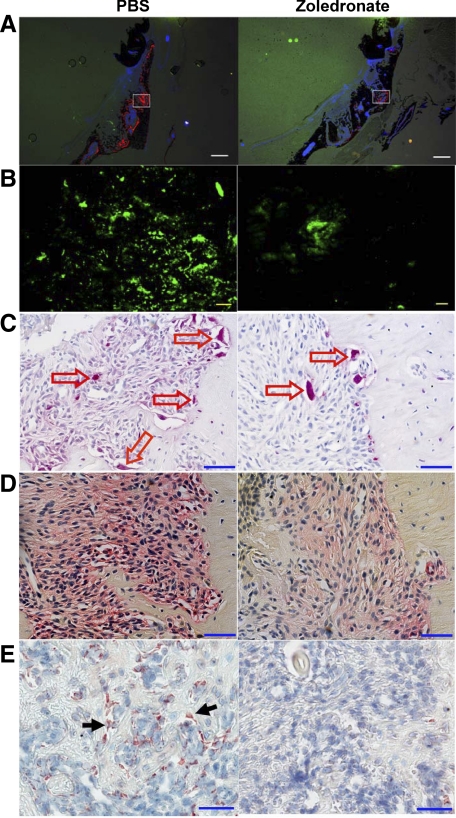
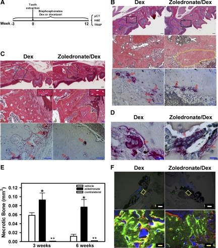
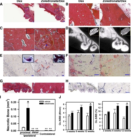
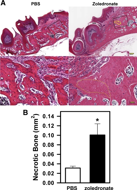
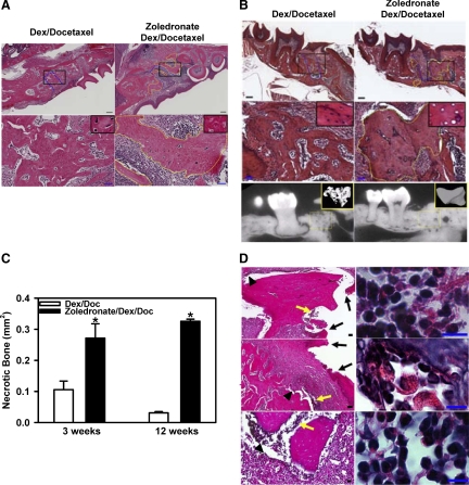
Bisphosphonate-associated osteonecrosis of the jaw (BONJ) is a morbid bone disease linked to long-term bisphosphonate use. Despite its broad health impact, mechanistic study is lacking. In this study, we have established a mouse model of BONJ-like disease based on the equivalent clinical regimen in myeloma patients, a group associated with high risk of BONJ. We demonstrate that the murine BONJ-like disease recapitulates major clinical and radiographical manifestations of the human disease, including characteristic features of osseous sclerosis, sequestra, avascular, and radiopaque alveolar bone in the jaw that persists beyond a normal course of wound healing following tooth extraction. We find that long-term administration of bisphosphonates results in an increase in the size and number of osteoclasts and the formation of giant osteoclast-like cells within the alveolar bone. We show that the development of necrotic bone and impaired soft tissue healing in our mouse model is dependent on long-term use of high-dose bisphosphonates, immunosuppressive and chemotherapy drugs, as well as mechanical trauma. Most importantly, we demonstrate that bisphosphonate is the major cause of BONJ-like disease in mice, mediated in part by its ability to suppress osseous angiogenesis and bone remodeling. The availability of this novel mouse model of BONJ-like disease will help elucidate the pathophysiology of BONJ and ultimately develop novel approaches for prevention and treatment of human BONJ.


Figures

Figure 1
Figure 1
Tooth injury leads to the development of necrotic bone and promotes osseous angiogenesis and bone remodeling. A: Three-dimensional reconstruction of μCT images of the left maxilla from mice without (left) and with (right) tooth extraction. B: H&E staining of the paraffin-embedded sections of the left maxilla from mice at one day after tooth extraction of the first left maxillary molar (top left; Scale bar = 200 μm). Blue dotted lines indicate the extraction socket. Higher magnification image shows the presence of amorphous thrombi (top right panel; asterisks) within the extraction tooth sockets and the dead bone with empty lacunae (bottom panel; yellow dotted line), respectively. Scale bar = 50 μm. C: H&E staining of the left maxilla three days after tooth extraction. The top panels show the low (left; Scale bar = 200 μm) and high (right; Scale bar = 50 μm) magnification images of the extraction socket. The bottom panels show the presence of abundant mesenchymal cells and capillary-like structures (left; blue arrows) within the provisional matrix and the presence of necrotic bone with empty lacunae (right; yellow arrows) adjacent to the amorphous thrombi. Scale bars = 50 μm. D: TRAP staining. Three days after extraction, abundant TRAP+ osteoclasts (purple) were found on the alveolar bone surface within the extraction socket (left; Scale bar = 200 μm). Higher magnification of the yellow dotted area was shown in the right panel (red arrows indicate osteoclasts; Scale bar = 50 μm). E: Bone and blood vessel formation. Alizarin complexone labeling on day one and FITC-BSA perfusion on day three following extraction reveal the formation of new bone (left; red fluorescence) and the presence of functional blood vessels (green fluorescence; yellow arrows). Inset shows newly formed bone adjacent to a blood vessel. Scale bar = 50 μm. Immunofluorescent staining in the right panel shows the presence of osteocalcin+ bone matrix (green) residing proximally to CD31+ capillaries (red). DAPI-stained nuclei are shown in blue. Scale bar = 20 μm.
Figure 2
Figure 2
Bisphosphonates suppress angiogenesis and bone remodeling in osseous tissues in response to injury. Mice were treated with PBS (left panels) or zoledronate (right panels) for one week following tooth extraction (no pretreatment), and then injected with alizarin 4 days and perfused with FITC-BSA 7 days later. A: Fluorescent images of the frozen sections of the left maxilla showed alizarin incorporation for bone formation (red fluorescence). Scale bars = 500 μm. B: Higher magnification images of the white squares in A show the presence of osseous angiogenesis (green), which was suppressed by zoledronate treatment (right). Scale bars = 50 μm. C: TRAP staining of the paraffin-embedded sections of the left maxilla showed the presence of TRAP+ osteoclasts (red arrows). D: ALP staining of the paraffin-embedded sections of the left maxilla showed the presence of osteoblasts and their precursor cells (red color). E: Immunohistochemistry shows the presence of osteocalcin+ mature osteoblasts (black arrows). Scale bars for to C–E: 50 μm.
Figure 3
Figure 3
Development of necrotic bone in zoledronate-treated mice following tooth extraction. A: Strategy for generating and analyzing zoledronate-induced ONJ-like disease in mice. C57BL6 mice were treated with zoledronate, Dex, docetaxel, or their combinations for three weeks before tooth extraction. The first left maxillary molar tooth was removed and these different drugs were administered for up to 12 weeks. Histological analysis was conducted on the maxilla from Dex-treated (left panels) or zoledronate/Dex-treated (right panels) mice at three weeks (B) and six weeks (C) after tooth extraction. Top panels show low magnification of H&E staining of the paraffin-embedded sections of the left maxilla (Scale bars = 200 μm). Blue dotted lines indicate the extraction socket. Middle panels show higher magnification of the areas indicated by the black squares in the top panel (Scale bars = 50 μm). A yellow dotted line circles the dead bone with empty lacunae. Bottom panels show the presence of TRAP+ osteoclasts (red arrows). Scale bars = 50 μm. Insets in the middle panels in C show lacunae with (left) or without (right) osteocytes. D: TRAP staining of the left maxilla shows the presence of giant osteoclast-like cells in zoledronate/Dex-treated (right) but not Dex-treated (left) mice three weeks after extraction. Scale bars = 10 μm. E: Quantification of the necrotic bone areas in the left or right (contralateral) maxilla of Dex- and zoledronate/Dex-treated mice at three weeks and six weeks after tooth extraction using ImageScope. Data are means ± SEM of three mice. *P < 0.05 versus Dex. **Necrotic bone area <0.0005 mm2. F: Dex- and zoledronate/Dex-treated mice were labeled with calcein (green) four weeks and alizarin (red) six weeks after extraction. Higher magnification of the yellow dotted areas in the top panels (Scale bars = 500 μm) is presented in the corresponding bottom panels (Scale bars = 50 μm).
Figure 4
Figure 4
Zoledronate-treated mice develop BONJ-like disease following tooth extraction. A: H&E staining of the paraffin-embedded sections of the left maxilla from Dex-treated (left panels) or zoledronate/Dex-treated (right panels) mice at 12 weeks after tooth extraction. Blue dotted lines indicate the extraction socket. Green squares and black squares indicate the areas distal and proximal to the site of extracted tooth, respectively. B: Higher magnification images of the distal areas (green squares in A) show the presence of viable alveolar bone. C: Higher magnification images of the proximal areas (black squares in A) show the development of BONJ-like disease, characterized by the presence of both sclerotic bone (yellow dotted line) and necrotic bone (white dotted lines) that were interspersed with viable bone (right panel). Insets show lacunae with (left) or without (right) osteocytes. Scale bars for A–C: 200 μm. D: μCT scan images of the left maxilla illustrates an overall increase in bone density. Insets show the three-dimensional reconstructed images of the jaw bone within the region of tooth extraction (dotted lines). E: TRAP staining (purple; red arrows) of the paraffin-embedded sections showed increased osteoclasts in zoledronate-treated mice (right). Insets show the presence of giant osteoclast-like cell in zoledronate-treated (right) but not control (left) mice. F: ALP staining (red; black arrows) of the paraffin-embedded sections shows osteoblasts. Scale bars for E and F: 50 μm. G and H: Contralateral maxilla. Left panels show low magnification images of H&E-stained (G) and TRAP-stained (H) paraffin-embedded sections of the right (contralateral) maxilla from zoledronate/Dex-treated mice at 12 weeks after tooth extraction. Right panels are the higher magnification of the area in the black square in left panels. Scale bars: left panel, 200 μm; right panel, 50 μm. I: Quantification of necrotic bone area in the distal (B) and proximal (C) regions of the left (ipsilateral) and right (contralateral) maxilla was done by ImageScope. Data are mean ± SEM of seven mice. *P < 0.05 versus vehicle. **Necrotic bone area <0.0005 mm2. J: Histomorphometric analysis. The number of osteoclasts per millimeter of bone surface (Oc.N/BS) and the percentage of osteoclast surface per bone surface (Oc.S/BS) were determined (*P < 0.01, n = 3–6). Open bars: vehicle; filled bars: zoledronate.
Figure 5
Figure 5
Bisphosphonates are a major cause of BONJ-like disease in mice. A: H&E staining of the paraffin-embedded sections of the left maxilla from PBS-treated (left) and zoledronate-treated (right) mice at 12 weeks after tooth extraction. Lower panels are the higher magnification of the area in the black square in the top panels. The yellow dotted lines indicate areas of necrotic bone. Scale bars: top panels, 200 μm; bottom panels, 50 μm. B: Quantification of the areas of necrotic bone in the left maxilla. Data are means ± SEM of three mice. *P < 0.05 versus PBS.
Figure 6
Figure 6
Docetaxel worsens BONJ-like disease in bisphosphonate-treated mice. H&E staining of the paraffin-embedded sections of the left maxilla at three weeks (A) and mandible at 12 weeks (B) after tooth extraction from Dex/docetaxel-treated (left panels) or zoledronate/Dex/docetaxel-treated (right panels) mice. Top panels: Low magnification of H&E staining of the paraffin-embedded sections of the left maxilla. Blue dotted lines indicate the extraction sockets. Yellow dotted lines indicate the areas of bone necrosis. Bottom panels in A and middle panels in B: Higher magnification of areas in the black squares shown in the top panels. Insets show lacuna with (left) or without (right) osteocytes. Scale bars: 200 μm (top panels) and 50 μm (bottom panels). Bottom panels in B: μCT images of the left maxilla from mice at 12 weeks after tooth extraction. Insets showed the three-dimensional reconstructed images of the extraction socket (yellow dotted squares). C: Quantification of the areas of necrotic bone in the left maxilla. Data are means ± SEM of 10 mice. *P < 0.05, zoledronate/Dex/docetaxel versus Dex/docetaxel. D: Persistent soft tissue defects in mice treated with zoledronate, Dex, and docetaxel at 12 weeks after tooth extraction. Low magnification (left panels) of H&E-stained paraffin-embedded sections of the left maxilla shows the disruption of epithelium (black arrows), osteomyelitis (yellow arrows), and separation of soft tissue from the necrotic bone (black arrowheads). High magnification (right panels) shows the presence of inflammatory cells within the areas of osteomyelitis. Scale bars: 50 μm (left panels) and 10 μm (right panels).

Similar articles

Cited by

References

    1. Bamias A, Kastritis E, Bamia C, Moulopoulos LA, Melakopoulos I, Bozas G, Koutsoukou V, Gika D, Anagnostopoulos A, Papadimitriou C, Terpos E, Dimopoulos MA. Osteonecrosis of the jaw in cancer after treatment with bisphosphonates: incidence and risk factors. J Clin Oncol. 2005;23:8580–8587. - PubMed
    1. Ruggiero SL, Woo SB. Bisphosphonate-related osteonecrosis of the jaws. Dent Clin North Am. 2008;52:111–128. - PubMed
    1. Kuehn BM. Reports of adverse events from bone drugs prompt caution. JAMA. 2006;295:2833–2836. - PubMed
    1. Woo SB, Hellstein JW, Kalmar JR. Systematic review: bisphosphonates and osteonecrosis of the jaws. Ann Intern Med. 2006;144:753–761. - PubMed
    1. Khosla S, Burr D, Cauley J, Dempster DW, Ebeling PR, Felsenberg D, Gagel RF, Gilsanz V, Guise T, Koka S, McCauley LK, McGowan J, McKee MD, Mohla S, Pendrys DG, Raisz LG, Ruggiero SL, Shafer DM, Shum L, Silverman SL, Van Poznak CH, Watts N, Woo SB, Shane E. Bisphosphonate-associated osteonecrosis of the jaw: report of a task force of the American Society for Bone and Mineral Research. J Bone Miner Res. 2007;22:1479–1491. - PubMed

Publication types

MeSH terms