. 2010 Jul;177(1):493-500.
doi: 10.2353/ajpath.2010.090480. Epub 2010 May 14.

Distinct infiltration of neutrophils in lesion shoulders in ApoE-/- mice

Affiliations

Distinct infiltration of neutrophils in lesion shoulders in ApoE-/- mice

Pierre Rotzius et al. Am J Pathol. 2010 Jul.

Abstract

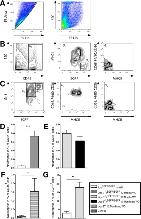
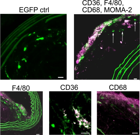
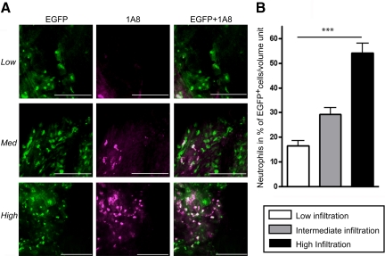
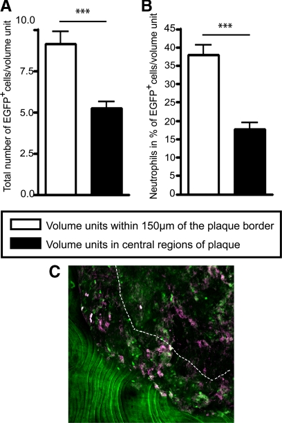
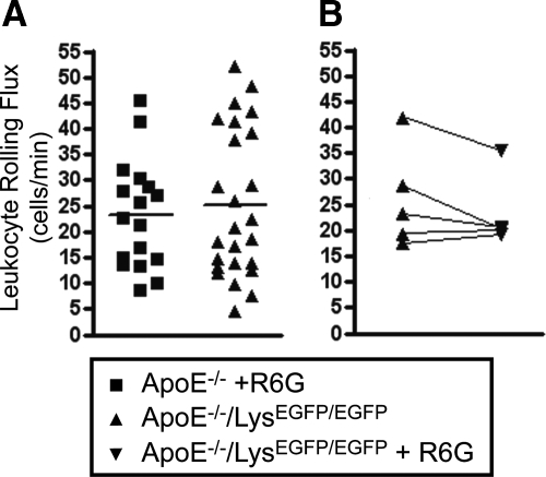
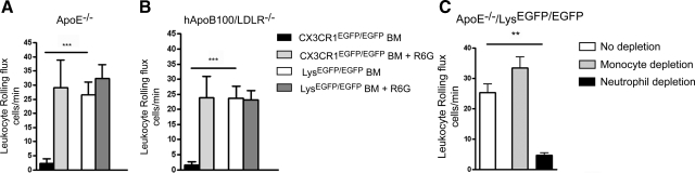
Inflammation and activation of immune cells are key mechanisms in the development of atherosclerosis. Previous data indicate important roles for monocytes and T-lymphocytes in lesions. However, recent data suggest that neutrophils also may be of importance in atherogenesis. Here, we use apolipoprotein E (ApoE)-deficient mice with fluorescent neutrophils and monocytes (ApoE(-/-)/Lys(EGFP/EGFP) mice) to specifically study neutrophil presence and recruitment in atherosclerotic lesions. We show by flow cytometry and confocal microscopy that neutrophils make up for 1.8% of CD45(+) leukocytes in the aortic wall of ApoE(-/-)/Lys(EGFP/EGFP) mice and that their contribution relative to monocyte/macrophages within lesions is approximately 1:3. However, neutrophils accumulate at sites of monocyte high density, preferentially in shoulder regions of lesions, and may even outnumber monocyte/macrophages in these areas. Furthermore, intravital microscopy established that a majority of leukocytes interacting with endothelium on lesion shoulders are neutrophils, suggesting a significant recruitment of these cells to plaque. These data demonstrate neutrophilic granulocytes as a major cellular component of atherosclerotic lesions in ApoE(-/-) mice and call for further study on the roles of these cells in atherogenesis.


Figures

Figure 1
Figure 1
Neutrophils are present in significant numbers in aortas from ApoE−/−/LysEGFP/EGFP mice. A: Representative gating algorithm with exclusion of double events (left) and FSC/SSC plotting of remaining events (right). B: Representative staining for CD45 (left), staining for MHC-II and EGFP on CD45+ cells (middle), and staining for CD68, F4/80, and CD36 on MHC-IIEGFPhighCD45+ cells (right). C: Representative staining for EGFP and Gr-1 (left) on CD45+ cells, and staining for CD68, F4/80, CD36, and MHC-II on Gr-1+EGFPlowCD45+ (middle) and Gr-1highEGFPhighCD45+ (right) cells. D–F: Contribution of neutrophils of all CD45+ cells identified by the protocol outlined in A and B in aortas from different groups of mice as indicated. G: Contribution of neutrophils of total EGFP+ cells in the indicated mice. The number of mice in each group was: ApoE−/−/LysEGFP/EGFP mice: 10 months chow, 3 mice, 12 months WD, 6 mice, 16 months WD, 4 mice. ApoE−/− mice: 12 months chow, 3 mice. LysEGFP/EGFP mice: 12 months chow, 10 mice. C57Bl/6 mice: 4 months chow, 4 mice. *P < 0.05, **P < 0.01 and ***P < 0.001.
Figure 2
Figure 2
Representative immunofluorescence images on sections from atherosclerotic lesions from ApoE−/−/LysEGFP/EGFP mice after staining for the indicated antigens. Images show overlay EGFP in green and immunofluorescence in red. In the top left, EGFP fluorescence without staining is shown. In the top right, a section from an atherosclerotic lesion stained for F4/80, CD68, CD36, and MOMA-2 is displayed. Arrows indicate EGFP+ cells negative for staining of these antigens. Bottom row shows staining for individual macrophage markers. Note the positive immunostaining for some, but not all, EGFP+ cells. Scale bar, 10 μm.
Figure 3
Figure 3
Neutrophil invasion varies between different regions of atherosclerotic lesions. A: Representative images from confocal microscopy of en face mounted atherosclerotic lesions from ApoE−/−/LysEGFP/EGFP mice after staining with the neutrophil specific mAb 1A8. Plaques were analyzed in 100×100×40 μm volume units. Volume units were characterized as low infiltration (≤5 EGFP+ cells), intermediate infiltration (6 to 15 EGFP+ cells), and high infiltration units (>15 EGFP+ cells). Scale bar, 100 μm. B: Quantification of the number of neutrophils in percentage of total EGFP+ cells in areas of different infiltration of EGFP+ cells. ***P < 0.001.
Figure 4
Figure 4
Neutrophils and monocytes invade lesion shoulders. Graphs show the quantitative contribution of EGFP+ cells (A) and relative neutrophil contribution (B) in shoulders and central regions of plaque. ***P < 0.0001. C: Representative image of an atherosclerotic lesion stained by 1A8 as Figure 3. The healthy aortic wall and the lesion border are clearly visible in the lower left. The area between the dashed line and the lesion border is 150 μm wide and represents the plaque shoulder.
Figure 5
Figure 5
Leukocytes rolling on atherosclerotic endothelium are primarily of myelomonocytic origin. A: Leukocyte rolling in rhodamine 6G (R6G)-treated ApoE−/− mice (n = 17) and untreated ApoE−/−/LysEGFP/EGFP mice (n = 25). Rolling was analyzed in a 100 × 100 μm endothelial surface area on shoulder regions of plaque (P = 0.62). B: Leukocyte rolling flux in ApoE−/−/LysEGFP/EGFP mice before and after treatment with R6G (n = 5, P = 0.59).
Figure 6
Figure 6
Neutrophils account for most leukocyte–endothelial interactions in shoulder regions on lesions. Leukocyte rolling flux in ApoE−/− mice (A) or hApoB100/LDLR−/− mice (B) after BMT with LysEGFP/EGFP (fluorescent neutrophils and monocytes, n = 6 and 4) or CX3CR1EGFP/EGFP (fluorescent monocytes, n = 7 and 4) bone marrow before and after treatment with R6G. C: Leukocyte rolling on atherosclerotic endothelium after depletion of circulating monocytes (n = 5) or neutrophils (n = 5). **P < 0.01, ***P < 0.0001.

References

    1. Hansson GK. Inflammation, atherosclerosis, and coronary artery disease. N Engl J Med. 2005;352:1685–1695. - PubMed
    1. Weber C, Zernecke A, Libby P. The multifaceted contributions of leukocyte subsets to atherosclerosis: lessons from mouse models. Nat Rev Immunol. 2008;8:802–815. - PubMed
    1. Edfeldt K, Agerberth B, Rottenberg ME, Gudmundsson GH, Wang XB, Mandal K, Xu Q, Yan ZQ. Involvement of the antimicrobial peptide LL-37 in human atherosclerosis. Arterioscler Thromb Vasc Biol. 2006;26:1551–1557. - PubMed
    1. Daugherty A, Dunn JL, Rateri DL, Heinecke JW. Myeloperoxidase, a catalyst for lipoprotein oxidation, is expressed in human atherosclerotic lesions. J Clin Invest. 1994;94:437–444. - PMC - PubMed
    1. van Leeuwen M, Gijbels MJ, Duijvestijn A, Smook M, van de Gaar MJ, Heeringa P, de Winther MP, Tervaert JW. Accumulation of myeloperoxidase-positive neutrophils in atherosclerotic lesions in LDLR-/- mice. Arterioscler Thromb Vasc Biol. 2008;28:84–89. - PubMed

Publication types

MeSH terms

Substances