. 2010 Jul 16;285(29):22676-88.
doi: 10.1074/jbc.M110.122929. Epub 2010 May 15.

Actin-depolymerizing factor cofilin-1 is necessary in maintaining mature podocyte architecture

Affiliations

Actin-depolymerizing factor cofilin-1 is necessary in maintaining mature podocyte architecture

Puneet Garg et al. J Biol Chem. .

Abstract

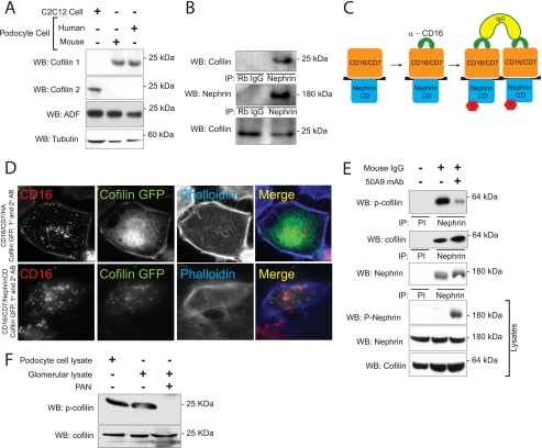
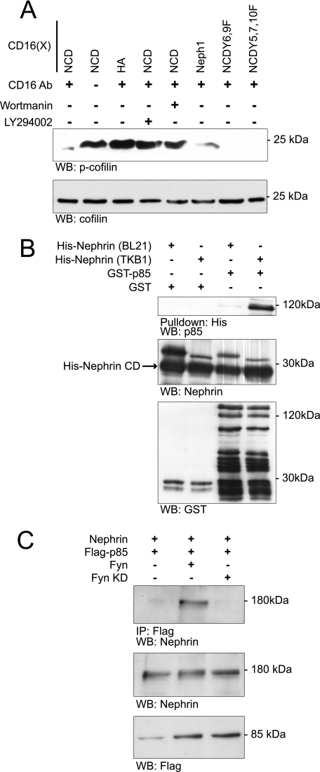
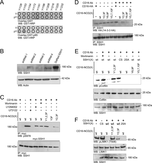
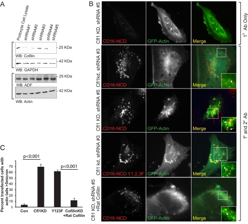
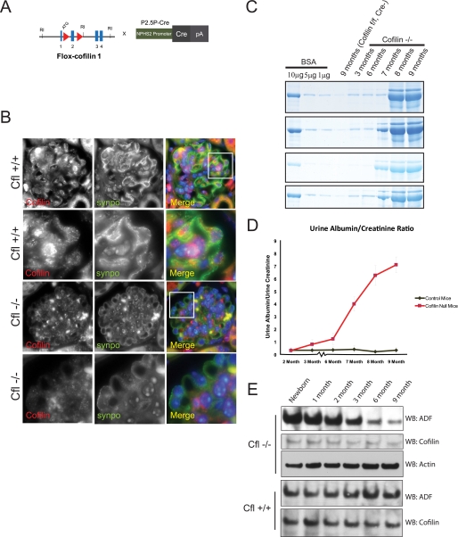
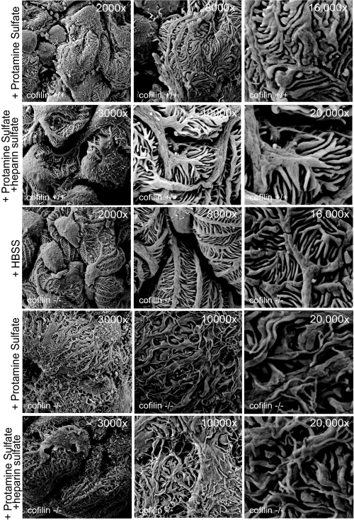
Actin dynamics determines podocyte morphology during development and in response to podocyte injury and might be necessary for maintaining normal podocyte morphology. Because podocyte intercellular junction receptor Nephrin plays a role in regulating actin dynamics, and given the described role of cofilin in actin filament polymerization and severing, we hypothesized that cofilin-1 activity is regulated by Nephrin and is necessary in normal podocyte actin dynamics. Nephrin activation induced cofilin dephosphorylation via intermediaries that include phosphatidylinositol 3-kinase, SSH1, 14-3-3, and LIMK in a cell culture model. This Nephrin-induced cofilin activation required a direct interaction between Nephrin and the p85 subunit of phosphatidylinositol 3-kinase. In a similar fashion, cofilin-1 dephosphorylation was observed in a rat model of podocyte injury at a time when foot process spreading is initially observed. To investigate the necessity of cofilin-1 in the glomerulus, podocyte-specific Cfl1 null mice were generated. Cfl1 null podocytes developed normally. However, these mice developed persistent proteinuria by 3 months of age, although they did not exhibit foot process spreading until 8 months, when the rate of urinary protein excretion became more exaggerated. In a mouse model of podocyte injury, protamine sulfate perfusion of the Cfl1 mutant mouse induced a broadened and flattened foot process morphology that was distinct from that observed following perfusion of control kidneys, and mutant podocytes did not recover normal structure following additional perfusion with heparin sulfate. We conclude that cofilin-1 is necessary for maintenance of normal podocyte architecture and for actin structural changes that occur during induction and recovery from podocyte injury.


Figures

FIGURE 1.
FIGURE 1.
Cofilin-1 is expressed in podocytes and interacts with Nephrin. A, expression of cofilin and its isoforms in cultured podocytes. Cell lysates from human and mouse podocyte immortalized cell lines were blotted with cofilin-1, cofilin-2, and ADF isoform-specific antibodies. Lysate from myoblast cell line was used as a positive control for cofilin-2. B, Nephrin and cofilin associate in vivo. Co-immunoprecipitation experiments performed on mouse glomerular lysate using antibodies against Nephrin and cofilin. C, schematic showing the CD16 clustering system. Chimeras with CD16 extracellular domain, CD7 transmembrane domain, and Nephrin cytosplasmic domain are clustered by the addition of anti-CD16 antibody and subsequently anti-mouse IgG, resulting in phosphorylation of Nephrin tyrosine residues. D, cofilin is recruited to the CD16/CD7/Nephrin CD cluster at the plasma membrane. Human podocytes expressing indicated CD16/CD7 chimeric proteins (red) and GFP-cofilin (green) were treated with anti-CD16 antibody (primary) and rhodamine-conjugated anti-IgG antibody (secondary) and then fixed and examined by confocal microscopy. CD16-HA represents a CD16/7 chimera in which the Nephrin cytoplasmic domain is replaced by an HA tag and serves as control. Data are representative of three separate experiments. Magnification was ×600. E, HEK293 cells expressing full-length human Nephrin and GFP-cofilin-1 were treated with 50A9 anti-Nephrin antibody followed by anti-mouse IgG. Lysates were immunoprecipitated with anti-Nephrin antibody and immunoblotted for phosphocofilin(phospho-Ser3). F, serine 3 phosphorylation of cofilin is attenuated in the PAN-induced podocyte injury model. Glomerular lysates from rats injected with PBS (control) or PAN were lysed and immunoblotted as shown. Immortalized human podocyte cell lysate were immunoblotted for the presence of cofilin and phosphocofilin as control. WB, Western blot; IP, immunoprecipitation; PI, preimmune serum.
FIGURE 2.
FIGURE 2.
Nephrin interacts with the p85 subunit of PI3K. A, Nephrin-mediated cofilin Ser3 dephosphorylation is abrogated by PI3K inhibitors. Human podocytes expressing indicated CD16-Nephrin cytoplasmic domain (CD16-NCD) or CD16-Neph1 cytoplasmic domain (CD16-Neph1) plasmids were pretreated with LY294002 and wortmannin before chimeric proteins were clustered and activated with anti-CD16 antibody and secondary antibody. CD16-NCD plasmids with the indicated tyrosine to phenylalanine mutations were used as controls because mutations at these sites were shown previously to interact with Src homology 2 domain adaptor proteins other than the p85 subunit of PI3K. Lysates from the experiment were divided into two equal samples and loaded on two separate gels. B, direct interaction between Nephrin and PI3K p85 subunit. Purified recombinant His-Nephrin cytoplasmic domain expressed in BL21 or TKB1 E. coli and GST-p85 were mixed and pulled down using cobalt-conjugated magnetic beads (Talon®, Invitrogen) and then immunoblotted with anti-p85 antibody. Control immunoblots demonstrate the presence of recombinant Nephrin, GST, and GST-p85. C, Nephrin interacts with p85 only in the presence of Fyn. Human podocytes expressing the indicated plasmids were lysed and immunoprecipitated with FLAG antibody and then blotted with Nephrin antibody. Lysates were also blotted to detect expression of Nephrin or FLAG-p85. Kinase-dead Fyn (Fyn KD) was used as a negative control. WB, Western blot; IP, immunoprecipitation; p-cofilin, phosphocofilin. Data are representative of four experiments.
FIGURE 3.
FIGURE 3.
Nephrin mediates cofilin dephosphorylation in a manner dependent on PI3K and Slingshot1. A, GST-p85 overlay. Arrayed Nephrin oligopeptides synthesized with and without phosphorylated tyrosines were incubated with GST-p85 and then probed with horseradish peroxidase-conjugated anti-GST antibody. B, stable knockdown of SSH1. Human podocyte cell lines were produced in which four different small hairpin RNAs were stably expressed; cells incorporating shRNA 1 (SSH1kd) were selected for additional studies. Western blot analysis examining SSH1 expression is shown. C, Nephrin ligation results in SSH1 dephosphorylation on its Ser978 residue. Human podocyte SSH1kd cells expressing Myc-SSH1 were co-transfected with plasmids encoding the indicated CD16-chimeric proteins. Where indicated, cells were pretreated with PI3K inhibitors LY294002 (lane 4) or wortmannin (lane 5). Phospholipase C inhibitor U73122 (lane 3) was used as a positive control because phospholipase activity is necessary for release of activated cofilin from a plasma membrane sequestered pool. Except where indicated, cells were treated with anti-CD16 antibody and anti-mouse IgG secondary antibody. Cell lysates were immunoprecipitated with Myc antibody and blotted with phospho-Ser978-specific antibody. Lysates from the experiment were divided into two equal samples and loaded on two separate gels. D, Nephrin clustering abrogates association between SSH1 and 14-3-3. Human podocytes expressing HA-tagged 14-3-3ζ were co-transfected with the indicated plasmids encoding CD16-Nephrin or its tyrosine mutants or CD16-HA, and cells were treated as indicated with clustering antibody. CD16-HA represents a CD16/7 chimera in which the Nephrin cytoplasmic domain is replaced by an HA tag and serves as control. Cell lysates were immunoprecipitated using SSH1 antibody and blotted with indicated antibodies. E, Nephrin mediates cofilin Ser3 dephosphorylation via PI3K and SSH1. Human podocyte SSH1kd cells expressing the indicated proteins were clustered using the CD16 antibody. Lysates were resolved using SDS-PAGE and blotted with indicated antibodies. SSH1(CS), phosphatase-dead mutant; SSH1(2SA), SSH1(S937A/S978A); wt, wild type SSH1. F, Nephrin-mediated SSH1 activation results in simultaneous LIMK1 dephosphorylation. SSH1kd cells expressing the indicated plasmids were clustered using the anti-CD16 antibody. Cell lysates were examined by Western blotting using the indicated antibodies. IP, immunoprecipitation; WB, Western blot; WT, wild type. Data are representative of three separate experiments.
FIGURE 4.
FIGURE 4.
Cofilin knockdown results in longer actin tails. A, stable knockdown of cofilin-1. Human podocyte cell lines were produced in which five different small hairpin RNA oligonucleotides were stably expressed; cells incorporating shRNA (Cfl1kd) were selected for additional study. After protein estimation, lysates were blotted for cofilin and ADF on two separate gels. Glyceraldehyde-3-phosphate dehydrogenase (GAPDH) and actin were used as loading controls for cofilin and ADF, respectively. B, human podocytes expressing the indicated plasmids were clustered as described and examined using confocal microscopy. The arrows indicate actin tails. Cells in the top panel were not treated with the secondary antibody. The rest of the panels show cells treated with both the primary anti-CD16 antibody and the anti-mouse IgG secondary antibody. C, percentage of cells with identifiable actin tails longer than 5 μm (p < 0.001). Cfl1KD, podocytes with stable knockdown using clone 5. As a control (Con), podocytes with infected with shRNA 3 was used. Image J software (National Institutes of Health) was used to determine the length of tail. Approximately 100 cells were analyzed in each condition. Data are representative of five separate experiments. Error bars, S.E.
FIGURE 5.
FIGURE 5.
Selective deletion of cofilin-1 in mouse podocyte. A, cofilin-flox mice with loxP sites flanking exon 2 were bred with NPHS2-Cre mice to generate conditional knockdown of cofilin-1 in podocytes. B, double immunofluorescence of mouse kidney section. Paraffin-embedded mouse kidney sections from Cfl1+/+ and Cfl1−/− mice were double-stained with cofilin-1 (red) and synaptopodin (green), showing podocyte specific deletion of Cfl1. Nuclei were stained with 4′,6-diamidino-2-phenylindole (DAPI). Rows 2 and 4 show higher magnification of the area in the white square. C, urine protein. Urine was collected from the same mouse for the indicated times. The indicated concentrations of bovine serum albumin (BSA) were run as a quantitative measure. One microliter of urine was run in each lane. Gel was stained using SimplyBlueTM Safe Stain. Data from four different Cfl1−/− and Cfl+/+ mice are shown. D, urine protein/creatinine ratio of Cfl−/− and Cfl+/+ mice indicating the appearance of proteinuria by 3 months of age in Cfl−/− mice. E, ADF and cofilin expression in glomerular lysates for Cfl1−/− and Cfl+/+ mice. Glomerular lysates from mouse kidneys were obtained at the indicated time points. Lysates were loaded equally after protein estimation. WB, Western blot. Error bars, S.E.
FIGURE 6.
FIGURE 6.
Podocyte-specific Cfl1−/− mice develop foot process spreading by 8 months of age. A, scanning EM of mouse kidney sections at the indicated postnatal age. There is evidence of foot process spreading by 8 months of age. There is evidence of fine finger-like projections from the podocyte cell bodies and processes at 6 months of age. B, transmission EM of Cfl1−/− and Cfl+/+ mice at 9 months. C, slit diaphragm frequency in wild type and Cfl−/− mice at the indicated ages. D, histologically, hematoxylin and eosin staining of kidney sections from Cfl1−/− and Cfl+/+ mice show no evidence of scarring even at 9 months of age, when the mouse are significantly proteinuric. E, serum creatinine concentration in mutant mice compared with control. NS, not significant. Error bars, S.E.
FIGURE 7.
FIGURE 7.
Cfl1 is necessary for restoration of podocyte morphology following injury with protamine sulfate. Three-month-old Cfl1−/− and Cfl+/+ mice were injected with HBSS and protamine sulfate where indicated. The bottom panel shows lack of recovery following heparin sulfate infusion. Data are representative of four or more mice for each experimental condition.

References

    1. Kestilä M., Lenkkeri U., Männikkö M., Lamerdin J., McCready P., Putaala H., Ruotsalainen V., Morita T., Nissinen M., Herva R., Kashtan C. E., Peltonen L., Holmberg C., Olsen A., Tryggvason K. (1998) Mol. Cell 1, 575–582 - PubMed
    1. Kim J. M., Wu H., Green G., Winkler C. A., Kopp J. B., Miner J. H., Unanue E. R., Shaw A. S. (2003) Science 300, 1298–1300 - PubMed
    1. Winn M. P., Conlon P. J., Lynn K. L., Farrington M. K., Creazzo T., Hawkins A. F., Daskalakis N., Kwan S. Y., Ebersviller S., Burchette J. L., Pericak-Vance M. A., Howell D. N., Vance J. M., Rosenberg P. B. (2005) Science 308, 1801–1804 - PubMed
    1. Brown E. J., Schlöndorff J. S., Becker D. J., Tsukaguchi H., Uscinski A. L., Higgs H. N., Henderson J. M., Pollak M. R., Tonna S. J. (2010) Nat. Genet. 42, 72–76 - PMC - PubMed
    1. Kaplan J. M., Kim S. H., North K. N., Rennke H., Correia L. A., Tong H. Q., Mathis B. J., Rodríguez-Pérez J. C., Allen P. G., Beggs A. H., Pollak M. R. (2000) Nat. Genet. 24, 251–256 - PubMed

Publication types

MeSH terms

LinkOut - more resources