. 2010 Jul 23;285(30):23296-308.
doi: 10.1074/jbc.M109.098129. Epub 2010 May 15.

Serine 34 phosphorylation of rho guanine dissociation inhibitor (RhoGDIalpha) links signaling from conventional protein kinase C to RhoGTPase in cell adhesion

Affiliations

Serine 34 phosphorylation of rho guanine dissociation inhibitor (RhoGDIalpha) links signaling from conventional protein kinase C to RhoGTPase in cell adhesion

Athanassios Dovas et al. J Biol Chem. .

Abstract

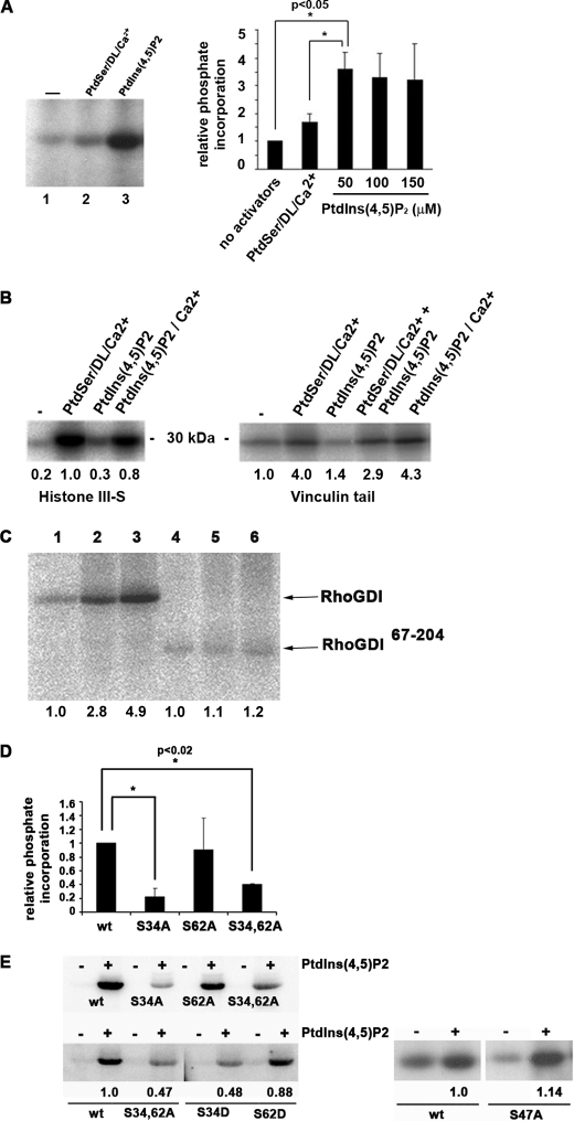
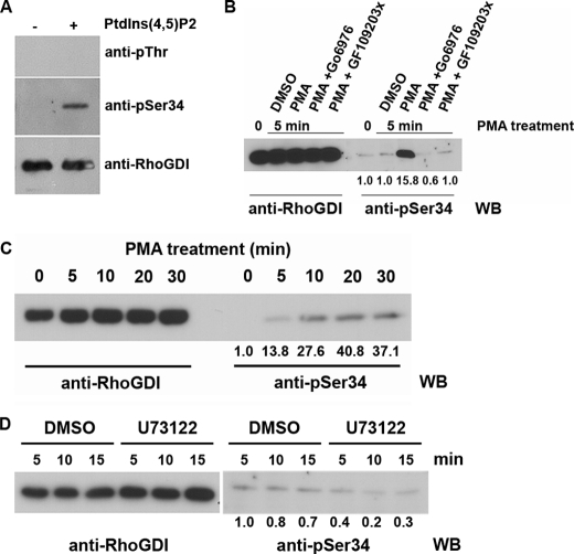
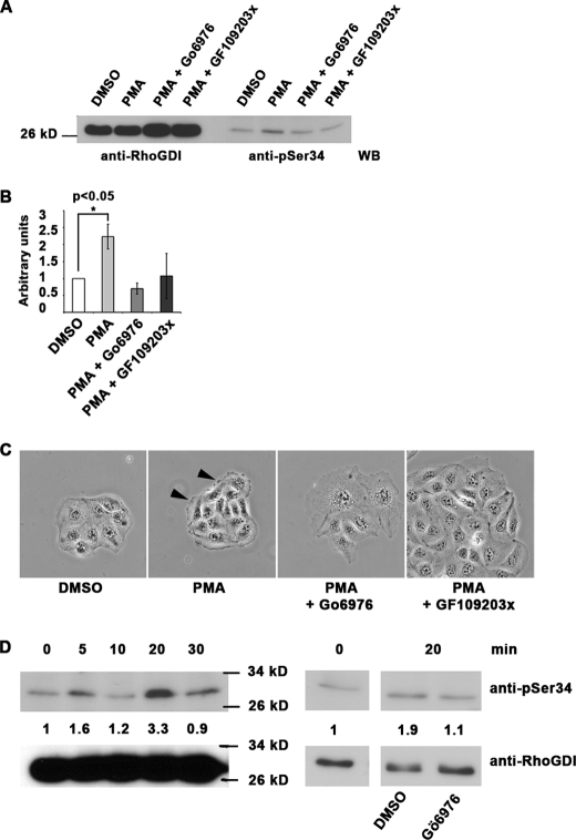
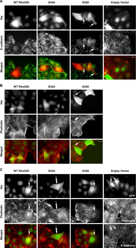
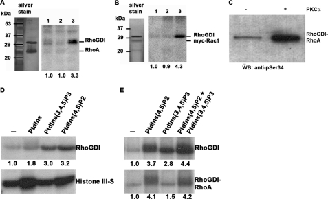
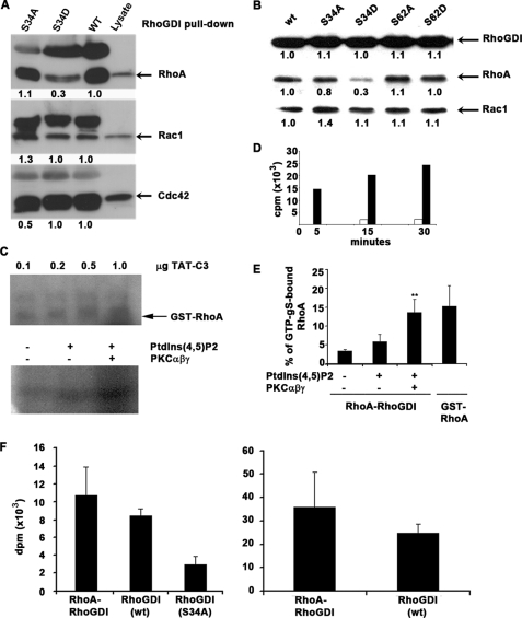
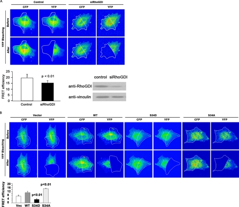
Conventional protein kinase C (PKC) isoforms are essential serine/threonine kinases regulating many signaling networks. At cell adhesion sites, PKCalpha can impact the actin cytoskeleton through its influence on RhoGTPases, but the intermediate steps are not well known. One important regulator of RhoGTPase function is the multifunctional guanine nucleotide dissociation inhibitor RhoGDIalpha that sequesters several related RhoGTPases in an inactive form, but it may also target them through interactions with actin-associated proteins. Here, it is demonstrated that conventional PKC phosphorylates RhoGDIalpha on serine 34, resulting in a specific decrease in affinity for RhoA but not Rac1 or Cdc42. The mechanism of RhoGDIalpha phosphorylation is distinct, requiring the kinase and phosphatidylinositol 4,5-bisphosphate, consistent with recent evidence that the inositide can activate, localize, and orient PKCalpha in membranes. Phosphospecific antibodies reveal endogenous phosphorylation in several cell types that is sensitive to adhesion events triggered, for example, by hepatocyte growth factor. Phosphorylation is also sensitive to PKC inhibition. Together with fluorescence resonance energy transfer microscopy sensing GTP-RhoA levels, the data reveal a common pathway in cell adhesion linking two essential mediators, conventional PKC and RhoA.


Figures

FIGURE 1.
FIGURE 1.
RhoGDIα is a substrate for PKCα on serine 34, in a PtdIns(4,5)P2-dependent manner. A, phosphorylation of RhoGDI by PKCαβγ. Kinase activators were phosphatidylserine/diolein/calcium or phosphatidylinositol 4,5-bisphosphate in the presence of radiolabeled ATP followed by SDS-PAGE and autoradiography. The graph shows quantitation of RhoGDIα phosphorylation by PKCα with increasing inositide levels. The mean ± S.D. of three independent experiments is shown. Phosphorylation in the presence of 50 μm PtdIns(4,5)P2 is significantly higher than that without activators or PtdSer/diolein/Ca2+ (*, p < 0.05). B, phosphorylation of histone IIIS (left) and vinculin tail (right) by recombinant PKCα in the presence of the shown activators and [32P]ATP. Proteins were resolved by SDS-PAGE and autoradiography. Unlike RhoGDIα, both substrates were phosphorylated inefficiently by PKCα in the presence of PtdIns(4,5)P2 unless calcium ions were present (25 μm). Mean values from triplicate experiments are shown. C, although wild-type RhoGDIα was phosphorylated by recombinant PKCα (lanes 1–3), a truncated form consisting of the immunoglobulin domain alone (residues 67–204; lanes 4–6) was not. Lanes 1 and 4, no activators; lanes 2 and 5, PtdSer/diolein/calcium; lanes 3 and 6, PtdIns(4,5)P2. Quantitation is shown, normalized to lanes 1 (for lanes 1–3) and lane 4 (for lanes 4–6). D, phosphorylation of RhoGDIα by PKCαβγ is mostly abrogated by a serine-alanine mutation at residue 34, but a similar mutation of serine 62 has no effect. A representative autoradiogram is shown, together with quantitation by a phosphorimager from triplicate experiments (mean ± S.D., *, p < 0.02 compared with wild type). wt = wild-type RhoGDIα. E, mutation of serine 34 to aspartate also reduced RhoGDIα phosphorylation by PKCαβγ, although an equivalent mutation of serine 62 had no effect. Quantitation from autoradiograms is shown below. S34A/S62A (S34,62A) is a double alanine mutant. Although serine 47 is a potential PKC phosphorylation target, its mutation to alanine had no effect on overall RhoGDIα phosphorylation.
FIGURE 2.
FIGURE 2.
Phosphorylation of RhoGDIα on serine 34 in cells occurs in a PKC-dependent manner. A, 25 ng of in vitro phosphorylated RhoGDIα was immunoblotted with a phosphothreonine antibody, an affinity-purified phosphospecific antibody (pSer-34), and an antibody against total RhoGDIα. B and C, K562 cells undergo rapid phosphorylation of RhoGDIα on serine 34 when treated with 800 nm PMA to promote cell adhesion but not if pretreated with 1 μm PKCα/β inhibitor (Gö69796) or a more general PKC inhibitor (GF109203X). C, phosphorylation is shown to persist for at least 30 min after 800 nm PMA treatment. Total RhoGDIα in the cell lysates is shown on the left as loading controls. Quantitation of the phosphorylation from triplicate Western blots (WB) is shown. D, RhoGDIα phosphorylation in rat embryo fibroblasts is not eliminated by inhibition of phospholipase Cγ. Cells were treated with 3 μm U73122 (or DMSO as vehicle control) for 5, 10, or 15 min, and phosphorylated RhoGDIα was detected by Western blotting with the phosphospecific antibody. Quantitation of triplicates is shown below the blots, and total RhoGDIα is shown in the left 6 lanes.
FIGURE 3.
FIGURE 3.
Serine 34 phosphorylation of RhoGDIα is a regulator of PKC-mediated adhesion modulation in MDCK cells. A–C, 10-min PMA treatment (200 nm) of MDCK cells leads to phosphorylation of RhoGDIα on serine 34 that is inhibited by 1 μm Gö6976 PKCαβ inhibitor or a broader spectrum PKC inhibitor (GF109203X). Total RhoGDIα is shown in the four left lanes. Quantitation of triplicates is shown in B, ± S.D. Phase contrast images (C) show the characteristic ruffling induced by 200 nm PMA (arrowheads), but this was not seen where PKC inhibitors (1 μm) were present. Scale bar, 50 μm. D, MDCK cells were treated with 50 ng/ml HGF for 0–30 min, and serine 34 phosphorylation of RhoGDIα was quantitated. Molecular mass marker positions are shown. On the right, sensitivity of this phosphorylation to PKCαβ inhibition at the 20-min time point is shown. Quantitation data for the experiment shown are presented but represent one of three repeated experiments. WB, Western blot.
FIGURE 4.
FIGURE 4.
RhoGDIα status regulates MDCK cytoskeletal organization. MDCK epithelial cells were transfected with empty vector or RhoGDI cDNAs encoding His-tagged wild-type (WT RhoGDI), S34A, or S34D mutations. Transfected cells were detected by His antibodies or green fluorescent protein co-transfected with the empty vector. Cultures were stained for E-cadherin as a marker of adherens junctions (A and control in C) or phalloidin for F-actin (B and C). Micrographs in C show cultures treated for 10 min with 200 nm phorbol ester, which triggers loss of adherens junctions and reorganization of the actin cytoskeleton, in a Rho-dependent manner. In each case single fluorescence and merged images are shown. A, E-cadherin is abundant in cell-cell contact sites in all cases except S34D, where the cells exhibit protrusions (arrows) and do not have extensive E-cadherin on their surface. Staining for the actin cytoskeleton (B) shows that all transfected and untransfected cells have some F-actin containing microfilament bundles. Protrusions in S34D cells are labeled with arrows. In phorbol ester treated cultures (C), microfilament bundles are lost or are condensed into small aggregates (arrows) in all cases except S34A cells, where bundles persist (double arrows). Increased cell spreading is also characteristic of phorbol ester-treated cells, again not seen in the S34A transfected cells. Scale bar, 10 μm.
FIGURE 5.
FIGURE 5.
RhoGDIα can be phosphorylated on serine 34 when complexed with RhoA or Rac1. A and B, RhoGDIα was prepared in complex with either RhoA or Rac1 (0.5 μg/assay) and subjected to phosphorylation by 3 ng of PKCα for 10 min. In both figures, a silver-stained SDS-polyacrylamide gel is shown on the left and an autoradiograph on the right. The positions of GDI and GTPase are marked. Lanes 1, no PKC activators; lanes 2, PtdSer/diolein/calcium (250/32/750 μm); and lanes 3, PtdIns(4,5)P2 (50 μm). Neither GTPase is phosphorylated detectably by PKCα. C, RhoGDIα complexed with RhoA (0.5 μg of protein) was mixed with (50 μm) PtdIns(4,5)P2 ± 3 ng of PKCα for 10 min and in a Western blot (WB) by a phosphospecific antibody recognizing Ser(P)-34 RhoGDI. The result shows that the phosphospecific antibody can recognize the RhoGDI even when complexed with RhoA. D and E, three inositides (50 or 25 μm of each where two were mixed) were compared as PKCαβγ activators for RhoGDIα, histone III-S. or RhoA/RhoGDIα complex phosphorylation. For complexed RhoGDIα, PtdIns(4,5)P2 was superior, with no additive effect of PtdIns(3,4,5)P3, although histone III-S phosphorylation was increased by all the inositides. Quantitation is shown below the autoradiograms.
FIGURE 6.
FIGURE 6.
Phosphomimetic RhoGDIα at residue 34 leads to decreased retention of RhoA but no change in binding of Rac1 or Cdc42. A, wild type (WT) and RhoGDIα mutated to alanine (S34A) or aspartate (S34D) were compared in GTPase pulldown assays with lysate from rat embryo fibroblasts. In each Western blot, the right lane shows the GTPase detected in the lysate. Quantitation shows that phosphomimetic RhoGDIα has decreased ability to bind RhoA, although binding to Rac1 and Cdc42 is unaffected. Nonspecifically reacting polypeptides are present in the upper portion of each Western blot. B, serine 34 and 62 mutants were compared in their abilities to bind RhoA or Rac1 from rat embryo fibroblast lysates. Although mutation of serine 62 to alanine (S62A) or aspartate (S62D) had no impact on GTPase binding, mutation of serine 34 to aspartate (S34D) reduced RhoA binding specifically. The corresponding alanine mutant at serine 34 (S34A) bound both GTPases similarly to the wild-type (wt) RhoGDIα. Quantitation of the Western blots is shown. C, C3 transferase ADP ribosylates RhoA once the RhoGDIα-RhoA complex has been phosphorylated by PKCαβγ. Varying amounts of TAT-C3 transferase were added to GST-RhoA (upper panel). In the lower panel, 500 ng of RhoA in complex with RhoGDIα were untreated (−) or incubated with 50 μmol of PtdIns(4,5)P2 ± PKCαβγ (lower panel). In both cases, 1 μCi of [32P]NAD was included in a 5-min reaction at room temperature, and autoradiographs are shown from one of four independent experiments. D, exchange of GDP for GTP in RhoA is high in free GTPase but low when complexed to RhoGDIα. Ten pmol of RhoA either alone (black bars) or complexed to RhoGDIα (white bars) was incubated with 30 pmol (0.5 μCi) of GTPγS for the indicated times, and radiolabel incorporation into the GTPase was measured by liquid scintillation spectroscopy. E, phosphorylation of RhoGDIα in complex with RhoA-GDP by PKCαβγ, in the presence of 50μmol PtdIns(4,5)P2, leads to increased nucleotide exchange for GTP. PKC phosphorylation of 10 pmol of RhoGDIα-RhoA complex for 1 h at room temperature was followed by a GTP exchange reaction in the presence of radiolabeled (0.5 μCi) GTPγS. The results are shown as mean ± S.D. from three independent experiments. ** Significantly more nucleotide exchange occurs with RhoGDIα phosphorylation (p < 0.05) than without. F, stoichiometry of RhoGDIα phosphorylation. Left panel, 10 pmol of wild-type RhoGDI complexed with RhoA (left), wild-type RhoGDI (center), or S34A RhoGDI alone (right) were incubated with 10 ng of PKCαβγ and 50 μmol of PtdIns(4,5)P2 for 1 h at room temperature, and radiolabel incorporation was measured. Values are mean ± S.D. from three experiments. Right panel, data from the left panel were recalculated to show stoichiometry of Ser-34 phosphorylation, subtracting background (S34A RhoGDI values) ± S.D. There was no significant difference in phosphorylation whether the RhoGDI was complexed with RhoA or not.
FIGURE 7.
FIGURE 7.
Serine 34 of RhoGDIα in fibroblasts regulates RhoA-GTP levels. A, rat embryo fibroblasts were cotransfected with pRaichu 1502 and either control siRNA or siRNA oligonucleotides for RhoGDIα. After 48 h, cells were lysed and immunoblotted with anti-RhoGDI antibody. Vinculin was used as a loading control. FRET efficiency was measured after acceptor photobleaching and was quantitated (n = 20). Efficiency was reduced after reduction in RhoGDI protein levels, indicative of higher GTP-RhoA levels. B, rat fibroblasts with endogenous levels of RhoGDIα reduced by siRNA were co-transfected with pRaichu1502 and one of His vector (Vec), His-tagged RhoGDI (WT), or His-tagged RhoGDI mutants (S34D or S34A). FRET efficiency was measured after acceptor photobleaching, and quantitation is shown (n = 20). Expression of phosphomimetic RhoGDIα leads to decreased FRET efficiencies and therefore elevated RhoGTP levels. The converse was measured after expression of the nonphosphorylatable S34A mutant. p < 0.01 indicates a FRET signal significantly different to the vector only control (t test).

References

    1. Ivaska J., Kermorgant S., Whelan R., Parsons M., Ng T., Parker P. J. (2003) Biochem. Soc. Trans. 31, 90–93 - PubMed
    1. Larsson C. (2006) Cell. Signal. 18, 276–284 - PubMed
    1. Suzuki A., Ohno S. (2006) J. Cell Sci. 119, 979–987 - PubMed
    1. Reyland M. E. (2009) Front. Biosci. 14, 2386–2399 - PMC - PubMed
    1. Mochly-Rosen D., Gordon A. S. (1998) FASEB J. 12, 35–42 - PubMed

Publication types

Substances