. 2010 Sep;51(9):2672-85.
doi: 10.1194/jlr.M007435. Epub 2010 May 15.

ABCA1 promotes the efflux of bacterial LPS from macrophages and accelerates recovery from LPS-induced tolerance

Affiliations

ABCA1 promotes the efflux of bacterial LPS from macrophages and accelerates recovery from LPS-induced tolerance

Patricia A Thompson et al. J Lipid Res. 2010 Sep.

Abstract

Macrophages play important roles in both lipid metabolism and innate immunity. We show here that macrophage ATP-binding cassette transporter A1 (ABCA1), a transporter known for its ability to promote apolipoprotein-dependent cholesterol efflux, also participates in the removal of an immunostimulatory bacterial lipid, lipopolysaccharide (LPS). Whereas monocytes require an exogenous lipoprotein acceptor to remove cell-associated LPS, macrophages released LPS in the absence of an exogenous acceptor by a mechanism that was driven, in part, by endogenous apolipoprotein E (apoE). Agents that increased ABCA1 expression increased LPS efflux from wild-type but not ABCA1-deficient macrophages. Preexposure of peritoneal macrophages to LPS for 24 h increased the expression of ABCA1 and increased LPS efflux with a requirement for exogenous apolipoproteins due to suppression of endogenous apoE production. In contrast, LPS preconditioning of ABCA1-deficient macrophages significantly decreased LPS efflux and led to prolonged retention of cell-surface LPS. Although the initial response to LPS was similar in wild-type and ABCA1-deficient macrophages, LPS-induced tolerance was greater and more prolonged in macrophages that lacked ABCA1. Our results define a new role for macrophage ABCA1 in removing cell-associated LPS and restoring normal macrophage responsiveness.


Figures

Fig. 1.
Fig. 1.
Macrophage differentiation increases LPS efflux in human macrophages: role of apolipoproteins. A: The release of [3H]LPS from the indicated cells into SFM-containing culture supernatants was measured after 15–60 min at 37°C as described in “Materials and Methods.” Data from several experiments performed in duplicate are shown as mean ± 95% CI (n = 2–18 for each time point). Asterisks denote significant differences from monocytes of each cell type at each time point. B: The release of [3H]LPS from cultured human macrophages was measured after 15 min in SFM in the presence of anti-apoE or control antibodies as described in “Materials and Methods.” In two independent experiments, the control cells released 33% and 47% of their cell-associated LPS, respectively. Error bars denote mean ± SD. Asterisks denote significant differences from the no antibody (No Ab) (***P = 0.0001) and control antibody (Ctrl Ab) (***P = 0.0003) groups. C: ApoE expression in cultured human macrophages was inhibited by apoE siRNA as described in “Materials and Methods.” ApoE protein was measured in the culture supernatants by ELISA, and [3H]LPS efflux was measured as described in (B). The data are expressed as the percent of LPS or apoE released by the cells that received control siRNA (Ctrl siRNA). Data from two (LPS) or three (apoE) independent experiments are shown as mean ± SD. Asterisks denote significant differences from the controls (***P < 0.001). The control cells released 29% and 46% of their cell-associated LPS in the respective experiments. D: The release of [3H]LPS from THP-1 macrophages was measured as described in “Materials and Methods” after incubating the cells for 30 min in SFM containing the indicated concentrations of purified apolipoproteins. Data from several experiments are shown as mean ± SD (n = 2–8 for each time point). Asterisks denote significant differences at each time point from cells that were incubated with apoE (*P < 0.05, **P < 0.01, ***P < 0.001). apo, apolipoprotein; LPS, lipopolysaccharide; SFM, serum-free medium; siRNA, small inhibitory RNA.
Fig. 2.
Fig. 2.
ABCA1 promotes LPS efflux from macrophages. Peritoneal macrophages from WT (ABCA1+/+) (A, C, E) and ABCA1-deficient (ABCA1−/−)(B, D, F) mice were incubated for 16–24 h in medium containing lipoprotein-deficient FBS to minimize LXR activation in the control (Ctrl) group; the medium also contained 5 µM T0901317 or vehicle (Ctrl). A, B: LPS efflux was determined by measuring the release of [3H]LPS into the culture supernatant as described in “Materials and Methods.” Protease-resistant (cell-surface) [3H]LPS (C, D) and protease-insensititve [3H]LPS (principally internalized LPS) (E, F) were measured as described in “Materials and Methods.” Data were compiled from several experiments and are shown as the percentage of the total initial cell-associated [3H]LPS. Error bars denote mean ± SEM (n = 4–8). Asterisks denote significant differences from the controls (*P < 0.05, **P < 0.01, ***P < 0.001). LPS, lipopolysaccharide; LXR, liver X receptor; WT, wild-type.
Fig. 3.
Fig. 3.
LPS preconditioning of macrophages increases ABCA1-dependent LPS efflux into exogenous apolipoproteins. Peritoneal macrophages from ABCA1+/+ and ABCA1−/− mice were preincubated for 24 h in complete medium containing 10% FBS without (NA) or with unlabeled LPS (preincubation). The cells were allowed to take up [3H]LPS for 10 min in the presence of 10% FBS and were then washed and incubated in SFM with or without apoE (5 µg/ml) for 60 min (assay). (A) LPS efflux, (B) cell-surface LPS, and (C) internalized LPS were measured as described in Fig. 2 and expressed as percentage of the total initial cell-associated LPS. Error bars denote mean ± SD for 3–4 experiments performed in duplicate. Asterisks denote significant differences as indicated (*P < 0.05, **P < 0.01, and ***P < 0.001). apo, apolipoprotein; LPS, lipopolysaccharide; SFM, serum-free medium.
Fig. 4.
Fig. 4.
LPS preconditioning of macrophages increases cellular ABCA1 and decreases cellular apoE. Peritoneal macrophages from ABCA1+/+ (A, B, C) and (ABCA1−/−) (C) mice were cultured overnight in medium containing 10% FBS in the presence or absence of 5 µM T0901317, where indicated, followed by the addition of LPS (10–100 ng/ml) for the indicated times. Total membrane fractions were prepared from cell lysates, and equal amounts of membrane protein were run on SDS-PAGE gels. The proteins were transferred and immunoblotted with antibodies to ABCA1 or apoE as described in “Materials and Methods.” Each panel represents a separate experiment derived from WT C57Bl/6 macrophages (A, B) and B6:129Sv ABCA1+/+ and ABCA1−/− macrophages (C). apo, apolipoprotein; LPS, lipopolysaccharide; WT, wild-type.
Fig. 5.
Fig. 5.
Diminished mRNA responses in LPS-challenged ABCA1−/− macrophages after LPS preconditioning. ABCA1+/+ and ABCA1−/− macrophages were preincubated in complete medium with (pre-cond) or without (naïve) 10 ng/ml LPS for 24 h as described in “Materials and Methods.” After washing, the cells were challenged with 10 ng/ml LPS for 2 h. mRNA levels were measured by real time PCR and normalized with 18S rRNA as described in “Materials and Methods.” Data for IL-1β (A) and IL-12a (B) are expressed as the fold change from the unchallenged naïve ABCA1+/+ cells. Ratios (ABCA1−/− to ABCA1+/+) of mRNA levels are shown above the bars. The responses of the LPS-preconditioned cells expressed as percentages of the naïve cell responses are shown on the right of each graph (Pre-cond/naïve). Error bars represent mean ± SEM of three experiments performed in duplicate representing a total of six ABCA1+/+ and seven ABCA1−/− animals. Significant differences between ABCA1−/− and ABCA1+/+ groups are denoted (*P < 0.05, **P < 0.01, ***P < 0.001). IL, interleukin; LPS, lipopolysaccharide.
Fig. 6.
Fig. 6.
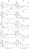
Delayed recovery from LPS-induced tolerance in ABCA1-deficient macrophages (mRNA responses). To measure the time course of recovery of LPS responsiveness after preconditioning, the experiments were performed as described in Fig. 5, except that the cells were washed twice after the 24 h LPS preincubation and incubated in fresh medium for 0–5 days before LPS challenge. The responses of the LPS-preconditioned cells are expressed as percentages of the naïve cell responses (Pre-cond/naïve) are represented as described in Fig. 5. Genes that were tolerized on day 0 are shown in column A, and nontolerized genes are shown in column B. COX-2, cyclooxygenase-2; Fpr1, formyl peptide receptor 1; IL, interleukin; LPS, lipopolysaccharide; MCP-1, monocyte chemotactic protein-1; TNF, tumor necrosis factor.
Fig. 7.
Fig. 7.
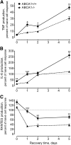
Delayed recovery from LPS-induced tolerance in ABCA1-deficient macrophages (cytokine production). ABCA1+/+ and ABCA1−/− macrophages were preincubated for 24 h with or without LPS as described in Fig. 5, washed twice, incubated in fresh complete medium for 0–5 days, and challenged with 10 ng/ml LPS for 6 h. TNF (A), IL-6 (B), and RANTES (C) were measured in the culture supernatants by ELISA as ng/100 µg total cellular protein. The responses of the LPS-preconditioned cells are expressed as percentages of the naïve cell responses. The ABCA1+/+ data were derived from 15 determinations from a total of 11 mice, and the ABCA1−/− data were from 8 determinations from 8 mice (cells from more than one mouse were mixed in some experiments). Error bars denote mean ± SEM. Asterisks denote significant differences between the groups at each time point (*P < 0.05, **P < 0.01, ***P < 0.001). IL, interleukin; LPS, lipopolysaccharide; TNF, tumor necrosis factor.

References

    1. Tang C., Oram J. F. 2009. The cell cholesterol exporter ABCA1 as a protector from cardiovascular disease and diabetes. Biochim. Biophys. Acta. 1791: 563–572. - PubMed
    1. Yvan-Charvet L., Wang N., Tall A. R. 2010. Role of HDL, ABCA1, and ABCG1 transporters in cholesterol efflux and immune responses. Arterioscler. Thromb. Vasc. Biol. 30: 139–143. - PMC - PubMed
    1. Francone O. L., Royer L., Boucher G., Haghpassand M., Freeman A., Brees D., Aiello R. J. 2005. Increased cholesterol deposition, expression of scavenger receptors, and response to chemotactic factors in Abca1-deficient macrophages. Arterioscler. Thromb. Vasc. Biol. 25: 1198–1205. - PubMed
    1. Koseki M., Hirano K. I., Masuda D., Ikegami C., Tanaka M., Ota A., Sandoval J. C., Nakagawa-Toyama Y., Sato S. B., Kobayashi T., et al. 2007. Increased lipid rafts and accelerated lipopolysaccharide-induced tumor necrosis factor-{alpha} secretion in Abca1-deficient macrophages. J. Lipid Res. 48: 299–306. - PubMed
    1. Yvan-Charvet L., Welch C., Pagler T. A., Ranalletta M., Lamkanfi M., Han S., Ishibashi M., Li R., Wang N., Tall A. R. 2008. Increased inflammatory gene expression in ABC transporter-deficient macrophages: free cholesterol accumulation, increased signaling via toll-like receptors, and neutrophil infiltration of atherosclerotic lesions. Circulation. 118: 1837–1847. - PMC - PubMed

Publication types

MeSH terms

LinkOut - more resources