. 2010 Jul;16(7):767-73.
doi: 10.1038/nm.2157. Epub 2010 May 16.

Rtp801, a suppressor of mTOR signaling, is an essential mediator of cigarette smoke-induced pulmonary injury and emphysema

Affiliations

Rtp801, a suppressor of mTOR signaling, is an essential mediator of cigarette smoke-induced pulmonary injury and emphysema

Toshinori Yoshida et al. Nat Med. 2010 Jul.

Abstract

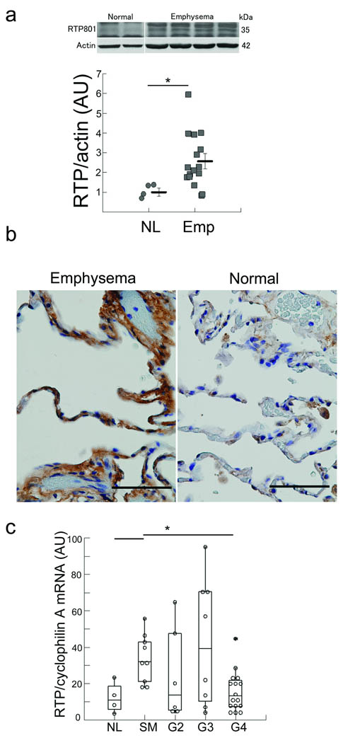
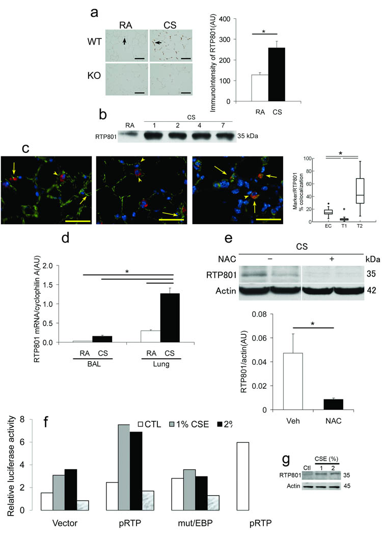
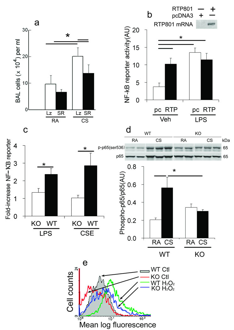
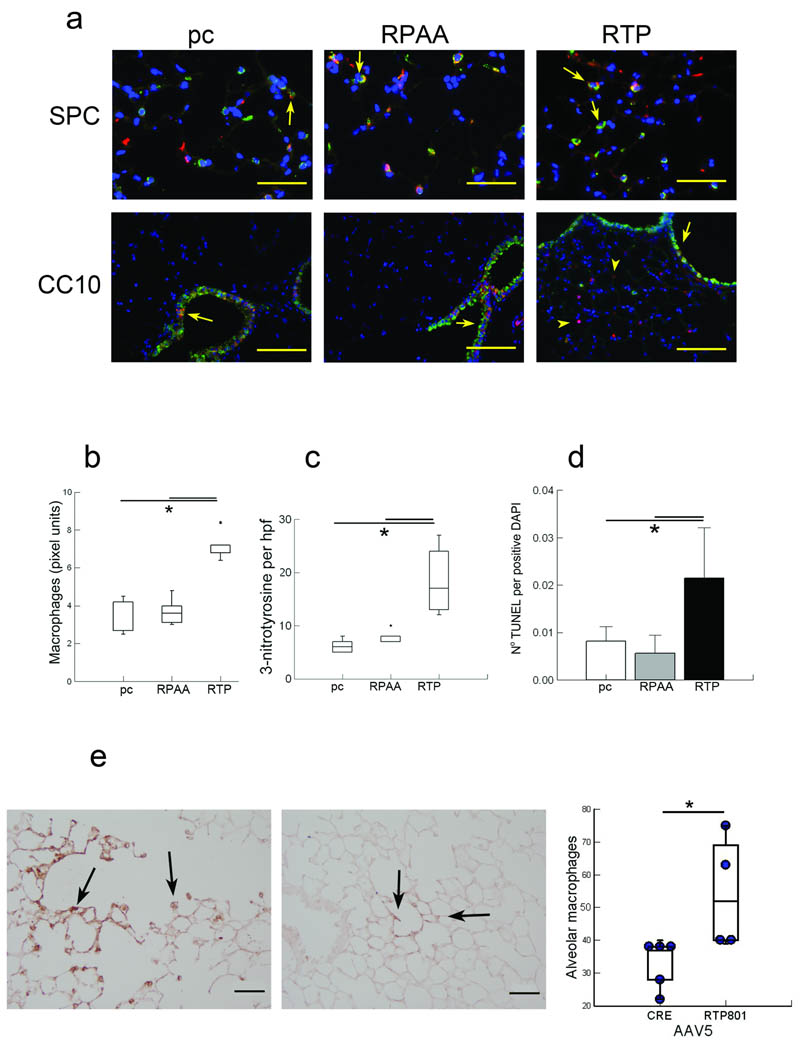
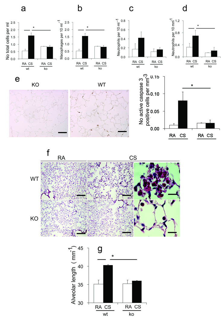
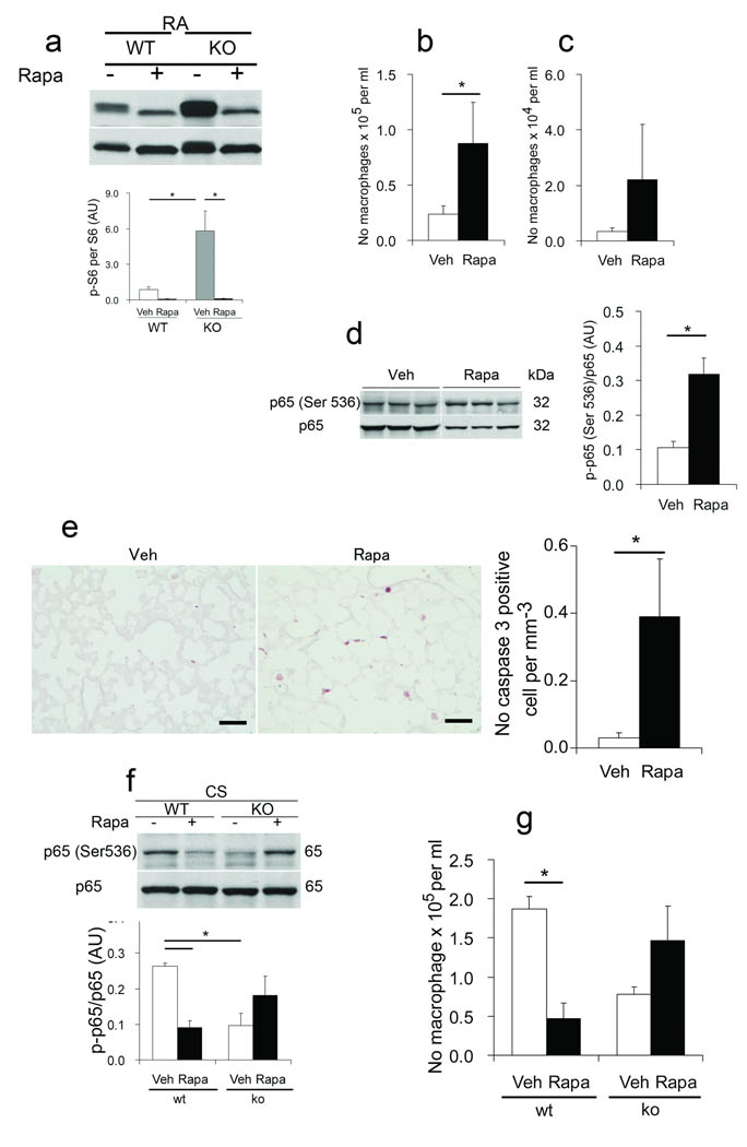
Rtp801 (also known as Redd1, and encoded by Ddit4), a stress-related protein triggered by adverse environmental conditions, inhibits mammalian target of rapamycin (mTOR) by stabilizing the TSC1-TSC2 inhibitory complex and enhances oxidative stress-dependent cell death. We postulated that Rtp801 acts as a potential amplifying switch in the development of cigarette smoke-induced lung injury, leading to emphysema. Rtp801 mRNA and protein were overexpressed in human emphysematous lungs and in lungs of mice exposed to cigarette smoke. The regulation of Rtp801 expression by cigarette smoke may rely on oxidative stress-dependent activation of the CCAAT response element in its promoter. We also found that Rtp801 was necessary and sufficient for nuclear factor-kappaB (NF-kappaB) activation in cultured cells and, when forcefully expressed in mouse lungs, it promoted NF-kappaB activation, alveolar inflammation, oxidative stress and apoptosis of alveolar septal cells. In contrast, Rtp801 knockout mice were markedly protected against acute cigarette smoke-induced lung injury, partly via increased mTOR signaling, and, when exposed chronically to cigarette smoke, against emphysema. Our data support the notion that Rtp801 may represent a major molecular sensor and mediator of cigarette smoke-induced lung injury.


Figures

Figure 1
Figure 1. Enhanced expression of Rtp801 in human emphysematous lungs
(a) Rtp801 expression in normal human lungs (lanes 1 and 2) or emphysematous lungs (GOLD 4) (lanes 3 – 6) (normalized by actin protein expression). (b) Histological sections showing increased expression of Rtp801 (brown) in a lung with emphysema (left) when compared with a normal lung (right) (arbitrary units (AU); n = 4 normal and 16 advanced emphysema lungs). (c) Determination of RTP801 mRNA expression in lungs of normal non smokers (n=8), normal smokers (n=13), and smoker patients with Gold stages 2 (n=12), 3 (n=12), and 4 (n=20) (normalized by cyclophilin A; signal intensity in arbitrary units (AU)).*: P < 0.05
Figure 2
Figure 2. Cigarette smoke – induced upregulation of Rtp801 expression occurs in lung septal but not resident or infiltrating inflammatory cells and relies on oxidative stress – dependent activation of the CCAAT promoter region
(a) Lung Rtp801 expression (brown, arrows) in wildtype (upper) or Rtp801 − / − (lower) mice exposed to RA (left) or CSk (right) for 7 days (× 50 µm) and expression levels (AU; n = 3 and 7, respectively). (b) Lung Rtp801 protein expression levels in mice exposed from 0 to 7 days to CSk (pooled n = 3 lungs in each time point). (c) Lungs costained with Rtp801 (red, arrowheads), nuclei (DAPI, bue), endothelial cells (thombomodulin, left), type I epithelial cells (T1α, middle), type II cells (ProSpC, right) (all in green) in mice exposed to CSk for 1 day (superimposed red plus green shown in yellow, arrows). Percent colocalization of alveolar cell specific markers (Marker) (thrombomodulin, T1α, or ProSPC over Rtp801 expression (10 fields, n = 3 lungs/marker; × 50 µm). (d) Rtp801 mRNA expression levels in Bal and lung tissue in wildtype mice exposed to CSk for 1 day or ambient air controls (RA) (AU, n = 3 – 4 mice in each group). (e) Rtp801 expression levels in lungs of Rtp801 wildtype mice treated with NAC (500 mg/kg, i.p.) or vehicle (veh) and exposed to CSk for 1 day (normalized by actin, AU; n = 4 – 5 mice in each group). (f, g) Activity of intact 2.5 kb Rtp801 promoter or with a point mutation within CCAAT binding site (mut/CEBP) or pGL3 plasmid (Vector) – Firefly luciferase in mouse lung fibroblasts (MLF) exposed to media alone (CTL), CSE (1 or 2%), or NAC (10 mM) (positive control: H2O2, 250 µM; pRTP+H2O2) for 12 h (normalized by Renilla luciferase; data representative of 2 independent experiments). (g) Expression of Rtp801 in cells from (f). *: P < 0.05
Figure 3
Figure 3. Rtp801 – dependent NF – κB activation by cigarette smoke
(a) Total Bal cells in Rtp801 wildtype mice intratracheally transduced with AAV5 – expressing IκB super repressor (SR) or β – gal (Lz) for 7 – 10 days in ambient air (RA) or exposed to CSk for 1 day (n = 5 – 6 mice in each group). (b) Effect of transfection with RTP801 – (RTP) or empty vector pcDNA3.1+ (pc) on NF – kB – dependent reporter Firefly luciferase activity (lower) in LPS, 1 µg ml−1 or vehicle – (veh) treated rat lung endothelial cells (normalized by Renilla luciferase activity, AU; n = 3 independent experiments), and RTP801 mRNA expression in transfected cells (upper). (c) NF – κB reporter activity in wildtype (WT) or Rtp801 − / − (KO) MLF treated with LPS – (1 ug ml−1) or 1% CSE (normalized AU; n = 3 independent experiments). (d) Expression levels of phosphorylated Ser536 p65 NF – κB (p – p65 NF – κB) in whole lung lysates of Rtp801 wildtype and Rtp801 − / − mice, exposed to CSk for 1 day (normalized by total p65, n = 3 – 5 mice in each group). (e) Vehicle (Ctl) and H2O2 induced (125 µM, 6 h) ROS levels in Rtp801 wildtype and Rtp801 − / − MEF. *: P < 0.05
Figure 4
Figure 4. Forced in vivo overexpression of humanRTP801 in mouse lungs enhances oxidative stress, inflammation, and alveolar cell apoptosis
Effect of human RTP801 – expressing (RTP, right), mutant Rtp801 – RPAA (RPAA, middle), or control pcDNA3.1 (pc, left) injected i.v. (24 h) in Rtp801 wildtype lungs costained (a) with RTP801 (red), type II cells (ProSPC, red, upper), or airway epithelial cells (CC10, green, lower) with coexpression highlighted by arrows (in yellow) (upper row: × 50 µm; lower row: × 200 µm). Numbers of infiltrating lung macrophages (b), 3 – nitrotyrosine positive cell profiles per high power field (hpf) (c), and alveolar cell apoptosis (TUNEL, normalized by DAPI – positive nuclei) (d) (10 fields, n = 5 – 10 mice in each group). (e) Infiltrating lung macrophages (brown, upper) in alveolar septa along an alveolar duct (arrows) in mice infected i.t. with AAV5 – RTP801 (left) or AAV5 – Cre (right) (negative control) for 4 weeks with quantification of numbers of alveolar macrophages per high power field (lower; 10 fields, n = 4 – 5 mice in each group). *: P < 0.05.
Figure 5
Figure 5. Rtp801 − / − mice are protected against cigarette smoke – induced pulmonary inflammation, apoptosis, and emphysema
Bal total cell counts (a), including numbers of macrophages (b), and neutrophils (c), infiltrating neutrophils in alveolar lung tissue (d) (µm−1 alveolar length), active caspase 3 – positive cells (brown, arrows) (upper) (× 50 µm) and numbers of active caspase 3 – positive cells (µm−1 alveolar septa) (lower) in Rtp801 wildtype and Rtp801 − / − mice kept in RA or exposed to CSk for 7 days (n = 3 and 7 mice, respectively). Alveolar morphology in Rtp801 wildtype CSk for 6 months (f) showing airspace enlargement and large clusters of alveolar macrophages containing smoking pigment in the cytoplasm when compared with Rtp801 − / − mouse lungs (× 250 µm, right and middle; × 25 µm, left). Alveolar length represent mean linear intercepts (g) (n = 5 to 7 mice in each group). *: P < 0.05
Figure 6
Figure 6. mTOR regulates lung homeostasis and inflammatory responses due to cigarette smoke in Rtp801 wildtype and Rtp801 − / − mice
Effect of vehicle ( – ) or rapamycin (rapa) treatment on phosphorylated serine 235/236 S6 (p – S6) protein expression (normalized by S6, AU) (a), on inflammatory cell counts of Bal macrophages (b) and neutrophils (c), lung p – p65 NF – κB protein expression (d) (normalized by p65, AU), on lung expression of active caspase 3 (e) (brown, left, × 50 µm), and total counts (right) of active caspase 3 – positive cellular profiles (normalized by µm alveolar length) in RA – exposed C57Bl6 mice. Effect of vehicle or rapa treatment on p – p65 NF – kB expression (f) (normalized by total p65, AU) and numbers of macrophages (g) in Bal fluid from lungs of wildtype and Rtp801 − / − mice exposed to CSk for 1 day (10 fields, n = 4/5 mice in each group). *: P < 0.05).

Comment in

References

    1. Yoshida T, Tuder RM. Pathobiology of cigarette smoke-induced chronic obstructive pulmonary disease. Physiol Rev. 2007;87:1047–1082. - PubMed
    1. Tuder RM, Yoshida T, Arap W, Pasqualini R, Petrache I. Cellular and Molecular Mechanisms of Alveolar Destruction in Emphysema: An Evolutionary Perspective. Proc Am Thorac Soc. 2006;3:503–510. - PMC - PubMed
    1. Tuder RM, et al. Oxidative stress and apoptosis interact and cause emphysema due to vascular endothelial growth factor receptor blockade. Am. J. Respir. Cell Mol. Biol. 2003;29:88–97. - PubMed
    1. Ryter SW, Chen ZH, Kim HP, Choi AM. Autophagy in chronic obstructive pulmonary disease: homeostatic or pathogenic mechanism? Autophagy. 2009;5:235–237. - PubMed
    1. Houghton AM, et al. Elastin fragments drive disease progression in a murine model of emphysema. J Clin Invest. 2006;116:753–759. - PMC - PubMed

Publication types

MeSH terms