. 2010 Jun;17(6):703-9.
doi: 10.1038/nsmb.1813. Epub 2010 May 16.

A'-form RNA helices are required for cytoplasmic mRNA transport in Drosophila

Affiliations

A'-form RNA helices are required for cytoplasmic mRNA transport in Drosophila

Simon L Bullock et al. Nat Struct Mol Biol. 2010 Jun.

Abstract

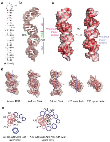
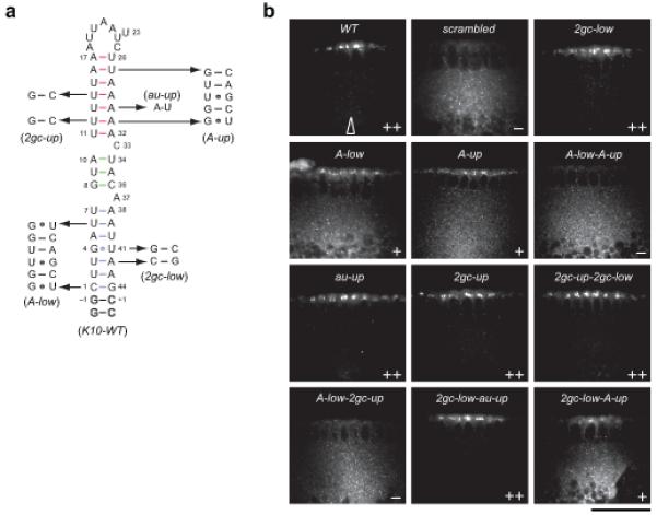
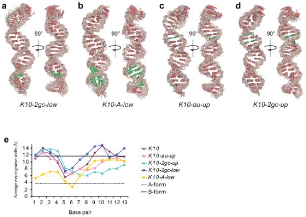
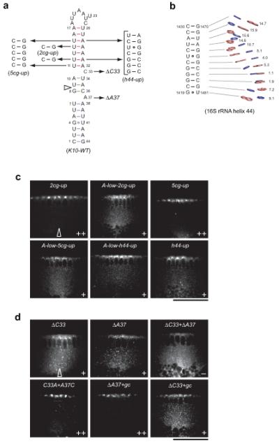
Microtubule-based mRNA transport is widely used to restrict protein expression to specific regions in the cell and has important roles in defining cell polarity and axis determination as well as in neuronal function. However, the structural basis of recognition of cis-acting mRNA localization signals by motor complexes is poorly understood. We have used NMR spectroscopy to describe the first tertiary structure to our knowledge of an RNA element responsible for mRNA transport. The Drosophila melanogaster fs(1)K10 signal, which mediates transport by the dynein motor, forms a stem loop with two double-stranded RNA helices adopting an unusual A'-form conformation with widened major grooves reminiscent of those in B-form DNA. Structure determination of four mutant RNAs and extensive functional assays in Drosophila embryos indicate that the two spatially registered A'-form helices represent critical recognition sites for the transport machinery. Our study provides insights into the basis for RNA cargo recognition and reveals a key biological function encoded by A'-form RNA conformation.


Figures

Figure 1
Figure 1
Solution structure of K10 TLS RNA. (a) Secondary structure of wild-type K10 TLS RNAs. Numbering according to ref.. Outlined nucleotides were added to improve transcription efficiency. The three helical segments are separated by single nucleotide bulges (C33 and A37). The lower helix (blue) comprises nt 1-7 and 38-44, the middle helix (green) nt 8-10 and 34-36, and the upper helix (red) nt 11-17 and 26-32, respectively. (b) Heavy-atom superposition of the 12 lowest-energy K10-WT RNAs refined with RDCs. Bases are red and the ribose-phosphate backbone is pink. The three helical regions are indicated beside the structures using the same color scheme as in a. C33 and A37 are shown in green. (c) Electrostatic surface potential of K10-WT RNA. Both the upper and lower helical regions display widened major grooves with a relative orientation of 90° along the helical axis. The widened major grooves are indicated beside the structures using the same color scheme as in a. (d) Representative structures of A-form and A′-form dsRNA and B-dsDNA compared to upper and lower helical regions of K10-WT RNA. All helices are shown from the major groove side to visualize the differences in inclination angles and groove widths. The helical axis is shown in blue and the inclination angle of base pairs is indicated by a dashed line. Bases are red and the ribose-phosphate backbone is pink. The PDB IDs are 1SDR (A-form RNA), 413D (A′-form RNA) and 1BNA (B-form DNA). (e) View down the lower and upper helix of K10-WT RNA displaying continuous stacking of purine bases. Five purine bases in the lower helix (A5-G44) and seven adenine bases in the upper helix (A17-A32) display continuous base-base stacking giving rise to A′-form inclination angles and widened major grooves (see Supplementary Tables 1 and 2). Pyrimidine bases are blue and purine bases are pink; ribose-phosphate backbone and chemical groups on the bases are omitted for clarity. Numbering according to a.
Figure 2
Figure 2
Localization activity of wild-type and lower and upper stem mutant K10 RNAs. (a) Secondary structure of wild-type and mutant K10 TLS RNAs. Numbering according to ref.. Outlined nucleotides were added to improve transcription efficiency. The sequences of K10 RNA mutations and the corresponding names used throughout the text are displayed. (b) Representative confocal images of blastoderm embryos injected with transcripts as indicated. TLS mutations were introduced within the context of a 2300 nt K10 sequence (see Online Methods). Transcripts were visualized by virtue of directly incorporated fluorochrome-coupled UTP. Arrow indicates the approximate site of injection in all experiments. Apical is to the top and basal is to the bottom in all images. Images of injections of additional transcripts are shown in Supplementary Fig. 5. Scale bar, 50μm.
Figure 3
Figure 3
Solution structure of mutant K10 TLS RNAs. (a-d) Heavy-atom superposition of the lowest-energy mutant K10 RNAs refined with RDCs. Bases are red and the ribose-phosphate backbone is pink. The mutated bases are green and the naming and sequence of each mutant corresponds to Fig. 2a. The widest opening of the major groove in the upper and lower helix is shown in the left and right view (rotation by 90° relative to the helical axis) of each ensemble. (e) Plot of the major groove width (Å) at each base pair in wild-type and mutant K10 RNAs. Base pairs are indicated corresponding to their 5′ nucleotide numbered according to Fig. 1a. Mean values are displayed for each RNA. Standard errors of the mean for each value are below 1.0 Å and summarized in Supplementary Table 2. Idealized A-form (dotted line) and B-form (solid line) values from ref. are also displayed. The corresponding mean base pair inclination angles are listed in Supplementary Table 1.
Figure 4
Figure 4
Localization activity of A′-form and bulge mutant K10 RNAs. (a) Secondary structure of wild-type and mutant K10 TLS RNAs. Numbering according to ref.. The sequences of K10 RNA mutations and the corresponding names used throughout the text are displayed; ΔC33 and ΔA37 denote deletion of the correspond nucleotides. Open arrowhead indicates position where an additional base pair is inserted in ΔC33+gc and ΔA37+gc. In the h44-up mutant 4 U-A base pairs are replaced by nucleotides 1420-1427 and 1473-1479 of the 16S rRNA helix 44 shown in b. (b) Secondary and tertiary structure of an A′-form helix 44 segment from 16S rRNA (PDB ID 2J00). Numbering according to (PDB ID 2J00). Pyrimidine bases are blue and purine bases are pink; ribose-phosphate backbone and chemical groups on the bases are omitted for clarity. The inclination angles (in degrees) are given for each base pair. (c and d) Representative confocal images of blastoderm embryos injected with transcripts as indicated. TLS mutations were introduced within the context of a 2300 nt K10 sequence (see Online Methods). Transcripts were visualized by virtue of directly incorporated fluorochrome-coupled UTP. Arrow indicates the approximate site of injection in all experiments. Apical is to the top and basal is to the bottom in all images. Images of injections of additional transcripts are shown in Supplementary Fig. 5. Scale bar, 50μm.

References

    1. Martin KC, Ephrussi A. mRNA localization: gene expression in the spatial dimension. Cell. 2009;136:719–30. - PMC - PubMed
    1. Holt CE, Bullock SL. Subcellular mRNA localization in animal cells and why it matters. Science. 2009;326:1212–6. - PMC - PubMed
    1. Wilkie GS, Davis I. Drosophila wingless and pair-rule transcripts localize apically by dynein-mediated transport of RNA particles. Cell. 2001;105:209–19. - PubMed
    1. Van De Bor V, Hartswood E, Jones C, Finnegan D, Davis I. gurken and the I factor retrotransposon RNAs share common localization signals and machinery. Dev Cell. 2005;9:51–62. - PubMed
    1. Snee MJ, Arn EA, Bullock SL, Macdonald PM. Recognition of the bcd mRNA localization signal in Drosophila embryos and ovaries. Mol. Cell. Biol. 2005;25:1501–10. - PMC - PubMed

References for online methods only

    1. Easton LE, Shibata Y, Lukavsky PJ. Rapid, nondenaturing RNA purification using weak anion-exchange fast performance liquid chromatography. RNA. 2010;16:647–653. - PMC - PubMed
    1. Lukavsky PJ, Puglisi JD. Large-scale preparation and purification of polyacrylamide-free RNA oligonucleotides. RNA. 2004;10:889–893. - PMC - PubMed
    1. Nikonowicz EP. Preparation and use of 2H-labeled RNA oligonucleotides in nuclear magnetic resonance studies. Methods Enzymol. 2001;338:320–41. - PubMed
    1. Scott LG, Tolbert TJ, Williamson JR. Preparation of specifically 2H- and 13C-labeled ribonucleotides. Methods Enzymol. 2000;317:18–38. - PubMed
    1. Hansen MR, Hanson P, Pardi A. Filamentous bacteriophage for aligning RNA, DNA, and proteins for measurement of nuclear magnetic resonance dipolar coupling interactions. Methods Enzymol. 2000;317:220–40. - PubMed

Publication types

MeSH terms