. 2010 Jul 8;29(27):3990-6.
doi: 10.1038/onc.2010.137. Epub 2010 May 17.

The viral tropism of two distinct oncolytic viruses, reovirus and myxoma virus, is modulated by cellular tumor suppressor gene status

Affiliations

The viral tropism of two distinct oncolytic viruses, reovirus and myxoma virus, is modulated by cellular tumor suppressor gene status

M Kim et al. Oncogene. .

Abstract

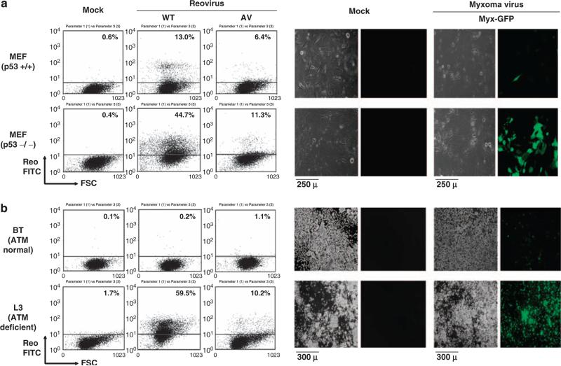
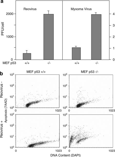
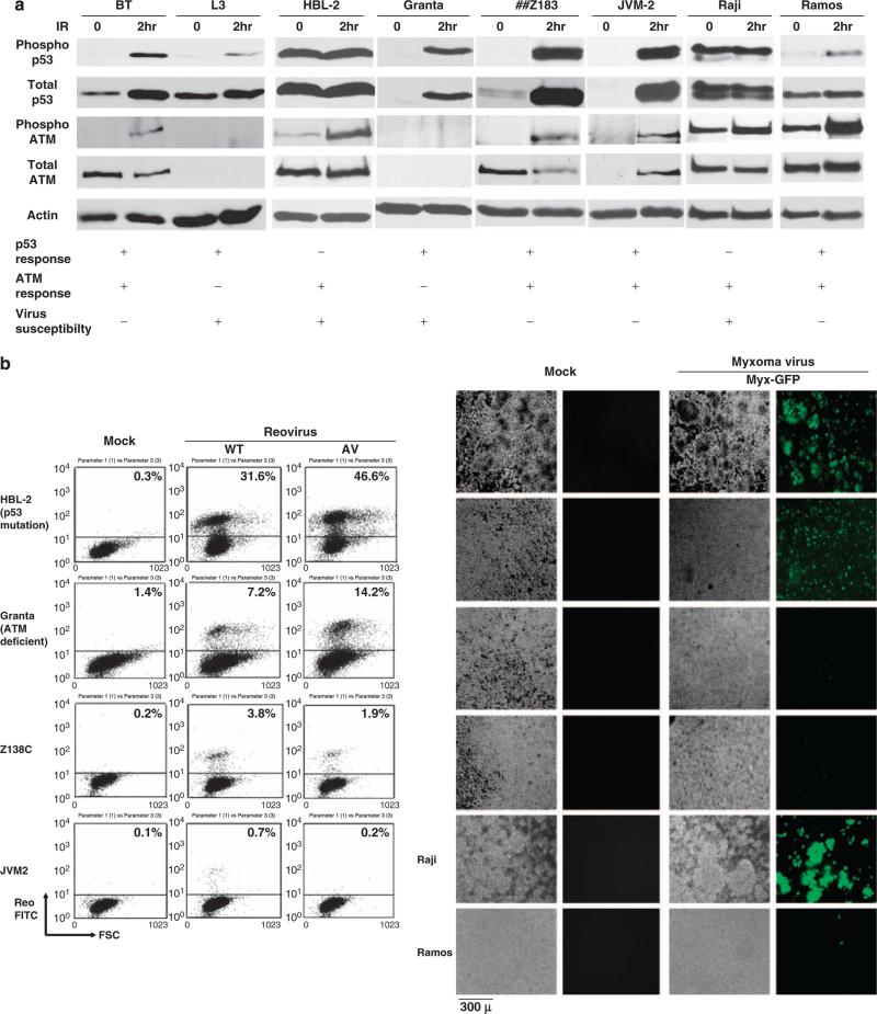
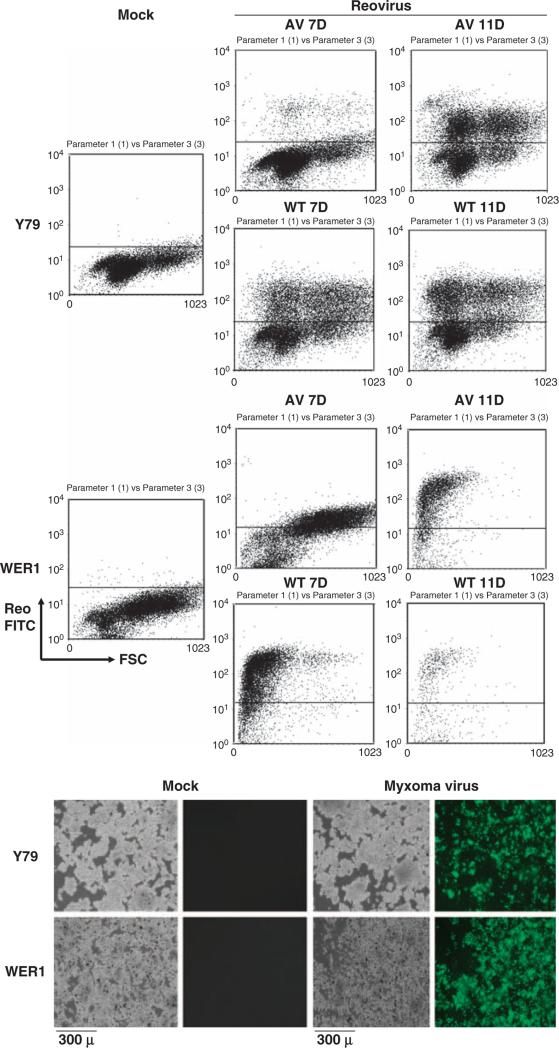
Replication-competent oncolytic viruses hold great potential for the clinical treatment of many cancers. Importantly, many oncolytic virus candidates, such as reovirus and myxoma virus, preferentially infect cancer cells bearing abnormal cellular signaling pathways. Reovirus and myxoma virus are highly responsive to activated Ras and Akt signaling pathways, respectively, for their specificity for viral oncolysis. However, considering the complexity of cancer cell populations, it is possible that other tumor-specific signaling pathways may also contribute to viral discrimination between normal versus cancer cells. Because carcinogenesis is a multistep process involving the accumulation of both oncogene activations and the inactivation of tumor suppressor genes, we speculated that not only oncogenes but also tumor suppressor genes may have an important role in determining the tropism of these viruses for cancer cells. It has been previously shown that many cellular tumor suppressor genes, such as p53, ATM and Rb, are important for maintaining genomic stability; dysfunction of these tumor suppressors may disrupt intact cellular antiviral activity due to the accumulation of genomic instability or due to interference with apoptotic signaling. Therefore, we speculated that cells with dysfunctional tumor suppressors may display enhanced susceptibility to challenge with these oncolytic viruses, as previously seen with adenovirus. We report here that both reovirus and myxoma virus preferentially infect cancer cells bearing dysfunctional or deleted p53, ATM and Rb tumor suppressor genes compared to cells retaining normal counterparts of these genes. Thus, oncolysis by these viruses may be influenced by both oncogenic activation and tumor suppressor status.


Figures

Figure 1
Figure 1
Reoviral and myxoma viral preferential infection of p53−/− MEF and ATM-defective L3 cells. (a) p53−/− and+/+ MEF (p53 knockout or p53 wild-type murine embryonic fibroblasts, respectively) were infected with WT/AV reovirus (multiplicity of infection (MOI) of 40) or GFP-expressing myxoma virus (Myx-GFP, which is a GFP-expressing version of the Lausanne (American Type Culture Collection, ATCC, Manassas, VA, USA) strain of MYXV, as described by Johnston et al. (2003); MOI of 5). GFP expression in the myxoma virus is driven by the poxviral synthetic E/L promoter; the GFP expression cassette is located between M135R and M136R by intergenic insertion (Johnston et al., 2003). At 3 days after infection with reovirus, cells were fixed/permeabilized and analyzed by FACS using reovirus antiserum and secondary FITC antiserum. The results show that both wild-type and attenuated reovirus were able to preferentially infect p53−/− MEF. For the myxoma infection study, cells were infected for 24 h and visualized under phase-contrast and fluorescence microscopy. GFP-expressing cells represent myxoma virus infection. Mock, mock infection. (b) BT (ATM normal lymphoblastoid C3ABR cells; Kozlov et al., 2003) and L3 (ATM-deficient lymphoblastoid cells; Kozlov et al., 2003) were infected with WT/AV reovirus or myxoma virus (Myx-GFP). At 3 days after infection with reovirus, cells were fixed/ permeabilized and analyzed by FACS using reovirus antiserum and secondary FITC antiserum. The results show that both wild-type and attenuated reovirus were able to preferentially infect ATM-deficient L3 cells. For myxoma infection, cell were infected for 48 h and visualized under phase-contrast and fluorescence microscopy. GFP-expressing cells represent myxoma virus infection. C35ABR (B3) and L3 cell lines were kindly provided by Dr M Lavin (Queensland Institute of Medical Research) and Dr Y Shiloh (Tel Aviv University), respectively. Mock, mock infection.
Figure 2
Figure 2
p53 deficiency enhances viral replication and virusinduced apoptosis. (a) MEF p53+/+ and −/− cells were infected with reovirus at multiplicity of infection (MOI) of 40 or myxoma virus at MOI of 5. At 3 days after infection, cells and supernatant are harvested and frozen and thawed three times. Viral titer was determined by plaque assays using L929 cells (reovirus) or RK13 cells (myxoma) and expressed as PFU per cell. (b) MEF p53+/+ and −/− cells were either mock infected or exposed to reovirus for 9 days, then stained for DNA content with DAPI and for apoptotic activity with 7AAP. MEF p53−/− cells showed extensive cellular fragmentation and apoptosis in response to reovirus, whereas MEF p53+/+ cells did not.
Figure 3
Figure 3
Reovirus and myxoma virus preferential infection of p53 and ATM dysfunctional human lymphomas. (a) p53 and ATM phosphorylation in response to IR. Mantle cell lymphomas (Granta-519, HBL-2 (p53 deletion, Tucker et al., 2006 and/or mutation, Beà et al., 2009), Z138, JVM-2) and Burkitt's lymphomas (Raji and Ramos) were irradiated at 2 Gy for 2 h and cells were harvested. Whole-cell lysates were produced by treatment with NET-N lysis buffer (1% NP-40) followed by sonication. 50 μg of protein was then separated on either 8% or 10% SDS–PAGE, transferred to nitrocellulose and probed with the indicated antibodies (Phospho-ATM; pSer1981 ATM and Phospho-p53; pSer15, and p53 were purchased from Rockland Immunochemicals (Philadelphia, PA, USA) and Cell Signaling Technology (Danvers, MA, USA); the ATM-specific rabbit polyclonal antibody 4BA was a kind gift from Dr M Lavin). BT and L3 cells were used as controls. (b) Cells were infected with WT/AV reovirus at multiplicity of infection (MOI) of 40 or myxoma virus (Myx-GFP) at MOI of 5. Previous work showed that Raji cells are susceptible and Ramos cells are resistant to reovirus (Alain et al., 2002). At 3 days after infection, cells were fixed/permeabilized and analyzed by FACS using reovirus antiserum and secondary FITC antiserum. The results showed that both reovirus and myxoma virus preferentially infect p53 and/or ATM nonresponsive lymphomas. For the myxoma infection study, cell were infected for 48h and visualized under phase-contrast and fluorescence microscopy. GFP-expressing cells represent myxoma virus infection. Mock, mock infection.
Figure 4
Figure 4
Reovirus and myxoma virus infection of retinoblastoma cells. Retinoblastoma cells (Y79 and WERI-Rb-1, purchased from ATCC) were infected with WT/AV reovirus at multiplicity of infection (MOI) of 40 or myxoma virus (Myx-GFP) at MOI of 5. At designated days after infection, cells were fixed/permeabilized and analyzed by FACS using reovirus antiserum and secondary FITC antiserum. For the myxoma infection study, cell were infected for 48 h and visualized under phase-contrast and fluorescence microscopy. GFP-expressing cells represent myxoma infection. Mock, mock infection.

References

    1. Abou El Hassan MA, van der Meulen-Muileman I, Abbas S, Kruyt FA. Conditionally replicating adenoviruses kill tumor cells via a basic apoptotic machinery-independent mechanism that resembles necrosis-like programmed cell death. J Virol. 2004;78:12243–12251. - PMC - PubMed
    1. Alain T, Hirasawa K, Pon KJ, Nishikawa SG, Urbanski SJ, Auer Y, et al. Reovirus therapy of lymphoid malignancies. Blood. 2002;100:4146–4153. - PubMed
    1. Beà S, Salaverria I, Armengol L, Pinyol M,V, Hartmann EM, et al. Uniparental disomies, homozygous deletions, amplifications, and target genes in mantle cell lymphoma revealed by integrative high-resolution whole-genome profiling. Blood. 2009;113:3059–3069. - PMC - PubMed
    1. Bischoff JR, Kirn DH, Williams A, Heise C, Horn S, Muna M, et al. An adenovirus mutant that replicates selectively in p53-deficient human tumor cells. Science. 1996;274:373–376. - PubMed
    1. Carroll PE, Okuda M, Horn HF, Biddinger P, Stambrook PJ, Gleich LL, et al. Centrosome hyperamplification in human cancer: chromosome instability induced by p53 mutation and/or Mdm2 overexpression. Oncogene. 1999;18:1935–1944. - PubMed

Publication types

MeSH terms