. 2010 Apr;14(2):91-7.
doi: 10.4196/kjpp.2010.14.2.91. Epub 2010 Apr 30.

Pyrithione-zinc Prevents UVB-induced Epidermal Hyperplasia by Inducing HIF-1alpha

Affiliations

Pyrithione-zinc Prevents UVB-induced Epidermal Hyperplasia by Inducing HIF-1alpha

Young-Suk Cho et al. Korean J Physiol Pharmacol. 2010 Apr.

Abstract

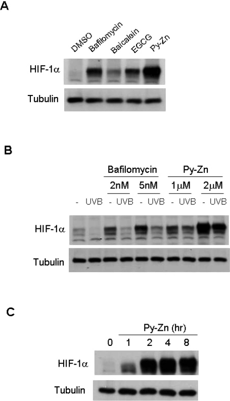
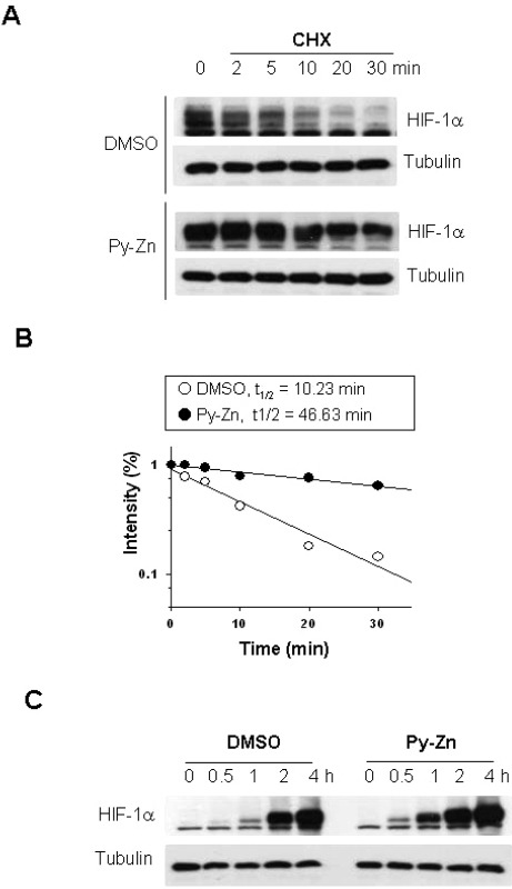
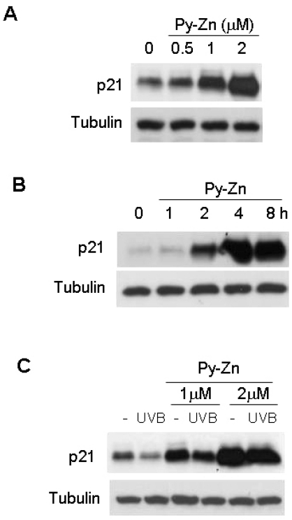
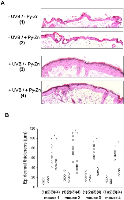
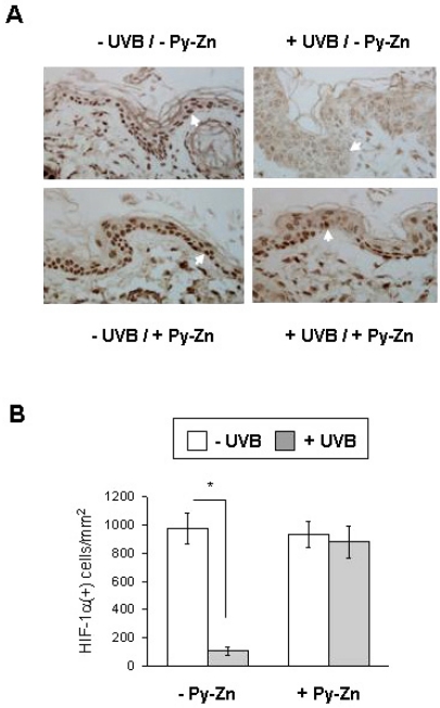
Epidermal keratinocytes overgrow in response to ultraviolet-B (UVB), which may be associated with skin photoaging and cancer development. Recently, we found that HIF-1alpha controls the keratinocyte cell cycle and thereby contributes to epidermal homeostasis. A further study demonstrated that HIF-1alpha is down-regulated by UVB and that this process is involved in UVB-induced skin hyperplasia. Therefore, we hypothesized that the forced expression of HIF-1alpha in keratinocytes would prevent UVB-induced keratinocyte overgrowth. Among several agents known to induce HIF-1alpha, pyrithione-zinc (Py-Zn) overcame the UVB suppression of HIF-1alpha in cultured keratinocytes. Mechanistically, Py-Zn blocked the degradation of HIF-1alpha protein in keratinocytes, while it did not affect the synthesis of HIF-1alpha. Moreover, the p21 cell cycle inhibitor was down-regulated after UVB exposure, but was robustly induced by Py-Zn. In mice repeatedly irradiated with UVB, the epidermis became hyperplastic and HIF-1alpha disappeared from nuclei of epidermal keratinocytes. However, a cream containing Py-Zn effectively prevented the skin thickening and up-regulated HIF-1alpha to the normal level. These results suggest that Py-Zn is a potential agent to prevent UVB-induced photoaging and skin cancer development. This work also provides insight into a molecular target for treatment of UVB-induced skin diseases.

Keywords: Hyperplasia; Hypoxia-inducible factor-1α; Pyrithione-zinc; Skin; Ultraviolet.


Figures

Fig. 1
Fig. 1
Chemical structures of the test compounds. (A) Bafilomycin A1, (B) baicalein, (C) epigallocatechin 3-gallate (EGCG), (D) pyrithione-zinc (Py-Zn).
Fig. 2
Fig. 2
HIF-1α levels in keratinocytes. (A) Effects of test compounds on HIF-1α expression. After HaCaT cells had been cultured to a cell density of 80~100%, the cells were treated with various compounds for 4 hours. The cells were harvested and subjected to immunoblotting for HIF-1α and β-tubulin. Concentration: bafilomycin A1, 5 nM; baicalein, 200 µM; EGCG, 500 µM; pyrithione-zinc (Py-Zn), 2 µM. (B) HIF-1α levels after UVB irradiation. HaCaT cells were pretreated with bafilomycin A1 or Py-Zn at the concentrations indicated 2 hours before UVB irradiation, and transiently irradiated with 15 mJ/cm2 of UVB. Two hours later, HaCaT cells were harvested for immunoblotting. (C) Time course of Py-Zn-induced HIF-1α expression. HaCaT cells were treated with 2 µM Py-Zn, harvested at the indicated time, and then subjected to immunoblotting.
Fig. 3
Fig. 3
Py-Zn stabilizes HIF-1α in keratinocytes. (A) HIF-1α protein stability. HaCaT cells were pretreated with DMSO vehicle or 2 µM Py-Zn for 1 hour, and then further treated with 60 µg/ml of cycloheximide (CHX). Cells were harvested at the indicated time, and HIF-1α levels were analyzed by immunoblotting. (B) Half-lives of HIF-1α proteins. The protein band densities were quantified using ImageJ and plotted as a function of time (first-order kinetics). (C) HIF-1α protein synthesis. HaCaT cells were treated with 60 µg/ml of cycloheximide for 30 min to remove remaining HIF-1α. To initiate HIF-1α synthesis, cells were washed with PBS and incubated in fresh media containing 10 µM MG132, and HIF-1α levels were determined at the indicated time by immunoblotting.
Fig. 4
Fig. 4
p21 levels in keratinocytes. (A) Effect of Py-Zn on p21 expression in keratinocytes. HaCaT cells were treated with Py-Zn at the concentrations indicated for 4 hours and then harvested for immunoblotting. (B) Time course of Py-Zn-induced p21 expression. HaCaT cells were treated with 2 µM Py-Zn, harvested at the indicated time, and then subjected to immunoblotting with anti-p21 antiserum. (C) p21 levels after UVB irradiation. HaCaT cells were pretreated with Py-Zn at the indicated concentrations 2 hours before UVB irradiation, and irradiated with 15 mJ/cm2 of UVB. Two hours later, HaCaT cells were harvested for immunoblotting.
Fig. 5
Fig. 5
In vivo effect of Py-Zn on UVB-induced skin hyperplasia. The back skin of each mouse was divided into four parts: the right lower quadrant for -UVB/-Py-Zn (1), the right upper quadrant for -UVB/+Py-Zn (2), the left lower quadrant for +UVB/-Py-Zn (3), the left upper quadrant for +UVB/+Py-Zn (4). Four mice were pretreated with PEG or 1% Py-Zn PEG cream 4 hours before UVB irradiation and then exposed to 460 mJ/cm2 of UVB once a day for four days. Back skins were excised, fixed, and embedded into paraffin blocks. (A) H&E staining of mouse skin. Skin specimens were obtained from 10 different sites per quadrant of each mouse and stained with H&E. (B) The thickness of the epidermis was analyzed at a magnification of 100×. The results were obtained from four mice (mouse 1~4). *Denotes p<0.05 between two groups.
Fig. 6
Fig. 6
In vivo effect of Py-Zn on UVB-induced HIF-1α suppression. (A) HIF-1α staining. The skin specimens were obtained as described in Fig. 5 and stained with anti-HIF-1α antibody. Nuclear HIF-1α was identified at a magnification of 100×. (B) Numbers of HIF-1α-positive keratinocytes. Three different slides per quadrant were stained and counted, and the data were collected from tissues of four mice (12 slides per group). *Denotes p<0.05 between two groups.

References

    1. Melnikova VO, Ananthaswamy HN. Cellular and molecular events leading to the development of skin cancer. Mutat Res. 2005;571:91–106. - PubMed
    1. Marrot L, Meunier JR. Skin DNA photodamage and its biological consequences. J Am Acad Dermatol. 2008;58:S139–S148. - PubMed
    1. Ouhtit A, Muller HK, Davis DW, Ullrich SE, McConkey D, Ananthaswamy HN. Temporal events in skin injury and the early adaptive responses in ultraviolet-irradiated mouse skin. Am J Pathol. 2000;156:201–207. - PMC - PubMed
    1. Lee JK, Kim JH, Nam KT, Lee SH. Molecular events associated with apoptosis and proliferation induced by ultraviolet-B radiation in the skin of hairless mice. J Dermatol Sci. 2003;32:171–179. - PubMed
    1. El-Abaseri TB, Putta S, Hansen LA. Ultraviolet irradiation induces keratinocyte proliferation and epidermal hyperplasia through the activation of the epidermal growth factor receptor. Carcinogenesis. 2006;27:225–231. - PubMed