. 2010 Nov;39(5):1507-19.
doi: 10.1007/s00726-010-0620-5. Epub 2010 May 16.

α-Helix peptides designed from EBV-gH protein display higher antigenicity and induction of monocyte apoptosis than the native peptide

Affiliations

α-Helix peptides designed from EBV-gH protein display higher antigenicity and induction of monocyte apoptosis than the native peptide

Mauricio Urquiza et al. Amino Acids. 2010 Nov.

Abstract

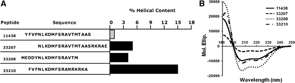
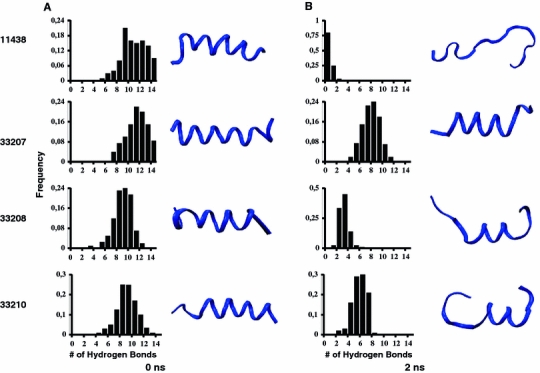
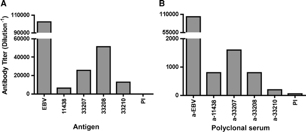
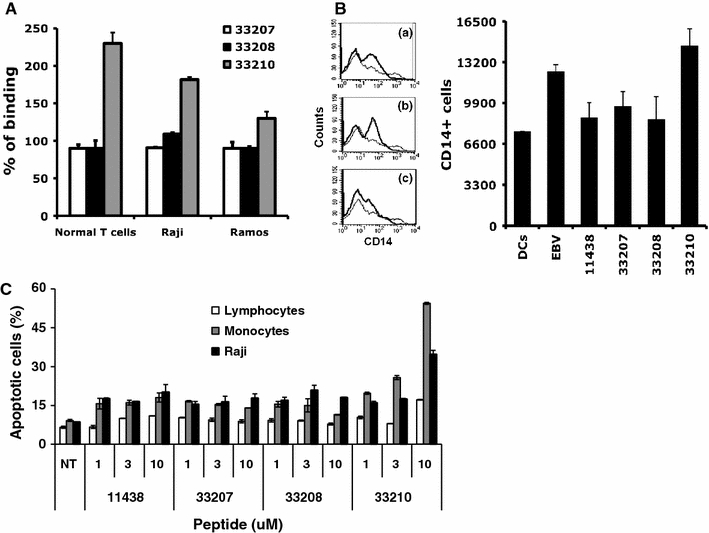
We tested the hypothesis that stabilizing α-helix of Epstein-Barr virus gH-derived peptide 11438 used for binding human cells will increase its biological activity. Non-stable α-helix of peptide 11438 was unfolded in an entropy-driven process, despite the opposing effect of the enthalpy factor. Adding and/or changing amino acids in peptide 11438 allowed the designing of peptides 33207, 33208 and 33210; peptides 33208 and 33210 displayed higher helical content due to a decreased unfolding entropy change as was determined by AGADIR, molecular dynamics and circular dichroism analysis. Peptides 33207, 33208 and 33210 inhibited EBV invasion of peripheral blood mononuclear cells and displayed epitopes more similar to native protein than peptide 11438; these peptides could be useful for detecting antibodies induced by native gH protein since they displayed high reactivity with anti-EBV antibodies. Anti-peptide 33207 antibodies showed higher reactivity with EBV than anti-peptide 11438 antibodies being useful for inducing antibodies against EBV. Anti-peptide 33210 antibodies inhibit EBV invasion of epithelial cells better than anti-peptide 11438 antibodies. Peptide 33210 bound to normal T lymphocytes and Raji cells stronger than peptide 11438 and also induced apoptosis of monocytes and Raji cells but not of normal T cells in a similar way to EBV-gH. Peptide 33210 inhibited the monocytes' development toward dendritic cells better than EBV and peptide 11438. In conclusion, stabilizing the α-helix in peptides 33208 and 33210 designed from peptide 11438 increased the antigenicity and the ability of the antibodies induced by peptides of inhibiting EBV invasion of host cells.


Figures

Fig. 1
Fig. 1
Helical content and circular dichroism analysis performed in EBV-gH-derived peptides. a Peptide 11438 was modified by adding or deleting amino acids at C- and N-terminal regions and the helical content was calculated by AGADIR analysis. The sequence of peptide 11438 is shown in the gray bar. b Circular dichroism of peptides selected from each group (33207, 33208 and 33210 from groups 1, 2 and 3, respectively) and peptide 11438 were dissolved in PBS with 30% TFE; the spectra were made on a JASCO spectrometer. The X-axis shows wavelength in nm and the Y-axis molar ellipticity per residue
Fig. 2
Fig. 2
Molecular dynamics performed in peptides 11438, 33207, 33208 and 33210. The α-helix of peptides 11438, 33207, 33208 and 33210 were built by using Quanta. After these peptides were hydrated and minimized the molecular dynamics were run during 2 ns at 310 K by using CHARMM. a Mean frequency of hydrogen bonds that are present in the peptide structure at 0.0 ns. On the right side are shown the ribbon structures acquired by these peptides during the molecular dynamics, which were built by using VMD program. In b is shown the same as in a but at 2 ns
Fig. 3
Fig. 3
Antigenicity and immunogenicity of peptides 11438, 33207, 33208 and 33210. Rabbits were immunized with EBV, 11438, 33207, 33208 or 33210. After three immunizations polyclonal serum was obtained and tested by ELISA. a Anti-EBV antibodies were tested for their reactivity against peptides to evaluate peptides’ antigenicity. b Reactivity of anti-peptides antibodies was tested against EBV to evaluate peptide’s immunogenicity. PI pre-immune sera
Fig. 4
Fig. 4
Effect of peptides and anti-sera of peptides 11438, 33207, 33208 and 33210 in EBV invasion of host cells. a PBMCs and HEK293 cells were treated with EBV and peptides 11438, 33207, 33208 or 33210 at different concentrations: 1 27 μM, 2 13 μM or 3 7 μM. After the incubation period DNA was isolated and PCR amplification of an EBV DNA fragment was performed. b Activity of anti-peptide sera at different dilutions in neutralizing EBV invasion of HEK293 cells
Fig. 5
Fig. 5
Capacity of peptides 11438, 33207, 33208 and 33210 to bind to PBMCs, Raji and Ramos cells, their effect in CD14 marker on dendritic cells and apoptosis in T cells, monocytes and Raji cells. a The capacity of peptides to bind to normal T cells, Raji and Ramos cells was tested by using flow cytometry. The percentage of binding compared with peptide 11438 was calculated as described in “Materials and methods”. b CD14 surface expression on monocytes exposed to GM-CSF and IL4 and incubated with EBV, peptides 11438, 33207, 33208 or 33210. The histogram data represents a typical example of cells not exposed to EBV or peptide (thick line) and exposed (dark line) to: a EBV, b peptide with similar effect to EBV (33210), c peptide without effect (33207). Number of CD14 positive cells shown on the right side. c Effect of peptides in apoptosis of PBMCs and Raji cells at 10 μM concentration. The percentage of apoptotic cells was determined by staining with Annexin V and PI. NT non-treated samples
Fig. 6
Fig. 6
Representation of peptide 11438 regions and its analogs related to their biological activity. We have found different regions that are important for the binding to the cells (blue-NLKDMFSRA), for keeping the α-helix structure (green-YFVP) and for the interaction with the anti-EBV antibodies (red-NLKDMFSRA)

Similar articles

References

    1. Bax A, Davis DG. MLEV-17-based two-dimensional homonuclear magnetization transfer spectroscopy. J Magn Reson. 1985;65:355–360.
    1. Borza C, Hutt-Fletcher L. Alternate replication in B cells and epithelial cells switches tropism of Epstein–Barr virus. Nat Med. 2002;8:594–599. doi: 10.1038/nm0602-594. - DOI - PubMed
    1. Chen P, Pan CC, Yang AH, Wang LS, Chiang H. Detection of Epstein–Barr virus genome within thymic epithelial tumours in Taiwanese patients by nested PCR, PCR in situ hybridization, and RNA in situ hybridization. J Pathol. 2002;197:684–688. doi: 10.1002/path.1141. - DOI - PubMed
    1. Chesnokova LS, Nishimura SL, Hutt-Fletcher LM. Fusion of epithelial cells by Epstein–Barr virus proteins is triggered by binding of viral glycoproteins gHgL to integrins alphavbeta6 or alphavbeta8. Proc Natl Acad Sci USA. 2009;106:20464–20469. doi: 10.1073/pnas.0907508106. - DOI - PMC - PubMed
    1. Delves PJ. Antibody production: essential techniques. Chichester, NY: Wiley and Sons; 1997.

MeSH terms