Review
. 2012 Jan;42(1):95-113.
doi: 10.1007/s00726-010-0603-6. Epub 2010 May 16.

Design of Mn porphyrins for treating oxidative stress injuries and their redox-based regulation of cellular transcriptional activities

Affiliations
Review

Design of Mn porphyrins for treating oxidative stress injuries and their redox-based regulation of cellular transcriptional activities

Ines Batinic-Haberle et al. Amino Acids. 2012 Jan.

Erratum in

  • Amino Acids. 2012 Jan;42(1):115-6

Abstract

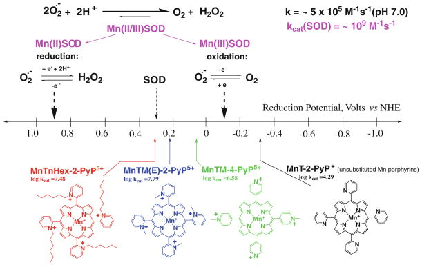
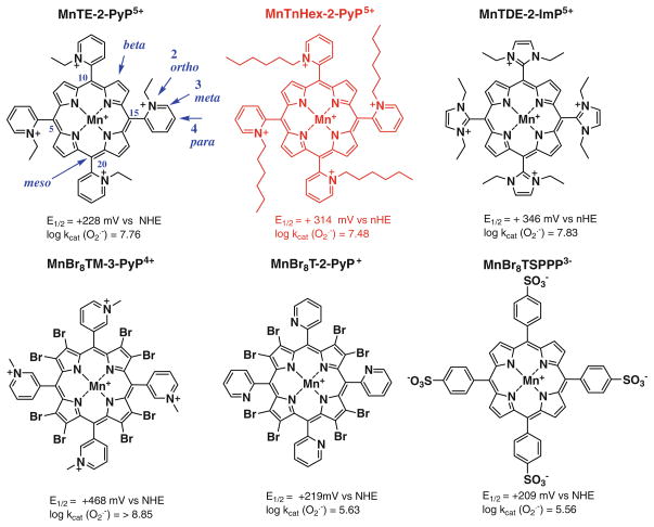
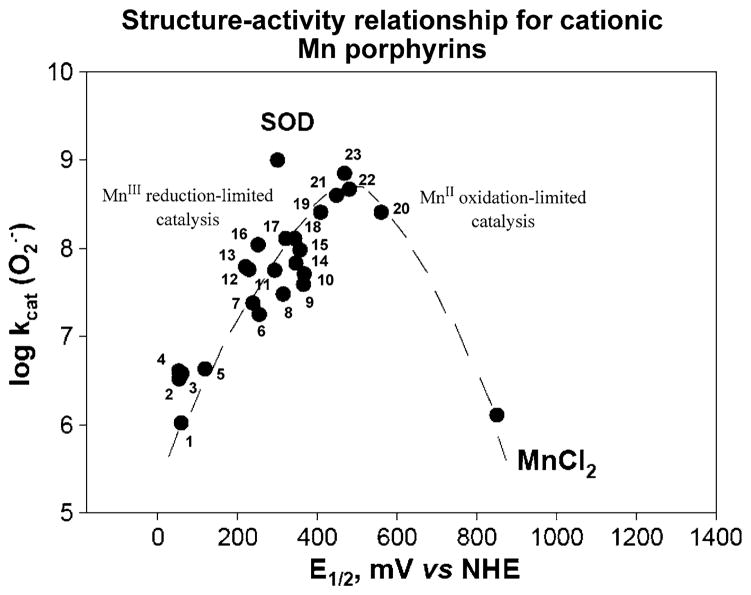
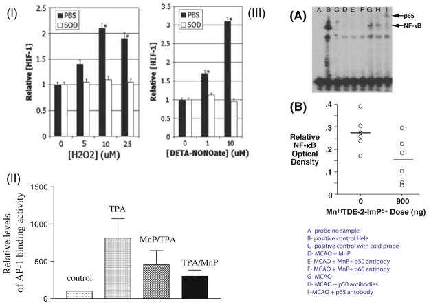
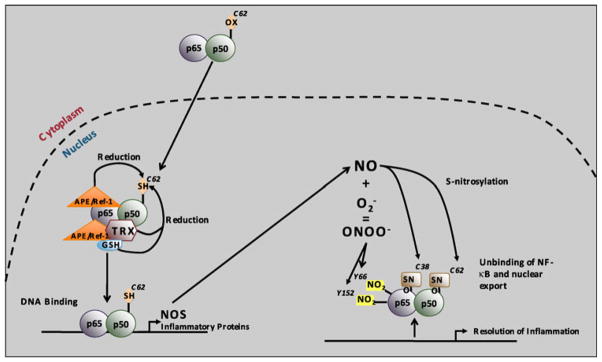
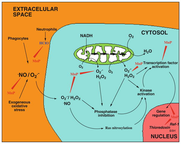
The most efficacious Mn(III) porphyrinic (MnPs) scavengers of reactive species have positive charges close to the Mn site, whereby they afford thermodynamic and electrostatic facilitation for the reaction with negatively charged species such as O (2) (•-) and ONOO(-). Those are Mn(III) meso tetrakis(N-alkylpyridinium-2-yl)porphyrins, more specifically MnTE-2-PyP(5+) (AEOL10113) and MnTnHex-2-PyP(5+) (where alkyls are ethyl and n-hexyl, respectively), and their imidazolium analog, MnTDE-2-ImP(5+) (AEOL10150, Mn(III) meso tetrakis(N,N'-diethylimidazolium-2-yl) porphyrin). The efficacy of MnPs in vivo is determined not only by the compound antioxidant potency, but also by its bioavailability. The former is greatly affected by the lipophilicity, size, structure, and overall shape of the compound. These porphyrins have the ability to both eliminate reactive oxygen species and impact the progression of oxidative stress-dependent signaling events. This will effectively lead to the regulation of redox-dependent transcription factors and the suppression of secondary inflammatory- and oxidative stress-mediated immune responses. We have reported on the inhibition of major transcription factors HIF-1α, AP-1, SP-1, and NF-κB by Mn porphyrins. While the prevailing mechanistic view of the suppression of transcription factors activation is via antioxidative action (presumably in cytosol), the pro-oxidative action of MnPs in suppressing NF-κB activation in nucleus has been substantiated. The magnitude of the effect is dependent upon the electrostatic (porphyrin charges) and thermodynamic factors (porphyrin redox ability). The pro-oxidative action of MnPs has been suggested to contribute at least in part to the in vitro anticancer action of MnTE-2-PyP(5+) in the presence of ascorbate, and in vivo when combined with chemotherapy of lymphoma. Given the remarkable therapeutic potential of metalloporphyrins, future studies are warranted to further our understanding of in vivo action/s of Mn porphyrins, particularly with respect to their subcellular distribution.


Figures

Fig. 1
Fig. 1
Design of Mn porphyrins based on thermodynamic considerations
Fig. 2
Fig. 2
Three most often in vivo studied ortho isomeric Mn(III) meso-tetrakis(N-ethyl(or n-hexyl)pyridinium-2-yl)porphyrins, MnTE-2-PyP5+ (AEOL10113), MnTnHex-2-PyP5+, and the N,N′-diethylimi-dazolium analog, MnTDE-2-ImP5+ (AEOL10150). The meso and beta positions on porphyrin ring are indicated as well as ortho, meta, and para positions of pyridyl nitrogens with respect to porphyrin meso positions. Also shown are three Mn porphyrins that are both meso- and beta-substituted. Such compounds are particularly valuable tool for the design of perspective therapeutics
Fig. 3
Fig. 3
Structure–activity relationship between logkcat(O2) and E1/2 (MnIIIP/MnIIP) in mV versus NHE for cationic Mn(III) porphyrins. Compounds at the plateau of the bell-shaped curve are those that bear both meso and beta substituents and have highest kcat. Yet, they are stabilized in Mn +2 oxidation state, lose readily Mn and are thus are only important for mechanistic purposes. The falling limb of the bell-shaped curve is more obvious in SARs of anionic and neutral porphyrins (Batinic-Haberle et al. 2010). Compounds are: 1 MnT(TFTMA)P5+, 2 MnBM-2-PyP5+, 3 MnTM-4-PyP5+, 4 MnTM-3-PyP5+, 5 MnTrM-2-PyP5+, 6 MnTnBu-2-PyP5+, 7 MnTnPr-2-PyP5+, 8 MnTnHex-2-PyP5+, 9 MnTDMOE-2-ImP5+, 10 MnTnOct-2-PyP5+, 11 MnClTE-2-PyP5+, 12 MnTE-2-PyP5+, 13 MnTM-2-PyP5+, 14 MnTDE-2-ImP5+, 15 MnTM,MOE-2-ImP5+, 16 MnTMOE-2-PyP5+, 17 MnTDM-2-ImP5+, 18 MnCl2TE-2-PyP5+, 19 MnCl3TE-2-PyP5+, 20 MnCl5TE-2-PyP5+, 21 MnCl4TE-2-PyP5+, 22 MnBr8TM-4-PyP4+, and 23 MnBr8TM-3-PyP4+. Data are taken from Batinic-Haberle et al. (2010) and Kachadourian et al. (1998). The names of porphyrins are listed in the list of abbreviations
Fig. 4
Fig. 4
Lipophilicity of MnPs increases tenfold by either (1) lengthening alkyl chains by each additional carbon atom, or (2) shifting alkyl groups from ortho (2) to meta 5 positions. The tenfold increased lipohilicity of meta ethyl analog, MnTE-3-PyP5+, resulted in its tenfold higher accumulation in the cytosol of E. coli as compared to ortho isomer, MnTE-2-PyP5+. Such enhanced accumulation compensated for a tenfold lower ability of MnTE-3-PyP5+ to dismute O2. In turn both isomers were equally able to substitute for the lack of cytosolic Cu,ZnSOD when E. coli grew in aerobic medium (Kos et al. 2009a, b)
Fig. 5
Fig. 5
MnP prevents activation of redox-active cellular transcription factors. In an in vitro 4T1 mouse breast cancer cell study, HIF-1α was activated by H2O2 and nitric oxide but blocked with equimolar concentrations of MnTE-2-PyP5+, indicated in I as SOD (Moeller et al. 2004). In II, the AP-1 activation in a mouse skin cancerogenesis model was inhibited with MnTE-2-PyP5+ given at 5 ng daily for 5 days a week for 4 weeks (Zhao et al. 2005), while in III, in a rat stroke 90-min middle cerebral artery (MCAO) occlusion model, the NF-κB activation at 6 h post-MCAO was inhibited with imidazolyl analog, MnTDE-2-ImP5+, given for 1 week intracerebroventricularly (ICV) at 900 ng bolus dose + 56 ng/h ICV infusion for a week starting at 90 min after 90-min MCAO (Sheng et al. 2009)
Fig. 6
Fig. 6
Redox-regulation of NF-κB DNA-binding. In resting cells, NF-κB is predominantly found in the cytoplasm of the cell, with an oxidized p50 cysteine 62. Upon activation, NF-κB translocates into the nucleus where p50 cysteine 62 is reduced by APE1/Ref-1, thereby allowing NF-κB DNA binding. APE1/Ref-1 can both act as a redox factor by directly reducing p50 or as a redox chaperone by promoting the reduction of p50 by TRX or GSH. NF-κB DNA binding leads to NO synthase (NOS) expression, thereby leading to nitric oxide (NO) production. NO can in turn modify p50 cysteine 62 and p65 cysteine 38 by S-nitrosylation, which unbinds NF-κB from the DNA and contributes to the resolution of inflammation. NO can also react with O2 to generate ONOO. ONOO induces tyrosine nitration of p65 on tyrosine 66 and tyrosine 152, thereby leading to its association with IκBα for nuclear export. Copy rights obtained to use Fig. 7 from Gloire and Piette (Antioxid Redox Signal, 11:2209–2222, 2009)
Fig. 7
Fig. 7
The pH-dependent reduction of MnTE-2-PyP5+ with gluta-thione. Spectra were taken in 0.1 M tris buffer with 10 μM MnTE-2-PyP5+ and 0.7 mM glutathione. The pH values were: 7.63 (1), 6.67 (2), and 8.14 (3). Spectra were taken at 1 min after the reaction had started. The MnIIITE-2-PyP5+ has the absorbance at 454 nm (spectrum 1) (Batinic-Haberle et al. 2002) and the reduced porphyrin MnIITE-2-PyP4+ at 438.4 nm (spectrum 3) (Spasojevic et al. 2000). The appearance of the shoulder at ~415 nm of the metal-free ligand (Batinic-Haberle et al. 2002) in spectrum 3 suggests that upon reduction a loss of Mn occurs to some extent. At higher pH > 9, GSH is predominantly in a form of more reactive thiolate species in solution. The reaction of MnP with GSH at higher pH may resemble the in vivo conditions where protein cysteines have presumably lower pKa values (Hill et al. 2009)
Fig. 8
Fig. 8
a The electrophoretic mobility shift assay (EMSA) of NF-κB in the presence of several different anionic and cationic Mn porphyrins, Mn(salen)+ (EUK-8) and MnCl2. The procedure was done as previously described (Tse et al. 2004). In brief, EMSAs with recombinant human NF-κB p50 (Promega) were performed in a cell-free system with the addition of anionic and cationic Mn porphyrins directly in the binding reaction for 15 min prior to the addition of 32P-labeled NF-κB consensus oligo. The following final concentrations of Mn porphyrins were added to the binding reaction: 13.5 μM Mn(salen)+, 11 μm MnTMOE-2-PyP5+, 21.3 μM MnhematoP, 10 μM MnCl2, 9.7 μM MnTM-3-PyP5+, 11.7 μM MnTnHex-2-PyP5+, 11 μM MnTnBu-2-PyP5+, 7.6 μM MnT (2,6-Cl2-3-SO3P)P3−, 11 μM MnTBAP3−, and 14.3 μM MnTE-2-PyP5+. Protein and DNA complexes were separated on a non-denaturing polyacrylamide gel and identified by autoradiography. b Unstimulated and LPS-stimulated macrophages were treated with 34 μM MnTE-2-PyP5+ for 1.25 h as described elsewhere (Tse et al. 2002). Using HPLC/fluorescence methodology (Spasojevic et al. 2008), the levels of MnP in the cytosol of unstimulated and LPS-stimulated macrophages were 35 and 44 ng/mg protein, respectively, while in the nucleus, the levels of MnP observed were 99 and 156 ng/mg protein, respectively. The study proved that MnTE-2-PyP5+ accumulates more readily in nucleus than in cytosol during macrophage activation
Fig. 9
Fig. 9
Mn porphyrins protect biomolecules by scavenging a variety of ROS/RNS, whereby eliminating a signal for transcription factors activation; such actions occur likely in cytosol. In addition to this antioxidant action, the data by Piganelli group strongly supports the notion that Mn porphyrins in nucleus act as pro-oxidants preventing p50 DNA binding. The drawing was inspired by McCord (2000) publication

References

    1. Al-Mutairi D, Craik JD, Batinic-Haberle I, Benov L. Inactivation of metabolic enzymes by photo-treatment with zinc meta N-methylpyridylporphyrin. Biochim Biophys Acta. 2007;1770:1520–1527. - PubMed
    1. Archibald FS, Fridovich I. The scavenging of superoxide radical by manganous complexes: in vitro. Arch Biochem Biophys. 1982;214:452–463. - PubMed
    1. Barnese K, Gralla EB, Cabelli DE, Valentine JS. Manganous phosphate acts as a superoxide dismutase. J Am Chem Soc. 2008;130:4604–4606. - PubMed
    1. Batinic-Haberle I, Benov L, Spasojević I, Fridovich I. The ortho effect makes manganese (III) meso-tetrakis(N-methylpy-ridinium-2-yl)porphyrin (MnTM-2-PyP) a powerful and potentially useful superoxide dismutase mimic. J Biol Chem. 1998;273:24521–24528. - PubMed
    1. Batinic-Haberle I, Benov L, Spasojević I, Hambright P, Crumbliss AL, Fridovich I. The relationship between redox potentials, proton dissociation constants of pyrrolic nitrogens, and in vitro and in vivo superoxide dismutase activities of manganese(III) and iron(III) cationic and anionic porphyrins. Inorg Chem. 1999;38:4011–4022.

Publication types

MeSH terms

LinkOut - more resources