. 2010 Jun 22;49(24):4968-76.
doi: 10.1021/bi100172c.

The stretching frequencies of bound alkyl isocyanides indicate two distinct ligand orientations within the distal pocket of myoglobin

Affiliations

The stretching frequencies of bound alkyl isocyanides indicate two distinct ligand orientations within the distal pocket of myoglobin

George C Blouin et al. Biochemistry. .

Abstract

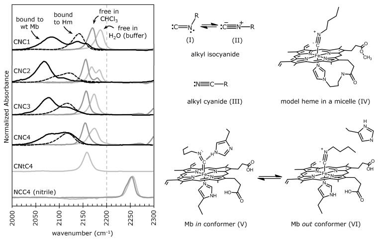
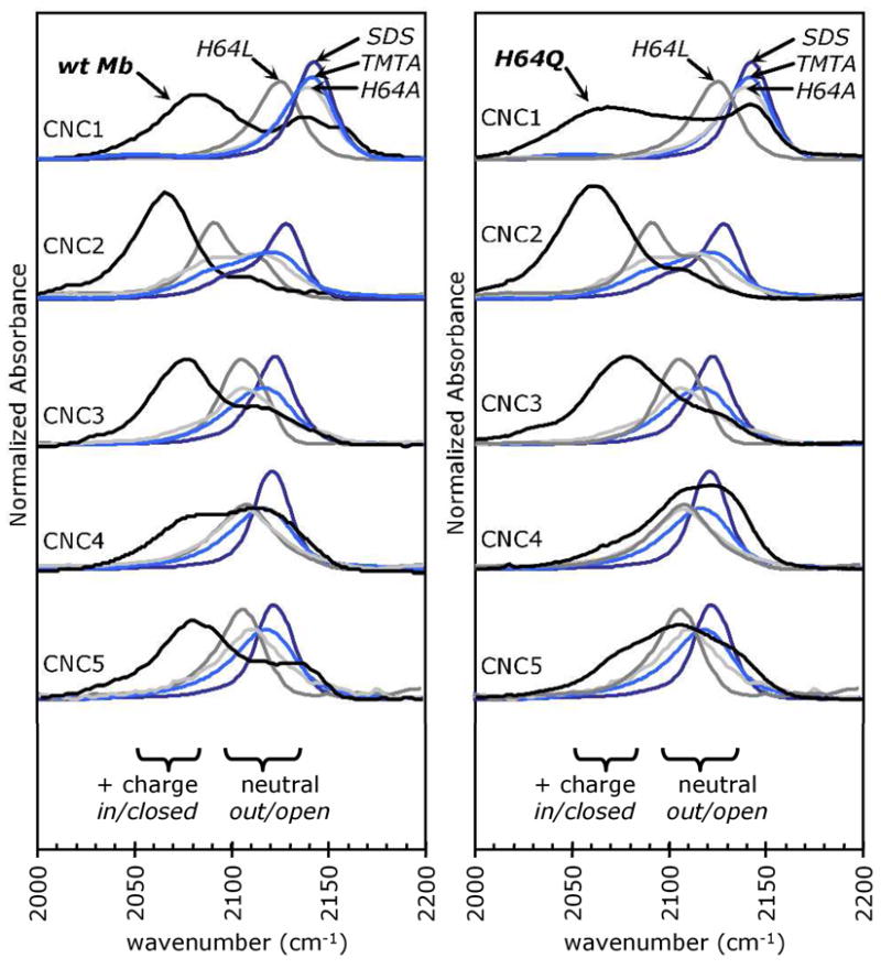
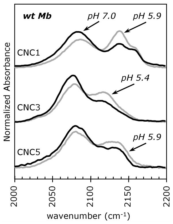
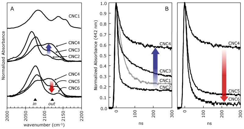
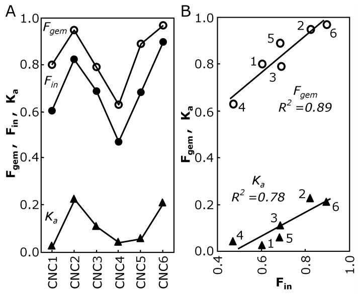
The FTIR spectra for alkyl isocyanides (CNRs) change from a single nu(CN) band centered at approximately 2175 cm(-1) to two peaks at approximately 2075 and approximately 2125 cm(-1) upon binding to sperm whale myoglobin (Mb). The low- and high-frequency peaks have been assigned to in and out conformations, respectively. In the in conformation, the ligand is pointing toward the protein interior, and the distal His64(E7) is in a closed position, donates a H-bond to the bound isocyano group, enhances back-bonding, and lowers the C-N bond order. In the out conformation, the ligand side chain points toward solvent through a channel opened by outward rotation of His64. Loss of positive polarity near the binding site causes an increase in C-N bond order. Support for this interpretation is threefold: (1) similar shifts to lower frequency occur for MbCO complexes when H-bond donation from His64(E7) occurs; (2) only one peak at approximately 2125 cm(-1), indicative of an apolar environment, is observed for CNRs bound to H64A or H64L Mb mutants or to chelated protoheme in soap micelles; and (3) the fraction of in conformation based on FTIR spectra correlates strongly with the fraction of geminate recombination after nanosecond laser photolysis. The in alkyl side chain conformation causes the photodissociated ligand to be "stuck" in the distal pocket, promoting internal rebinding, whereas the out conformation inhibits geminate recombination because part of the ligand is already in an open E7 channel, poised for rapid escape.


Figures

Figure 1
Figure 1
Dependence of the isocyano stretching frequency, νCN in cm−1, on solvent environment, chelation to a model heme, and binding to wt Mb. The solid black line spectra represent CNRs bound to wt sperm whale Mb. The two absorbance bands in the MbCNR spectra are interpreted to be structures V and VI in the right panel. The black dashed spectra represent CNRs bound to protoheme mono-3-(1-imidazoyl) propylamide mono-methyl ester (Hm) solubilized in trimethyl(tetradecyl)ammonium bromide (TMTA) micelles. The light and dark gray spectra represent free CNRs and n-butyl nitrile in 100 mM potassium phosphate, 1 mM EDTA, pH 7 aqueous buffer and in chloroform (CHCl3), respectively. Splitting of the νCN peak for free and Hm-bound CNC2 is due to Fermi resonance (see Supplement). The FTIR spectra are normalized by total area, except for the wt Mb spectra, which are shown at 2x area to aid in visual comparisons. The spectra were collected at room temperature (22–25°C). CNtC4 refers to tert-butyl isocyanide and NCC4 to n-butyl nitrile.
Figure 2
Figure 2
The high frequency νCN peak in the MbCNR spectra is due to a population of the bound ligands in an apolar environment. The black lines represent FTIR spectra for the homologous series methyl isocyanide (CNC1) through pentyl isocyanide (CNC5) bound to wt Mb (left panel) and H64Q Mb (right panel). These spectra were compared to those of CNRs complexed to H64A (light gray) and H64L (dark gray) Mb and to Hm in SDS (dark blue) and TMTA (light blue) micelles, all of which have apolar binding environments. Note that splitting of the νCN peak for CNC2 bound to H64A Mb, H64L Mb, and Hm is due to Fermi resonance (see Supplement). The wt and H64Q Mb spectra are shown at 2x their normalized area to allow better comparisons with the spectra containing narrow single peaks.
Figure 3
Figure 3
FTIR spectra for CNC1, CNC3 and CNC5 bound to Mb at neutral and low pH. The spectra for the complexes at pH 7.0 (black lines) are overlaid with those at low pH (5.9, 5.4 and 5.9, respectively; gray lines). Acidic pH increases the percentage of protonated His(E7) side chains and causes the imidazole side chain to rotate away from the bound ligand and into solution (39, 43). The samples contained 3–4 mM MbCNR in 0.1 M potassium phosphate buffer and were analyzed at room temperature.
Figure 4
Figure 4
A. FTIR spectra of the wt MbCNR complexes. The blue arrow shows the increase in intensity of the high frequency absorbance for the CNC2 to CNC4 complexes and the red arrow shows the reverse trend for the CNC4 to CNC6 complexes. FTIR samples contained 3–4 mM MbCNR in 0.1 M potassium phosphate buffer at pH 7.0, 20–22°C. B. Geminate recombination of MbCNRs following photolysis by a 7 ns YAG laser flash. Photodissociated CNRs either rebind geminately on 300 ns time scales (as shown) or escape to solvent and then rebind in a bimolecular process on μs to ms timescales (not shown). The blue arrow shows that Fgem decreases for the series CNC2-CNC4 and the red arrows shows that this trend reverses for the series CNC4-CNC6 (red arrow). These samples contained 0.1 mM Mb and 1 mM CNR in the same buffer.
Figure 5
Figure 5
A. Dependence of Fgem (open circles), Fin (filled circles) and Ka (filled triangles) on alkyl isocyanide length for wt MbCNR complexes. The equilibrium association constants, Ka, were taken from (10) and (7). All three parameters have the same undulating pattern, which arises from a combination of steric pressure and hydrophobic forces acting on the alkyl side chain. B. Correlations between Fin and Fgem, and between Fin and Ka. The three independent measurements and the strong correlations among them can be interpreted in terms of an equilibrium between the in and out conformations for each bound CNR and the hypothesis that non-covalently bound CNRs only escape rapidly from the distal binding pocket when the alkyl side chain is pointing out and the His(E7) gate is open (see text).

References

    1. Lein A, Pauling L. The Combining Power of Myoglobin for Alkyl Isocyanides and the Structure of the Myoglobin Molecule. Proc Natl Acad Sci U S A. 1956;42:51–54. - PMC - PubMed
    1. St George RC, Pauling L. The combining power of hemoglobin for alkyl isocyanides, and the nature of the heme-heme interactions in hemoglobin. Science. 1951;114:629–634. - PubMed
    1. Talbot B, Brunori M, Antonini E, Wyman J. Studies on the reaction of isocyanides with haemproteins. I. Equilibria and kinetics of the binding to the isolated chains of human haemoglobin. J Mol Biol. 1971;58:261–276. - PubMed
    1. Brunori M, Talbot B, Colosimo A, Antonini E, Wyman J. Studies on the reaction of isocyanides with haemproteins. II. Binding to normal and modified human haemoglobins. J Mol Biol. 1972;65:423–434. - PubMed
    1. Stetzkowski F, Cassoly R, Banerjee R. Binding of alkylisocyanides with soybean leghemoglobin. Comparisons with sperm whale myoglobin. J Biol Chem. 1979;254:11351–11356. - PubMed

Publication types