. 2010 Jun 22;49(24):4987-97.
doi: 10.1021/bi1001745.

Alkyl isocyanides serve as transition state analogues for ligand entry and exit in myoglobin

Affiliations

Alkyl isocyanides serve as transition state analogues for ligand entry and exit in myoglobin

George C Blouin et al. Biochemistry. .

Abstract

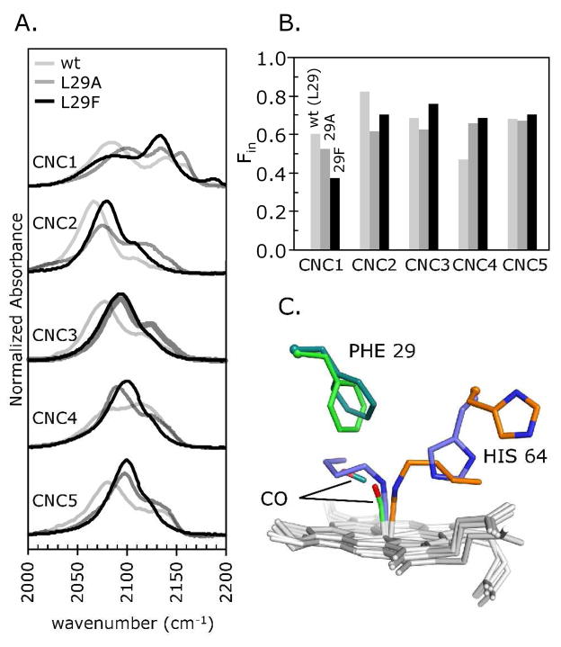
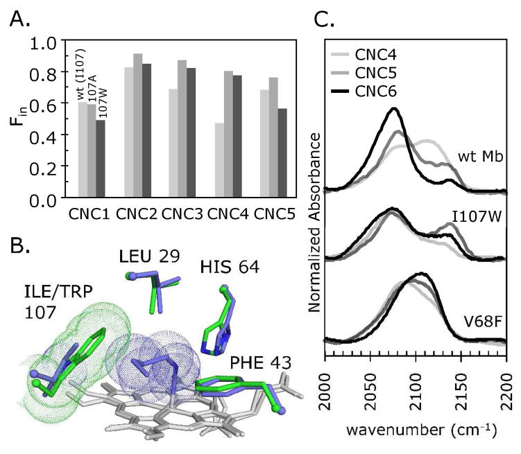
Alkyl isocyanides (CNRs) identify pathways for diatomic ligand movement into and out of Mb, with their side chains acting as transition state analogues. The bound alkyl groups point either into the back of the distal pocket (in conformation, nu(CN) approximately 2070-2090 cm(-1)), which allows hydrogen bond donation from His64(E7) to the isocyano group, or toward solvent through an open His(E7) channel (out conformation, nu(CN) approximately 2110-2130 cm(-1)), which prevents polar interactions with the isocyano atoms. Fractions of the in conformer (F(in)) were measured by FTIR spectroscopy for methyl through n-pentyl isocyanide bound to a series of 20 different distal pocket mutants of sperm whale myoglobin and found to be governed by the ease of rotation of the His(E7) side chain, distal pocket volume and steric interactions, and, for the longer isocyanides, the unfavorable hydrophobic effect of placing their terminal carbon atoms into the solvent phase in the out conformation. There are strong correlations between the fraction of in conformer, F(in), for long-chain MbCNR complexes measured by FTIR spectroscopy, the fraction of geminate recombination of photodissociated O(2), and the bimolecular rates of O(2) entry into the distal pocket. These correlations indicate that alkyl isocyanides serve as transition state analogues for the movement of O(2) into and out of the binding pocket of Mb.


Figures

Figure 1
Figure 1. Stereo view of amino acids selected for mutation near bound CNRs
The binding pockets are shown for native (pH 7) and wt (pH 9) MbCNC4 (104m and 111m, respectively, (1)) and are globally aligned by their Cα atoms. Mutation of the residues shown in dark blue are expected to affect the free energy of CNC4 in the in conformation (slate blue) by increased or decreased steric hindrance, whereas those in brown should selectively affect the out conformer of CNC4 (orange). The heme groups are in white, and van der Waals spheres for CNC4 are shown to provide an indication of residue/ligand packing. The alkyl groups of bound CNRs may act as transitions state analogs for CO in the photodissociated B state or during entry into the binding pocket through the His64 “gate” (marked as CO not observable). CO in the dissociated B state has been observed directly in low temperature and time-resolved crystallography experiments, whereas, to date, ligand movement through the E7 gate has not been observed directly.
Figure 2
Figure 2. Effect of mutating residues Arg45 and Phe46
A. The νCN peaks in the FTIR spectra of methyl through pentyl isocyanides (CNC1-CNC5) bound to wt, R45K, R45E, and F46V Mbs. B. The fraction of in conformers (Fin) for each mutant in panel A as a function of CNR size. These values indicate that the distal histidine is pushed outward more easily in the order wt < R45K < R45E ≈ F46V by the alkyl tail of each ligand. C. Structures of native (orange) and F46V (slate blue) MbCNC4 (104M and 101M, respectively; (1)), globally aligned by their Cα atoms. The smaller Val46 side chain allows the His64 imidazole to rotate away from the terminal carbon of the ligand and relieve steric hindrance of the CNR in the out conformation.
Figure 3
Figure 3. The CNR out conformation acts as a transition state analog for diatomic ligand entry
A. Correlations between the fraction of geminate recombination of photolyzed O2, Fgem,O2, and the fraction of bound CNC5 that adopts the in conformation, Fin,CNC5, for Mb mutants with substitutions near the His(E7) gate, including R45K, R45E and F46V. Fgem,O2 values were taken from (4). B. Correlations for wt, R45K, R45E, and F46V Mbs between the logs of k′entry,O2 or k′NO and Fin,CNC5.
Figure 4
Figure 4. Effect of mutating residue V68
A. FTIR spectra of methyl through pentyl isocyanides (CNC1-CNC5) bound to wt and V68A, V68F, and V68I Mbs. B. Fin for each mutant-isocyanide pair as a function of ligand size. C. Native (orange) and wt (slate blue) MbCNC4 and V68I MbCO (green) globally aligned by their Cα atoms (PDB IDs 104m, 111m (1) and 1mlm (18), respectively). The V68I MbCO structure has two Ile68 χ2 rotomers (A and B). Hemes are in white. D. The same structural overlay as in panel C, except V68I MbCO is replaced by V68F MbCNC4 (PDB ID 107m; (1)).
Figure 5
Figure 5. Effect of mutating residue Leu29
A. FTIR spectra of methyl through pentyl isocyanides (CNC1-CNC5) bound to wt, L29A, and L29F Mbs. B. Fin for each mutant-isocyanide pair as a function of ligand size. C. Structures of native (orange) and wt (slate blue) MbCNC4, and L29F MbCO (light green) and L29F Mb:CO photoproduct (dark green), globally aligned by their Cα atoms (PDB IDs 104m, 111m (1), 2g0r and 2g0v (23), respectively; hemes in light gray). The benzyl side chain at residue 29 rotates to accommodate photodissociated CO in the B-state. A similar rotation may explain how the large CNR alkyl groups are accommodated within the L29F Mb binding pocket.
Figure 6
Figure 6. Effect of side chain size at residue 107 and blocking the Xe4 pocket
A. Fin plotted against ligand size for methyl through pentyl isocyanides (CNC1-CNC5) bound to wt, I107A and I107W Mbs. B. Native MbCNC4 (slate blue; pH 9 structure; PDB ID 105m; (1)) and I107W Mb (green; ferric form; PDB ID 2ohb; (5)) globally aligned by their Cα atoms, with van der Waals spheres shown for CNC4 and the Trp107 side chain. The Fin value for the actual I107W MbCNC4 complex is unexpectedly high (panel A). C. FTIR spectra of CNC4-CNC6 bound to wt, I107W and V68F Mbs. Mutations that block access to the Xe4 site inhibit inward movement of longer CNRs, as shown by increases in the high-frequency out conformation band at ~2125 cm−1.
Figure 7
Figure 7. Correlations between the Fin values for MbCNC5 complexes and parameters describing the binding kinetics of O2 for internal distal pocket mutants of Mb
Fgem and the logarithms of k′entry and kbond, but not kescape, measured for the reaction of O2 with Leu29, Val68, and Ile107 Mb mutants show moderate correlations with the Fin values for bound CNC5. k′NO (gray circles, upper right panel) serves as an independent measure of the entry rate of diatomic gases. The O2 and NO kinetics data were taken from Scott et al. (4).
Figure 8
Figure 8. The bimolecular association rate (k′) for O2 and CNC1-4 binding to Mb mutants with varied residue sizes at the E7 gate
There is only a small dependence of k′O2 on the size of the residue at 64(E7). The association rate constants for the larger CNRs, however, are highly dependent on the amino acid size in the channel normally occupied by the E7 gate. The k′ values are from this work (W64), Rohlfs et al. (11) and Olson et al. (26).
Figure 9
Figure 9
Left panel – structure of the native out MbCNC4 conformer (PDB ID 104m; (1)) with CO atoms placed at the terminal carbon atoms of the alkyl side chain. Right panel – structure of wt in MbCNC4 conformer (PDB ID 111m; (1)) with CO placed in the position found in the structure of the room temperature B state photoproduct of wt MbCO (PDB ID 1abs; (27)). All structures were globally aligned by Cα atoms and rendered in the same orientation in Pymol. The right panel represents ligand entry through the E7 channel and the left panel, represents ligand capture in the side path model of ligand binding first described in detail by (4).

References

    1. Smith RD, Johnson KA, Blouin GC, Phillips GN, Jr, Olson JS. Straight-Chain Alkyl Isocyanides Open the Distal Histidine Gate in Crystal Structures of Myoglobin. Biochemistry. 2010 to be co-published with this article. - PMC - PubMed
    1. Blouin GC, Olson JS. The Stretching Frequencies of Bound Alkyl Isocyanides Indicate Two Distinct Ligand Orientations within the Distal Pocket of Myoglobin. Biochemistry. 2010 to be co-published with this article. - PMC - PubMed
    1. Scott EE, Gibson QH. Ligand migration in sperm whale myoglobin. Biochemistry. 1997;36:11909–11917. - PubMed
    1. Scott EE, Gibson QH, Olson JS. Mapping the pathways for O2 entry into and exit from myoglobin. J Biol Chem. 2001;276:5177–5188. - PubMed
    1. Olson JS, Soman J, Phillips GN., Jr Ligand pathways in myoglobin: a review of Trp cavity mutations. IUBMB Life. 2007;59:552–562. - PubMed

Publication types