. 2010 Jun 29;49(25):5176-87.
doi: 10.1021/bi100399e.

Increasing the conformational entropy of the Omega-loop lid domain in phosphoenolpyruvate carboxykinase impairs catalysis and decreases catalytic fidelity

Affiliations

Increasing the conformational entropy of the Omega-loop lid domain in phosphoenolpyruvate carboxykinase impairs catalysis and decreases catalytic fidelity

Troy A Johnson et al. Biochemistry. .

Abstract

Many studies have shown that the dynamic motions of individual protein segments can play an important role in enzyme function. Recent structural studies of the gluconeogenic enzyme phosphoenolpyruvate carboxykinase (PEPCK) demonstrate that PEPCK contains a 10-residue Omega-loop domain that acts as an active site lid. On the basis of these structural studies, we have previously proposed a model for the mechanism of PEPCK catalysis in which the conformation of this mobile lid domain is energetically coupled to ligand binding, resulting in the closed conformation of the lid, necessary for correct substrate positioning, becoming more energetically favorable as ligands associate with the enzyme. Here we test this model by introducing a point mutation (A467G) into the center of the Omega-loop lid that is designed to increase the entropic penalty for lid closure. Structural and kinetic characterization of this mutant enzyme demonstrates that the mutation has decreased the favorability of the enzyme adapting the closed lid conformation. As a consequence of this shift in the equilibrium defining the conformation of the active site lid, the enzyme's ability to stabilize the reaction intermediate is weakened, resulting in catalytic defect. This stabilization is initially surprising, as the lid domain makes no direct contacts with the enolate intermediate formed during the reaction. Furthermore, during the conversion of OAA to PEP, the destabilization of the lid-closed conformation results in the reaction becoming decoupled as the enolate intermediate is protonated rather than phosphorylated, resulting in the formation of pyruvate. Taken together, the structural and kinetic characterization of A467G-PEPCK supports our model of the role of the active site lid in catalytic function and demonstrates that the shift in the lowest-energy conformation between open and closed lid states is a function of the free energy available to the enzyme through ligand binding and the entropic penalty for ordering of the 10-residue Omega-loop lid domain.


Figures

Figure 1
Figure 1
The sequence of the active site lid domain (residues 462-476). The site of introducing the glycine (467) is indicated above the sequence.
Figure 2
Figure 2
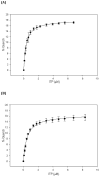
Binding isotherms illustrating the binding of ITP to (A) Wild-type and (B) A467G PEPCK as measured by intrinsic protein fluorescence quenching and described in the methods.
Figure 3
Figure 3
Structures of the OAA and PEP substrates and the enolate intermediate alongside the corresponding analogue utilized in these studies.
Figure 4
Figure 4
The structure of molecule A of the A467G-PEPCK-oxalate-GTP complex. The partial occupancy (0.7) of the closed lid in this molecule is illustrated by the 2Fo−Fc density rendered at 1.3 σ as a blue mesh with the corresponding lid domain rendered as a green stick model colored by atom type. The active site manganese ion and the bound oxalate molecule are rendered as a grey sphere and green stick model, respectively. The location of the A467G mutation is also labeled. Figures were generated using CCP4MG (47).
Figure 5
Figure 5
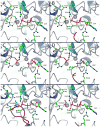
A comparison of the active sites of WT (PDB ID: 3DT2, 3DT7, 3DTB) and A467G PEPCK (PDB ID: 3MOE, 3MOF, 3MOH) in the PEPCK-Mn2+-βSP-Mn2+GTP (A: WT, B: A467G), PEPCK-Mn2+-PGA-Mn2+GDP (C: WT, D: A467G) and PEPCK-Mn2+-oxalate-Mn2+GTP (E: WT, F: A467G) complexes. All catalytic residues are rendered as ball-and-stick models colored by atom type whereas the ligands and nucleotides (βSP, PGA, oxalate, GTP, and GDP) are rendered as thick sticks colored by atom type and labeled. Potential hydrogen bonds between the ligands and the active site residues are indicated with black dashed lines. The active site and nucleotide associated manganese ions are rendered as grey spheres and the lid domain is rendered in dark red.
Figure 6
Figure 6
Minimal kinetic schemes for the PEPCK catalyzed decarboxylation of OAA in the presence of GDP. In red is shown those steps corresponding to the different steady state kinetic complexes and constants. Eo and Ec represent the lid-open and lid-closed forms of the enzyme, respectively.
Figure 7
Figure 7
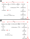
Minimal kinetic schemes for the PEPCK catalyzed reaction. In the (A) OAA→PEP direction and the (B) PEP→OAA direction. In red is shown those steps corresponding to the different steady state kinetic complexes and constants. Eo and Ec represent the lid-open and lid-closed forms of the enzyme, respectively.
Figure 8
Figure 8
Changes in the rotomeric state of Y235 and the water structure surrounding oxalate, in the A) lid-closed WT PEPCK-Mn2+-oxalate-Mn2+GTP (9) and B) lid-open A467G-Mn2+-oxalate-Mn2+GTP complexes. In A, the dashed lines represent potential hydrogen bonds with distances (a) 2.7 Å (b) 2.4 Å (c) 2.5 Å (d) 2.7 Å (e) 2.8 Å (f) 2.6 Å and (g) 3.0 Å. Similarly, in (B) the potential hydrogen bond distances are (a) 3.3 Å (b) 2.5 Å (c) 2.7 Å (d) 3.4 Å. Based upon structural work by Cotelesage et al (48), we propose that the water molecules separated by distance ‘c’ in panel A define the CO2 binding site. As illustrated in B, this binding site for CO2 is only present in the lid-closed conformation shown in A.
Figure 9
Figure 9
Cartoon reaction coordinate for WT (blue solid line) and A467G (orange dashed line) PEPCK based upon the differential inhibition of the two enzyme forms by the enolate intermediate analogue, oxalate.
Scheme 1
Scheme 1
The PEPCK catalyzed interconversion of OAA and PEP.

Similar articles

Cited by

References

    1. Lee MH, Hebda CA, Nowak T. The Role of Cations in Avian Liver Phosphoenolpyruvate Carboxykinase Catalysis. Activation and Regulation. J Biol Chem. 1981;256:12793–12801. - PubMed
    1. Colombo G, Carlson GM, Lardy HA. Phosphoenolpyruvate Carboxykinase (Guanosine 5′-Triphosphate) from Rat Liver Cytosol. Dual-Cation Requirement for the Carboxylation Reaction. Biochemistry. 1981;20:2749–2757. - PubMed
    1. Hebda CA, Nowak T. The Purification, Characterization, and Activation of Phosphoenolpyruvate Carboxykinase from Chicken Liver Mitochondria. J Biol Chem. 1982;257:5503–5514. - PubMed
    1. Hebda CA, Nowak T. Phosphoenolpyruvate Carboxykinase. Mn2+ and Mn2+ Substrate Complexes. J Biol Chem. 1982;257:5515–5522. - PubMed
    1. Yang J, Kalhan SC, Hanson RW. What Is the Metabolic Role of Phosphoenolpyruvate Carboxykinase? J Biol Chem. 2009;284:27025–27029. - PMC - PubMed

Publication types

Substances