. 2008 Aug;2(4):363-84.
doi: 10.2217/17520363.2.4.363.

Tau as a biomarker of neurodegenerative diseases

Affiliations

Tau as a biomarker of neurodegenerative diseases

Susanna Schraen-Maschke et al. Biomark Med. 2008 Aug.

Abstract

The microtubule-associated protein Tau is mainly expressed in neurons of the CNS and is crucial in axonal maintenance and axonal transport. The rationale for Tau as a biomarker of neurodegenerative diseases is that it is a major component of abnormal intraneuronal aggregates observed in numerous tauopathies, including Alzheimer's disease. The molecular diversity of Tau is very useful when analyzing it in the brain or in the peripheral fluids. Immunohistochemical and biochemical characterization of Tau aggregates in the brain allows the postmortem classification and differential diagnosis of tauopathies. As peripheral biomarkers of Alzheimer's disease in the cerebrospinal fluid, Tau proteins are now validated for diagnosis and predictive purposes. For the future, the detailed characterization of Tau in the brain and in peripheral fluids will lead to novel promising biomarkers for differential diagnosis of dementia and monitoring of therapeutics.


Conflict of interest statement

Disclosure Statement

The authors have reported no conflict of interest. Eugeen Vanmechelen is employee from Innogenetics NV, Gent, Belgium.

Figures

Figure 1
Figure 1. Stages in the neuropathology of Alzheimer’s disease. Implication for the use of Tau as biomarker during the course of the disease
AD lesions, namely neurofibrillary degeneration, follow a stereotyped, sequential, hierarchical pathway. The progression is categorized into 10 stages according to the brain regions affected: transentorhinal cortex (S1), entorhinal (S2), hippocampus (S3), anterior temporal cortex (S4), inferior temporal cortex (S5), medium temporal cortex (S6), polymodal association areas (prefrontal, parietal inferior, temporal superior) (S7), unimodal areas (S8), primary motor (S9a) or sensory (S9b, S9c) areas, and all neocortical areas (S10). Up to stage 6, the disease can be asymptomatic or paucisymptomatic (MCI): the CSF levels of pTau are however already altered and are useful for predictive diagnosis. In the more advanced stages when clinical criteria of AD are fulfilled CSF-tTau and CSF-pTau are altered and stable during disease course. Only the post-mortem analysis of brain with the presence of the two characteristic lesions of AD (amyloid plaques and NFD) allows to obtain the definite diagnosis of AD. The electrophoretic analysis of PHF-Tau aggregates is a useful tool for differential diagnosis of AD in brain tissue.
Figure 2
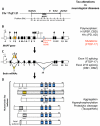
Figure 2. Microtubule-associated Tau gene, RNAs and human brain isoforms
(A) Genomic architecture of the 17q21.31 region encompassing MAPT gene and flanked by low copy repeats (rectangles) that are susceptible to chromosomal rearrangements such as deletions, duplications or inversion. Dotted lines illustrate the breakpoints responsible for the inversion of a ~900kb segment resulting in the H1/H2 polymorphism. (B) Tau gene MAPT spans more than 130 kb and is composed of 16 exons. The most studied polymorphism associated to the H1/H2 haplotype are 8 SNP (◆), 1 (TG)n microsatellite ({}) and a 238pb insertion/deletion (◇).H1 haplotype is associated with PSP and CBD. The three most frequent mutations responsible of FTDP-17 are in red. (C) Several mRNAs are generated by alternative splicing of exons 2, 3, 4A and 10. (D) In the human brain, six major Tau isoforms are generated from the alterative splicing of exons 2, 3 and 10. Exon 3 is always included with exon 2. The exon 10 encodes an additional microtubule-binding motif numbered R1 to R4. Half of Tau proteins contain three microtubule-binding motifs and the other half has four microtubule-binding motifs.
Figure 3
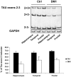
Figure 3. Semi-quantitative analysis of Tau exon 2 in control and DM1 brains
Reduced Tau exon 2 inclusion in DM1 brains. Tau exons 2 and 3 are alternatively spliced. Splicing of these two exons generated 3 Tau transcripts named 2+3+, 2+3- and 2-3-. The proportion of Tau transcripts including exon 2 and 3 was analysed by RT-PCR in three brain area (hippocampus, temporal and frontal cortex) in two control and in one DM1 patients. (B) Histogram of the percentage of transcripts including exon 2. The bands were quantified and the results expressed as the percentage of transcripts including exon 2. The mean and standard deviation were calculated from three individual experiments. Histograms are representative of the mean +/- standard deviation of three independent experiment.
Figure 4
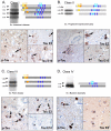
Figure 4. The “Bar code” of neurodegenerative diseases
Aggregated Tau proteins from the brain tissue of patients suffering from differents neurodegenerative disorders were resolved by 1D gels. Four main patterns of Tau bands were observed and the isoform content was determined using Tau exon-specific antibodies or two-dimensional gel electrophoresis. A classification is proposed according to Tau isoforms composing each of four biochemical patterns. A. Class I, which encompasses the largest number of degenerative diseases with Tau aggregation, is characterized by the aggregation of six Tau isoforms. This class is characterized by the occurrence on the brain samples of the hallmarks of Alzheimer’s disease i.e 1) intraneuronal somatic neurofibrillary tangles (NFT) as shown on the left panel by an antibody against pSer396-404 of Tau (AD2, arrows), 2) neuropile threads (NT), corresponding to neuritic processes filled of aggregated Tau (asterisks) and 3) neuritic plaques (NP). Antibodies raised against the sequences coded either by exon 2 (Tau E2, upper right panel) or by exon 10 (Tau E10, lower right) stain similarly NFTs(arrows), NTs (asterisks) and NPs (arrowhead) in AD and in other class I tauopathies. B. Only Tau isoforms containing four microtubule-binding domains aggregate in Class II disorders. They are represented here by a PSP case. AD2 anti-pTau labels the so-called astrocytic tufts, corresponding to intra-cytoplasmic Tau aggregation in astrocytes (arrow, left panel). Tau E2 and Tau E10 show glial Tau pathology as well, astrocytic (upper right, arrow) or oligodendrial (arrowhead, lower right). The “globoïd” neurobibrillary tangles are seen here in the substantia nigra (upper right, asterisk) and are stained by both Tau E2 and Tau E10. C. Tau isoforms with three microtubule-binding domains are found in Pick’s disease and few FTDP-17, corresponding to Class III diseases. Pick bodies are stained by anti-pTau and Tau E2 (arrows showing spherical 10μm bodies) but not by Tau E10 (in the lower right panel, a Pick body is readily seen but unstained, asterisk) D. At last, Class IV is characterized by the aggregation of Tau isoforms lacking sequences encoded by exons 2 and 3. Uptoday, the only known class IV diseases DM1 and DM2. On the left panel aggregates of hyperphosphorylated Tau are labelled with AD2. On the right panel, only unphosphorylated, non aggregated normal Tau are stained by anti-Tau E2 which in turn labels no aggregate.
Figure 5
Figure 5. Characterization of Tau in cerebrospinal fluid
Sequence of the largest form of brain-derived Tau, containing all alternatively spliced exons, is shown. Boxes in colour are the alternatively spliced exons and the microtubule-binding repeats: yellow, exon2; green exon 3, light blue exon 10 and the boxes in blue are the four tubulin-binding repeat domains Boxes indicate the epitopes of some antibodies commonly used i) in immuno-assays to quantify Tau in human CSF: for total Tau HT7, AT120 and BT2 are used, HT7-AT270, Tau-1-PHF-1 and Tau-1/CP27 are used in phospho-specific assays. [116, 166] ii) in brain tissue analysis : AD2, AT100 or 12E8 for Tau phospho-epitopes. Letters in red indicate the 8 sequences identified using an optimized immunoprecipitation (BT2) mass spectrometry analysis of 16 peptides [157].

References

    1. Buee L, Bussiere T, Buee-Scherrer V, Delacourte A, Hof PR. Tau protein isoforms, phosphorylation and role in neurodegenerative disorders. Brain Res Brain Res Rev. 2000;33(1):95–130. - PubMed
    1. Stefansson H, Helgason A, Thorleifsson G, Steinthorsdottir V, Masson G, Barnard J, Baker A, Jonasdottir A, Ingason A, Gudnadottir VG, et al. A common inversion under selection in Europeans. Nature genetics. 2005;37(2):129–137. - PubMed
    1. Schraen-Maschke S, Dhaenens CM, Delacourte A, Sablonniere B. Microtubule-associated protein tau gene: a risk factor in human neurodegenerative diseases. Neurobiology of disease. 2004;15(3):449–460. - PubMed
    1. Pittman AM, Myers AJ, Duckworth J, Bryden L, Hanson M, Abou-Sleiman P, Wood NW, Hardy J, Lees A, de Silva R. The structure of the tau haplotype in controls and in progressive supranuclear palsy. Human molecular genetics. 2004;13(12):1267–1274. - PubMed
    1. Andreadis A. Tau gene alternative splicing: expression patterns, regulation and modulation of function in normal brain and neurodegenerative diseases. Biochim Biophys Acta. 2005;1739(2-3):91–103. - PubMed