. 2010 Aug;9(4):592-606.
doi: 10.1111/j.1474-9726.2010.00586.x. Epub 2010 May 10.

AMP-activated protein kinase deficiency exacerbates aging-induced myocardial contractile dysfunction

Affiliations

AMP-activated protein kinase deficiency exacerbates aging-induced myocardial contractile dysfunction

Subat Turdi et al. Aging Cell. 2010 Aug.

Abstract

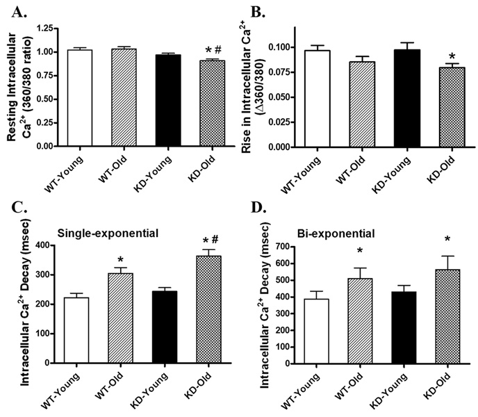
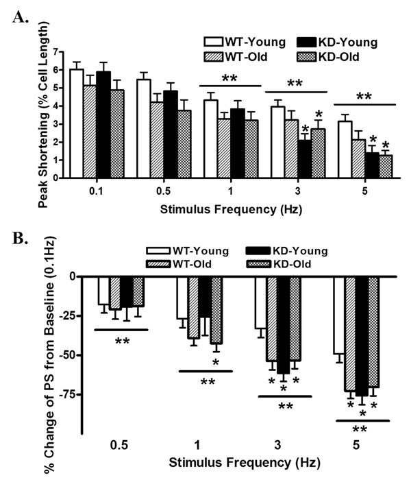
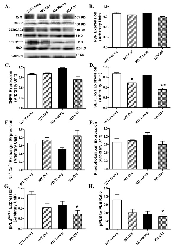
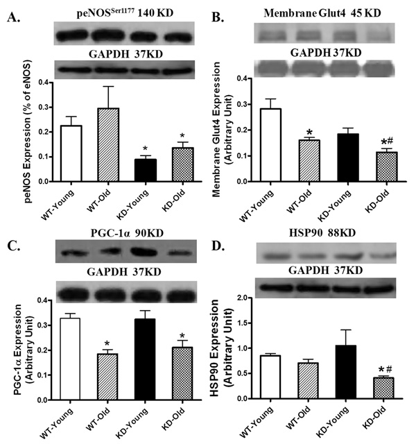
Aging is associated with myocardial dysfunction although the underlying mechanism is unclear. AMPK, a key cellular fuel sensor for energy metabolism, is compromised with aging. This study examined the role of AMPK deficiency in aging-associated myocardial dysfunction. Young or old wild-type (WT) and transgenic mice with overexpression of a mutant AMPK alpha(2) subunit (kinase dead, KD) were used. AMPK alpha isoform activity, myocardial function and morphology were examined. DCF and JC-1 fluorescence probes were employed to quantify reactive oxygen species (ROS) and mitochondrial membrane potential (DeltaPsim), respectively. KD mice displayed significantly reduced alpha(2) but not alpha(1) AMPK isoform activity at both ages with a greater effect at old age. Aging itself decreased alpha(1) isoform activity. Cardiomyocyte contractile function, intracellular Ca(2+) handling, and SERCA2a levels were compromised with aging, the effects of which were exacerbated by AMPK deficiency. H&E staining revealed cardiomyocyte hypertrophy with aging, which was more pronounced in KD mice. TEM micrographs displayed severe disruption of mitochondrial ultrastructure characterized by swollen, irregular shape and disrupted cristae in aged KD compared with WT mice. Aging enhanced ROS production and reduced DeltaPsim, the effects of which were accentuated by AMPK deficiency. Immunoblotting data depicted unchanged Akt phosphorylation and a significant decrease in mitochondrial biogenesis cofactor PGC-1alpha in aged groups. AMPK deficiency but not aging decreased the phosphorylation of ACC and eNOS. Expression of membrane Glut4 and HSP90 was decreased in aged KD mice. Moreover, treatment of the AMPK activator metformin attenuated aging-induced cardiomyocyte contractile defects. Collectively, our data suggest a role for AMPK deficiency in aging-induced cardiac dysfunction possibly through disrupted mitochondrial function and ROS production.


Figures

Fig. 1
Fig. 1
α-isoform-specific AMPK activity of left ventricular tissues from young or old wild-type (WT) and AMPK deficient KD mice. Mean ± SEM, n = 6–8 hearts per group, *p < 0.05 vs. WT young group, #p < 0.05 vs. WT- and KD-old groups.
Fig. 2
Fig. 2
Cardiomyocyte mechanical function in left ventricular myocytes from young or old wild-type (WT) and AMPK deficient KD mice. (A) Resting cell length; (B) Peak shortening (PS, normalized to resting cell length); (C) Maximal velocity of shortening (+ dL/dt); (D) Maximal velocity of relengthening (−dL/dt); (E) Time-to-PS (TPS); and (F) Time-to-90% relengthening (TR90). Mean ± SEM, n= 95 – 96 cells from 5 mice per group, *p < 0.05 vs. respective young group, #p < 0.05 vs. WT-old group.
Fig. 3
Fig. 3
Intracellular Ca2+ property in cardiomyocytes from young or old wild-type (WT) and AMPK deficient KD mice. (A) Baseline intracellular Ca2+ levels (360/380 ratio); (B) Rise in intracellular Ca2+ levels in response to electrical stimulus (Δ360/380 ratio); (C) Single exponential intracellular Ca2+ decay rate; and (D) Bi-exponential intracellular Ca2+ decay rate. Mean ± SEM, n = 73 – 74 cells from 5 mice per group, *p < 0.05 vs. respective young group, #p < 0.05 vs. WT-old group.
Fig. 4
Fig. 4
Peak shortening (PS) amplitude of cardiomyocytes from young or old wild-type (WT) and AMPK deficient KD mice at different stimulus frequencies (0.1 – 5.0 Hz). (A) Absolute peak shortening (normalized to resting cell length) amplitude at various stimulus frequencies; (B) Normalized PS value at various stimulus frequencies (PS was shown as % change from PS value obtained at 0.1 Hz from the same cell). Mean ± SEM, n = 17 – 24 cells from 5 mice per group, *p < 0.05 vs. WT-young group, **p < 0.05 vs. respective baseline value (0.1 Hz).
Fig. 5
Fig. 5
Western blot analysis depicting protein expression of ryanodine receptor (RyR), L-type Ca2+ channel dihydropyridine receptor (DHPR), sarco(endo)plasmic reticulum Ca2+-ATPase (SERCA2a), Na+/Ca2+ exchanger (NCX), phospholamban (PLB) and phosphorylated PLB (pPLB, Ser16) in myocardium from young or old wild-type (WT) and AMPK deficient KD mice. (A) Representative gel blots of RyR, DHPR, SERCA2a, NCX, PLB, pPLB and GAPDH (loading control) using specific antibodies; (B) RyR expression; (C) DHPR expression; (D) SERCA2a expression; (E) NCX expression; (F) PLB expression; (G) pPLB (Ser16) expression and (H) pPLB-to-PLB ratio. Mean ± SEM, n = 3 – 6 mice per group, *p < 0.05 vs. respective young group, #p < 0.05 vs. WT-old group.
Fig. 6
Fig. 6
Histological examination of hearts from (A) WT-young; (B) KD-young; (C) WT-old; and (D) KD-old mice stained with hematoxylin and eosin (H&E). Panel E/F displays representative transmission electron micrographs of cardiomyocytes from WT-old (E) and KD-old (F) mouse hearts. Scale bars = 50 µm (A–D) and 1 µm (E and F); G: Quantitative analysis of cardiomyocyte cross-sectional area in young or old WT and AMPK KD mice. Data were obtained by averaging areas of at least 200 nucleated myocytes per section from each mouse (3–5 mice per group).
Fig. 7
Fig. 7
Measurement of ROS production (A) and mitochondrial membrane potential (Ψm, B) in cardiomyocytes from young or old wild-type (WT) and AMPK deficient KD mice. Cells were loaded with the intracellular fluoroprobe CM-H2DCFDA (1 µM) at 37°C for 30 min prior to ROS detection. Quantification of Ψm was expressed as ratio between monomer and J-aggregate fluorescence (Red/Green). Mean ± SEM, n = 4 – 6 isolations per group, *p – 0.05 vs. respective young group, # p – 0.05 vs. WT-old group.
Fig. 8
Fig. 8
Expression of Akt, pAkt, AMPK and pACC in myocardium from young or old wild-type (WT) and AMPK deficient KD mice. A: Akt; B: pAkt; C: AMPK; and D: pACC (ser79). Inset: Representative gel blots of Akt, pAkt, AMPK and pACC using specific antibodies. GAPDH was used as the loading control. Mean ± SEM, n = 3 – 6 hearts per group, *p – 0.05 vs. respective young group, #p – 0.05 vs. WT-old group.
Fig. 9
Fig. 9
Cardiomyocyte contractile function in left ventricular myocytes from young or old wild-type (WT) mice with or without treatment of the AMPK agonist metformin (300 mg/kg b.w., for 5 days). (A) Resting cell length; (B) Peak shortening (PS, normalized to resting cell length); (C) Maximal velocity of shortening (+ dL/dt); (D) Maximal velocity of relengthening (− dL/dt); (E) Time-to-PS (TPS); and (F) Time-to-90% relengthening (TR90). Mean ± SEM, n = 63 – 69 cells from 3 – 4 mice per group, *p < 0.05 vs. WT young group, #p < 0.05 vs. WT-old group.
Fig. 10
Fig. 10
Expression of peNOS (Ser1177, A), membrane fraction of Glut4 (B), PGC1α (C) and HSP90 (D) in myocardium from young or old wild-type (WT) and AMPK deficient KD mice. Inset: Representative gel blots of peNOS, Glut4, PGC1α and HSP90 using specific antibodies. GAPDH was used as the loading control. Mean ± SEM, n = 4 – 6 mice per group, * p < 0.05 vs. respective young group, # p < 0.05 vs. WT-old group.

References

    1. Arad M, Seidman CE, Seidman JG. AMP-Activated Protein Kinase in the Heart: Role During Health and Disease. Circ Res. 2007;100:474–488. - PubMed
    1. Bhashyam S, Parikh P, Bolukoglu H, Shannon AH, Porter JH, Shen YT, Shannon RP. Aging Is Associated With Myocardial Insulin Resistance and Mitochondrial Dysfunction. Am J Physiol Heart Circ Physiol. 2007;293:H3063–H3071. - PubMed
    1. Calvert JW, Gundewar S, Jha S, Greer JJ, Bestermann WH, Tian R, Lefer DJ. Acute Metformin Therapy Confers Cardioprotection Against Myocardial Infarction Via AMPK-ENOS-Mediated Signaling. Diabetes. 2008;57:696–705. - PubMed
    1. Ceylan-Isik AF, Guo KK, Carlson EC, Privratsky JR, Liao SJ, Cai L, Chen AF, Ren J. Metallothionein Abrogates GTP Cyclohydrolase I Inhibition-Induced Cardiac Contractile and Morphological Defects: Role of Mitochondrial Biogenesis. Hypertension. 2009;53:1023–1031. - PMC - PubMed
    1. Chan AY, Soltys CL, Young ME, Proud CG, Dyck JR. Activation of AMP-Activated Protein Kinase Inhibits Protein Synthesis Associated With Hypertrophy in the Cardiac Myocyte. J Biol Chem. 2004;279:32771–32779. - PubMed

Publication types

MeSH terms