Review
. 2010 Aug;14(8):2066-72.
doi: 10.1111/j.1582-4934.2010.01088.x. Epub 2010 May 14.

The embryonic epicardium: an essential element of cardiac development

Affiliations
Review

The embryonic epicardium: an essential element of cardiac development

R Carmona et al. J Cell Mol Med. 2010 Aug.

Abstract

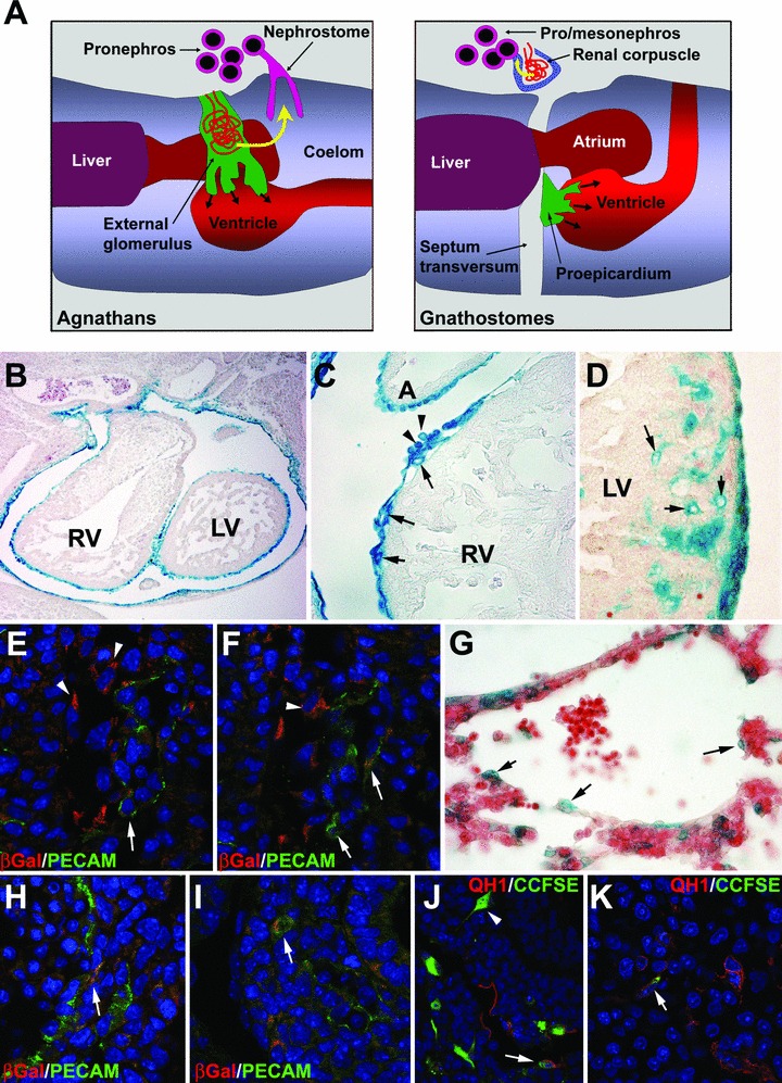
The epicardium has recently been identified as an active and essential element of cardiac development. Recent reports have unveiled a variety of functions performed by the embryonic epicardium, as well as the cellular and molecular mechanisms regulating them. However, despite its developmental importance, a number of unsolved issues related to embryonic epicardial biology persist. In this review, we will summarize our current knowledge about (i) the ontogeny and evolution of the epicardium, including a discussion on the evolutionary origins of the proepicardium (the epicardial primordium), (ii) the nature of epicardial-myocardial interactions during development, known to be essential for myocardial growth and maturation, and (iii) the contribution of epicardially derived cells to the vascular and connective tissue of the heart. We will finish with a note on the relationships existing between the primordia of the viscera and their coelomic epithelial lining. We would like to suggest that at least a part of the properties of the embryonic epicardium are shared by many other coelomic cell types, such that the role of epicardium in cardiac development is a particular example of a more general mechanism for the contribution of coelomic and coelomic-derived cells to the morphogenesis of organs such as the liver, kidneys, gonads or spleen.


Figures

Fig 1
Fig 1
(A) Hypothesis about the origin of the proepicardium of the gnathostomes from an ancestral pronephric external glomerulus. In agnathans, the external glomerulus filters the blood of the vascular network and releases the filtrate (yellow arrow) into the coelomic cavity, where it is aspirated by the nephrostome and eliminated through the pronephric tubules. Before its differentiation, the glomerular primordium from lamprey larvae originates the epicardial cells that spread over the heart surface. In gnathostomes, the excretory glomeruli have become internal (within the renal corpuscles), the pro and mesonephros are located caudal to the heart, but the primordium of the ancestral external glomerulus (in green), now located on the pericardial side of the septum transversum, is still providing the heart with epicardial cells as the proepicardium. (B–D) Labelling of the epicardium and the cells of the epicardial lineage in a transgenic mouse model (Wt1cre/Rosa26) that expresses β-Gal in cells that have previously expressed Wt1. We can see in (B) the lining of the heart from an E11.5 mouse embryo. In (C), EPDC appear in the subepicardial space by E11.5 (arrows). It is still possible to see some cells presumably released by the proepicardium and attached to the epicardium (arrowheads). In (D) we can see epicardially derived, intramyocardial coronary vessels by E15.5 (arrows). (E), (F) Early endothelial differentiation of EPDC in the atrioventricular groove of a Wt1cre/Rosa26 mouse embryo. β-Gal co-localizes with the endothelial marker PECAM-1 (arrows). Epicardial cells are shown by arrowheads. (G) Contribution of coelomic-derived cells to the liver sinusoids in a E10.5 mouse embryo. This is a different transgenic model expressing β-Gal under control of a Wt1 promoter (described in reference 37). Some presumptive endothelial cells are positive for this marker (arrows). (H), (I) Wt1cre/Rosa26 embryos of E11.5. Co-localization of β-Gal with the endothelial marker PECAM-1 is observed in organs other than the heart, such as the intestine (H) or the lung (I). (J), (K) The endothelial differentiation of coelomic-derived cells is also shown by this experiment of staining of the visceral coelomic epithelium of quail embryos (stage HH15) with the fluorescent tracer CCFSE. After 24 (J) and 48 hrs (K), fluorescence can be observed in cells of the stomach wall that are positive for the endothelial marker QH1 (arrows). Note the presence of CCFSE-labelled cells that area apparently migrating within the tissue (arrowhead in J).

References

    1. Mikawa T, Fischman DA. Retroviral analysis of cardiac morphogenesis: discontinuous formation of coronary vessels. Proc Natl Acad Sci USA. 1992;89:9504–8. - PMC - PubMed
    1. Pérez-Pomares JM, Macías D, García-Garrido L, et al. The origin of the subepicardial mesenchyme in the avian embryo, an immunohistochemical and quail-chick chimera study. Dev Biol. 1998;200:57–68. - PubMed
    1. Männer J, Pérez-Pomares JM, Macías D, et al. The origin, formation and developmental significance of the epicardium, a review. Cells Tiss Org. 2001;169:89–103. - PubMed
    1. Schulte I, Schlueter J, Abu-Issa R, et al. Morphological and molecular left-right asymmetries in the development of the proepicardium: a comparative analysis on mouse and chick embryos. Dev Dyn. 2007;236:684–95. - PubMed
    1. Van Wijk B, Van Den Berg G, Abu-Issa R, et al. Epicardium and myocardium separate from a common precursor pool by crosstalk between bone morphogenetic protein- and fibroblast growth factor-signaling pathways. Circ Res. 2009;105:431–41. - PMC - PubMed

Publication types