. 2010 May 17:7:99.
doi: 10.1186/1743-422X-7-99.

Interactions of SARS coronavirus nucleocapsid protein with the host cell proteasome subunit p42

Affiliations

Interactions of SARS coronavirus nucleocapsid protein with the host cell proteasome subunit p42

Qin Wang et al. Virol J. .

Abstract

Background: Severe acute respiratory syndrome-associated coronavirus (SARS-CoV) spreads rapidly and has a high case-mortality rate. The nucleocapsid protein (NP) of SARS-CoV may be critical for pathogenicity. This study sought to discover the host proteins that interact with SARS-CoV NP.

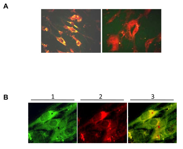
Results: Using surface plasmon resonance biomolecular interaction analysis (SPR/BIA) and matrix-assisted laser desorption/ionization time of flight (MALDI-TOF) mass spectrometry, we found that only the proteasome subunit p42 from human fetal lung diploid fibroblast (2BS) cells bound to SARS-CoV NP. This interaction was confirmed by the glutathione S-transferase (GST) fusion protein pulldown technique. The co-localization signal of SARS-CoV NP and proteasome subunit p42 in 2BS cells was detected using indirect immunofluorescence and confocal microscopy. p42 is a subunit of the 26S proteasome; this large, multi-protein complex is a component of the ubiquitin-proteasome pathway, which is involved in a variety of basic cellular processes and inflammatory responses.

Conclusion: To our knowledge, this is the first report that SARS-CoV NP interacts with the proteasome subunit p42 within host cells. These data enhance our understanding of the molecular mechanisms of SARS-CoV pathogenicity and the means by which SARS-CoV interacts with host cells.


Figures

Figure 1
Figure 1
Expression and Identification of SNP22b (SARS-CoV NP). (A) Supernatants (lanes 1 and 10) and pellets (lanes 2 and 6) of E. coli BL21 containing the pET22b-SNP22b vector expressing SARS-CoV NP, without IPTG induction. Supernatants (lanes 3 and 7) and pellets (lanes 4 and 8) of E. coli BL21 containing the pET22b-SNP22b vector and expressing SARS-CoV NP, induced with IPTG. Pellets of E. coli BL21 containing pET22b induced with IPTG (lane 5). Molecular weight markers (lane 9). The position of SARS-CoV NP (~47 kDa) is indicated by an arrow. (B) Western blot analysis of SARS-CoV NP using an anti-SARS-CoV NP horse polyclonal antibody. Lysate of E. coli BL21 expressing SARS-CoV NP, induced by IPTG (lane 1). Lysate of E. coli BL21 containing pET22b induced by IPTG (lane 2). Molecular weight markers (lane 3). The position of SARS-CoV NP (~47 kDa) is indicated by an arrow.
Figure 2
Figure 2
Identification of SARS-CoV NP-associated cellular protein(s). (A) Interaction of SARS-CoV NP with 2BS cell lysate proteins. The maximum reaction intensity was 1120 RU (manual injection). Baseline (a), injection of host cell lysate (b), RU value of flow cell after injection of host cell lysate (reactive amount = RUc-RUa) (c), eluted and recovered captured proteins (d), regeneration of flow cell (e). (B) Proteins captured from 2BS cell lysate (lane 1), blank controls (lane 2), and negative controls (lane 3). Molecular weight markers (lane 4). Arrows indicate the position of captured proteins.
Figure 3
Figure 3
Expression and identification of GST and the p42-GST fusion protein. (A) Lysate of E. coli BL21 containing the pGEX-5X-1-p42 vector, expressing p42-GST fusion protein without IPTG induction (lanes 1, 5) or induced by IPTG (lanes 2, 4). Lysate of E. coli BL21 containing the pGEX-5X-1 vector expressing GST with IPTG induction (lane 6) or without IPTG (lane 7). Molecular weight markers (lane 3). (B) Western blot analysis of p42-GST fusion protein and GST expression using an anti-GST mAb. Lysates of GST-expressing E. coli BL21 with IPTG induction (lanes 1, 2). Lysates of p42-GST fusion protein-expressing E. coli BL21 with IPTG induction (lanes 6, 7). Supernatants of E. coli BL21 expressing GST lysed using lysozyme (lane 3). Supernatants of E. coli BL21 expressing p42-GST fusion protein lysed by lysozyme (lane 5). Molecular weight markers (lane 4).
Figure 4
Figure 4
Western blot analysis of the interaction between p42-GST fusion protein and SARS-CoV NP in vitro. Bead + lysis buffer + NP (lane 1), bead + lysis buffer (lane 2), bead + GST lysate + NP (lane 3), bead + GST lysate (lane 4). Molecular weight markers (lane 5). SARS-CoV NP-SNP22b (positive control; lane 6), bead + p42-GST lysate (lane 7), bead + p42-GST lysate + NP (lane 8), p42-GST fusion protein bacterial lysate (lane 9). The position of SARS-CoV NP is indicated by an arrow.
Figure 5
Figure 5
Co-localization of SARS-CoV NP and p42 in 2BS cells. (A) SARS-CoV NP was expressed in 2BS cells transfected with recombinant -pcDNA3.0+SNP22b (left panel), 2BS cells transfected with pcDNA3.0 (right panel; negative control). (B) Proteasome subunit p42 in 2BS cells localized with anti-p42 antibody and anti-rabbit IgG-FITC conjugate, excitation at 488 nm (lane 1). SARS-CoV NP expressed in 2BS cells, localized using an anti-SARS-CoV NP mAb and anti-mouse IgG-TRITC conjugate, excitation at 568 nm (lane 2). Co-localization was detected by merging lanes 1 and 2 (lane 3).

Similar articles

Cited by

References

    1. Peiris JS, Lai ST, Poon LL, Guan Y, Yam LY, Lim W, Nicholls J, Yee WK, Yan WW. Coronavirus as a possible cause of severe acute respiratory syndrome. The Lancet. 2003;361:1319–1326. doi: 10.1016/S0140-6736(03)13077-2. - DOI - PMC - PubMed
    1. Ksiazek TG, Erdman D, Goldsmith CS. A Novel Coronavirus Associated with Severe Acute Respiratory Syndrome. N Engl J Med. 2003;348:1953–1966. doi: 10.1056/NEJMoa030781. - DOI - PubMed
    1. Tsui PT, Kwok ML, YUEN H. Severe acute respiratory syndrome: clinical outcome and prognostic correlates. Emerg Infect Dis. 2003;9:1064–1069. - PMC - PubMed
    1. Nicholl JM, Poon LL, Lee KC. Lung pathology of fatal severe acute respiratory syndrome. Lancet. 2003;361:1773–1778. doi: 10.1016/S0140-6736(03)13413-7. - DOI - PMC - PubMed
    1. Chen ZL, Pei DC, Jiang LX, Song YJ. Antigenicity analysis of different regions of the Severe Acute Respiratory Syndrome Coronavirus nucleocapsid protein. Clin Chem. 2004;50:988–995. doi: 10.1373/clinchem.2004.031096. - DOI - PMC - PubMed

Publication types

MeSH terms

Substances