. 2010 May 17:5:14.
doi: 10.1186/1747-1028-5-14.

Stat3: linking inflammation to epithelial cancer - more than a "gut" feeling?

Affiliations

Stat3: linking inflammation to epithelial cancer - more than a "gut" feeling?

Andrew Jarnicki et al. Cell Div. .

Abstract

Inflammation is an important environmental factor that promotes tumourigenesis and the progression of established cancerous lesions, and recent studies have started to dissect the mechanisms linking the two pathologies. These inflammatory and infectious conditions trigger immune and stromal cell release of soluble mediators which facilitate survival and proliferation of tumour cells in a paracrine manner. In addition, (epi-)genetic mutations affecting oncogenes, tumour-suppressor genes, chromosomal rearrangements and amplifications trigger the release of inflammatory mediators within the tumour microenvironment to promote neoplastic growth in an autocrine manner. These two pathways converge in tumour cells and result in activation of the latent signal transducer and activator of transcription 3 (Stat3) which mediates a transcriptional response favouring survival, proliferation and angiogenesis. The abundance of cytokines that activate Stat3 within the tumour microenvironment, which comprises of members of the interleukin (IL) IL6, IL10 and IL17/23 families, underpins a signaling network that simultaneously promotes the growth of neoplastic epithelium, fuels inflammation and suppresses the host's anti-tumour immune response. Accordingly, aberrant and persistent Stat3 activation is a frequent observation in human cancers of epithelial origin and is often associated with poor outcome.Here we summarize insights gained from mice harbouring mutations in components of the Stat3 signaling cascade and in particular of gp130, the shared receptor for the IL6 family of cytokines. We focus on the various feed-back and feed-forward loops in which Stat3 provides the signaling node in cells of the tumour and its microenvironment thereby functionally linking excessive inflammation to neoplastic growth. Although these observations are particularly pertinent to gastrointestinal tumours, we suggest that the tumour's addiction to persistent Stat3 activation is likely to also impact on other epithelial cell-derived cancers. These insights provide clues to the judicious interference of the gp130/Stat3 signaling cascade in therapeutically targeting cancer.


Figures

Figure 1
Figure 1
Hematopoietic cell infiltration and STAT3 hyperactivation in gastrointestinal tumours. (A) Haematopoietic cell infiltration (green) in gastric tumours of gp130Y757F mutant mice visualized following adoptive transfer of bone marrow from GFP-transgenic mice. Metaplastic gastric epithelium is visualized following antibody binding to the intestine-specific epithelial cells surface marker gpA33 (red) [164]. (B) Immunohistological stain for activated STAT3 in sections of spontaneous arising gastric and CAC-induced colonic adenomas in gp130Y757F mutant mice, and from human gastric and colonic adenocarcinomas using an antibody directed against tyrosine phosphorylated STAT3.
Figure 2
Figure 2
Regulation of intracellular Stat3 signalling. Stat3 signalling is induced by various kinases (green) in a phosphorylation dependent manner (green arrows), and counteracted by several regulatory proteins (red). For instance, Stat3 activation occurs in response to gp130 homodimerization following binding of IL6 or IL11 (blue) to their specific transmembrane receptor α-subunits (white) [165]. Phosphorylation of four membrane distal tyrosine (Y, black) residues by constitutively associated JAK-family tyrosine kinases (TK) is sufficient to enable src-homology domain (SH)-2 mediated binding of STAT3 and, to a lesser extent, of STAT1. Once tyrosine phosphorylated, STAT1/3 form homo- and heterodimers, which translocate and trans-activate target genes, including the negative regulator SOCS3. STAT3 can also be phosphorylated by certain cytosolic TKs and receptor TK. Meanwhile, serine threonine kinases (STK) mediate serine-phosphorylation that maximizes the transcriptional activity of STAT3 and enables its mitochondrial targeting. Gp130 also engages the Ras/ERK pathway through binding of the tyrosine phosphatase SHP2 to the membrane proximal phospho-YxxV consensus sequence (red, where V is valine and × any amino acid). This phosphor-tyrosine also provides the binding site for SOCS3 to mediate proteasomal degradation of ligand-occupied receptor complexes. Gp130 signaling is also attenuated by the Y-phosphatase activity of SHP2, while cytoplasmic PIAS3 protein sequesters Y-phosphorylated STAT3 from homodimerization, nuclear translocation and target gene activation. Representative examples of different types of bona-fide Stat3 target genes are listed [166].
Figure 3
Figure 3
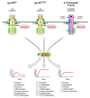
Strength and duration of STAT3 activation determines cellular responses. STAT3 activation patterns and associated outcomes differ between IL6 and IL10 family cytokine-induced signaling. In response to binding of ligand (blue), signaling from gp130 is transient, because the phosphorylated, membrane proximal tyrosine residue (Y, red) provides a binding site for the negative regulator SOCS3. Although SOCS3 is also induced in response to activation of the IL10 receptor family, STAT3 signaling remains sustained, because the IL10 receptor chains lack the corresponding YxxV motif. Similarly, signaling from the mutant gp130Y757F receptor is sustained due to phenylalanine (F) substitution of the Y757 residue (Y759 in human GP130), resulting in exaggerated activation of its down-stream signaling molecules. The range of target genes activated differs between acute and sustained STAT3 activation, most evident in macrophages where the former promotes and the latter suppresses inflammatory response.
Figure 4
Figure 4
Stat3 links inflammation to cancer. Inflammation and cancer are functionally linked by intrinsic, Stat3-dependent autocrine feedback loops in neoplastic epithelium (red arrows) and extrinsic, feedforward and often reciprocal interactions between tumour, inflammatory and stromal cells that collectively make up the microenvironment (green arrows) [167]. The ubiquitous expression of gp130 and the capacity of STAT3 to stimulate its own transcription as well as that of gp130 ligands (in particular IL6 and IL11) also provides numerous amplification loops between the different cell types. Furthermore, limited responsiveness to IL6 and IL11 imposed by restricted expression of the ligand-specific receptor α-subunits can be overcome by IL6-transsignaling [90]. Excessive cell-intrinsic STAT3 activation is also triggered by oncogenic events from (epi-)genetic activation of positive regulators (i.e. receptor (-associated) TKs) and loss of function mutations of negative regulators (i.e. SOCS3). Expression of IL6 is also directly induced by NF-κB and indirectly through a feedback mechanism including lin28 and the let7-microRNA, while latent Stat3 in conjunction with NF-κB triggers non-canonical IL6 expression. Epithelial NF-κB and STAT3 are activated in response to the abundantly present inflammatory cytokines IL1, TNFα and IL6 which are released from TLR-activated myeloid cell (macrophages), with IL6 and IL11 also contributed by tumour-associated stromal fibroblast and myoepithelial cells. Meanwhile, release of IL17 and IL22 from mature Th17 cells provide an additional extrinsic link which results in excessive STAT3 activation in tumour cells.

Similar articles

Cited by

References

    1. Coussens LM, Werb Z. Inflammation and cancer. Nature. 2002;420:860–867. doi: 10.1038/nature01322. - DOI - PMC - PubMed
    1. Pollard JW. Tumour-educated macrophages promote tumour progression and metastasis. Nat Rev Cancer. 2004;4:71–78. doi: 10.1038/nrc1256. - DOI - PubMed
    1. Dunn GP, Bruce AT, Ikeda H, Old LJ, Schreiber RD. Cancer immunoediting: from immunosurveillance to tumor escape. Nat Immunol. 2002;3:991–998. doi: 10.1038/ni1102-991. - DOI - PubMed
    1. zur Hausen H. Papovaviruses and human tumors. Haematol Blood Transfus. 1983;28:289–292. - PubMed
    1. Munoz N, Bosch FX, de Sanjose S, Herrero R, Castellsague X. Epidemiologic classification of human papillomavirus types associated with cervical cancer. N Engl J Med. 2003;348:518–527. doi: 10.1056/NEJMoa021641. - DOI - PubMed

LinkOut - more resources