. 2010;12(3):R90.
doi: 10.1186/ar3017. Epub 2010 May 18.

Monocyte surface expression of Fcgamma receptor RI (CD64), a biomarker reflecting type-I interferon levels in systemic lupus erythematosus

Affiliations

Monocyte surface expression of Fcgamma receptor RI (CD64), a biomarker reflecting type-I interferon levels in systemic lupus erythematosus

Yi Li et al. Arthritis Res Ther. 2010.

Abstract

Introduction: More than half of systemic lupus erythematosus (SLE) patients show evidence of excess type I interferon (IFN-I) production, a phenotype associated with renal disease and certain autoantibodies. However, detection of IFN-I proteins in serum is unreliable, and the measurement of interferon-stimulated gene (ISG) expression is expensive and time consuming. The aim of this study was to identify a surrogate marker for IFN-I activity in clinical samples for monitoring disease activity and response to therapy.

Methods: Monocyte surface expression of Fcgamma receptors (FcgammaRs), chemokine receptors, and activation markers were analyzed with flow cytometry in whole blood from patients with SLE and healthy controls. FcgammaR expression also was measured in peripheral blood mononuclear cells (PBMCs) from healthy controls cultured with Toll-like receptor (TLR) agonists, cytokines, or serum from SLE patients. Expression of ISGs was analyzed with real-time PCR.

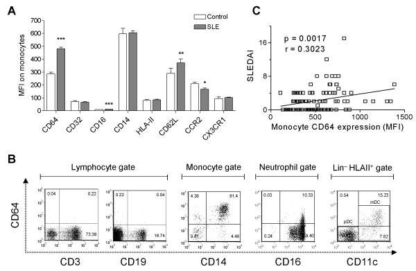
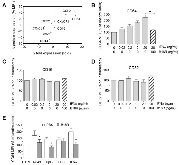
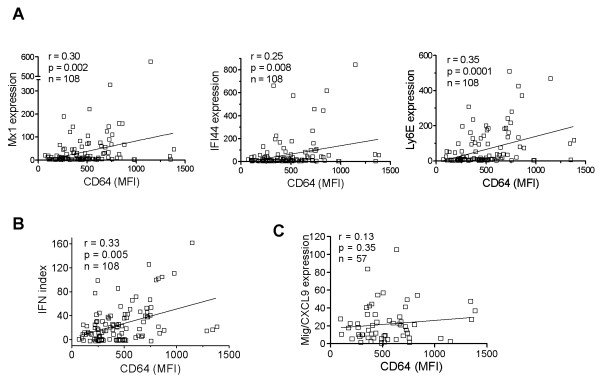
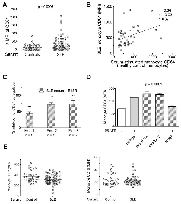
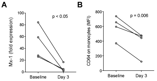
Results: Circulating CD14+ monocytes from SLE patients showed increased surface expression of FcgammaRI (CD64). The mean fluorescent intensity of CD64 staining correlated highly with the ISG expression (MX1, IFI44, and Ly6E). In vitro, IFN-I as well as TLR7 and TLR9 agonists, induced CD64 expression on monocytes from healthy controls. Exposure of monocytes from healthy controls to SLE sera also upregulated the expression of CD64 in an IFN-I-dependent manner. Decreased CD64 expression was observed concomitant with the reduction of ISG expression after high-dose corticosteroid therapy.

Conclusions: Expression of CD64 on circulating monocytes is IFN-I inducible and highly correlated with ISG expression. Flow-cytometry analysis of CD64 expression on circulating monocytes is a convenient and rapid approach for estimating IFN-I levels in SLE patients.


Figures

Figure 1
Figure 1
CD64 expression on monocytes is increased in SLE. (a) Flow-cytometry analysis of monocyte markers in SLE patients (n = 108) and healthy controls (n = 83). Bars represent the average mean fluorescent intensity on CD14+ monocytes, and error bars denote standard error. *P < 0.05; **P < 0.01; ***P < 0.001. (b) Representative flow cytometry of CD64 expression on peripheral blood cells from a lupus patient, including CD3+ T cells, CD19+ B cells, CD14+ monocytes, CD16+ neutrophils, and CD11c+ dendritic cells (primarily myeloid dendritic cells). Lymphocytes, monocytes, and neutrophils were gated based on their forward/sideward scatter characteristics. Dendritic cells were first gated on Lin-, HLA-DR+ cells, and then further identified as myeloid dendritic cells (CD123-, CD11c+) with flow cytometry. (c) Bivariate analysis of CD64 expression on monocytes (MFI, determined with flow cytometry) and SLEDAI (n = 108). Correlation coefficient was calculated by using Spearman's rank correlation (P = 0.0017; r = 0.301).
Figure 2
Figure 2
CD64 expression is inducible by IFN-I and TLR agonists. (a) Effects of recombinant IFN-α (4 ng/ml) on mRNA and protein expression of monocyte markers. Mean differences (fold increase/decrease versus control) in mRNA expression in PBMCs treated with PBS or IFN-α (n = 4 per group) were determined with real-time PCR. Changes in cell-surface protein levels on monocytes (MFI) were measured with flow cytometry. Positive values denote increased expression after IFN-α treatment compared with controls (PBS). (b through d) Dose-response analysis of CD64 (b), CD16 (c), and CD32 (d) expression in vitro on IFN-α-stimulated monocytes from healthy controls. In some groups, B18R (0.1 μg/ml) was added 1 hour before stimulation with IFN-α. (e) Induction of monocyte CD64 surface expression by R848 (1 μg/ml), CpG-DNA (10 ng/ml), LPS (1 μg/ml), or IFN-α (4 ng/ml) in the presence/absence of B18R (added 1 hour before stimulation). Flow-cytometry analysis was performed 19 hours after stimulation. Values represent the mean ± SEM from three independent experiments. *P < 0.05 (Student's t test).
Figure 3
Figure 3
CD64 expression correlates with the interferon signature in SLE. Bivariate analysis of CD64 expression on monocytes (MFI, determined with flow cytometry; n = 108) and (a) the expression of the ISGs MX1, IFI44, and Ly6E (determined with real-time PCR), (b) the composite IFN score derived from the three ISGs, or (c) expression of the IFN-γ-inducible gene CXCL9. Spearman's correlation was used for all analyses.
Figure 4
Figure 4
SLE serum induces surface expression of CD64 on monocytes. (a) Effect of sera from SLE patients (n = 65) and healthy controls (n = 44) on CD64 expression by healthy control monocytes. PBMCs from healthy donors were cultured in the presence of 25% serum for 19 hours before flow cytometry. ΔMFI was calculated by subtracting baseline CD64 MFI from donor monocytes cultured in autologous serum from the MFI of CD64 expression after incubation with serum from healthy controls or SLE patients. A positive ΔMFI indicates an upregulation of CD64 expression compared with the baseline levels. (b) Bivariate analysis of SLE serum-induced upregulation of CD64 on healthy control monocytes and CD64 expression on monocytes from the SLE serum donors (n = 37; r = 0.36; P < 0.05; Spearman's correlation). (c) Effect of B18R pretreatment on SLE serum-induced CD64 upregulation on healthy control monocytes. Three independent experiments, each using five or more serum samples from SLE patients are depicted. ** P < 0.01; *** P < 0.001 compared with the levels of CD64 induced by SLE-serum without B18R present (Student's t test). (d) Effect of anti-IFN-γ or anti-IL-12 neutralizing antibodies, or isotype control antibody (mouse IgG1) on SLE serum-induced upregulation of CD64 on healthy control monocytes. Bars represent the mean of four independent experiments. Difference in CD64 expression in the presence/absence of B18R was analyzed with Student's t test. (e) Flow-cytometry analysis of CD32 and CD16 expression on healthy control monocytes after incubation with sera from SLE patients or healthy controls, as described in (a).
Figure 5
Figure 5
Effect of therapy on CD64 fluorescence intensity. (a) Expression of MX1 in PBMCs (measured with real-time PCR) and (b) surface expression of CD64 on monocytes (measured with flow cytometry) in four patients before (day 0) and after (day 3) therapy with high-dose methylprednisolone (1 g IV daily for 3 days). Differences were analyzed by using Student's paired t test.

Comment in

Similar articles

Cited by

References

    1. Bennett L, Palucka AK, Arce E, Cantrell V, Borvak J, Banchereau J, Pascual V. Interferon and granulopoiesis signatures in systemic lupus erythematosus blood. J Exp Med. 2003;197:711–723. doi: 10.1084/jem.20021553. - DOI - PMC - PubMed
    1. Baechler EC, Batliwalla FM, Karypis G, Gaffney PM, Ortmann WA, Espe KJ, Shark KB, Grande WJ, Hughes KM, Kapur V, Gregersen PK, Behrens TW. Interferon-inducible gene expression signature in peripheral blood cells of patients with severe lupus. Proc Natl Acad Sci USA. 2003;100:2610–2615. doi: 10.1073/pnas.0337679100. - DOI - PMC - PubMed
    1. Feng X, Wu H, Grossman JM, Hanvivadhanakul P, FitzGerald JD, Park GS, Dong X, Chen W, Kim MH, Weng HH, Furst DE, Gorn A, McMahon M, Taylor M, Brahn E, Hahn BH, Tsao BP. Association of increased interferon-inducible gene expression with disease activity and lupus nephritis in patients with systemic lupus erythematosus. Arthritis Rheum. 2006;54:2951–2962. doi: 10.1002/art.22044. - DOI - PubMed
    1. Kirou KA, Lee C, George S, Louca K, Papagiannis IG, Peterson MG, Ly N, Woodward RN, Fry KE, Lau AY, Prentice JG, Wohlgemuth JG, Crow MK. Coordinate overexpression of interferon-alpha-induced genes in systemic lupus erythematosus. Arthritis Rheum. 2004;50:3958–3967. doi: 10.1002/art.20798. - DOI - PubMed
    1. Zhuang H, Narain S, Sobel E, Lee PY, Nacionales DC, Kelly KM, Richards HB, Segal M, Stewart C, Satoh M, Reeves WH. Association of anti-nucleoprotein autoantibodies with upregulation of type I interferon-inducible gene transcripts and dendritic cell maturation in systemic lupus erythematosus. Clin Immunol. 2005;117:238–250. doi: 10.1016/j.clim.2005.07.009. - DOI - PubMed

Publication types

MeSH terms