. 2010 May 14;141(4):583-94.
doi: 10.1016/j.cell.2010.04.020.

A temporarily distinct subpopulation of slow-cycling melanoma cells is required for continuous tumor growth

Affiliations

A temporarily distinct subpopulation of slow-cycling melanoma cells is required for continuous tumor growth

Alexander Roesch et al. Cell. .

Abstract

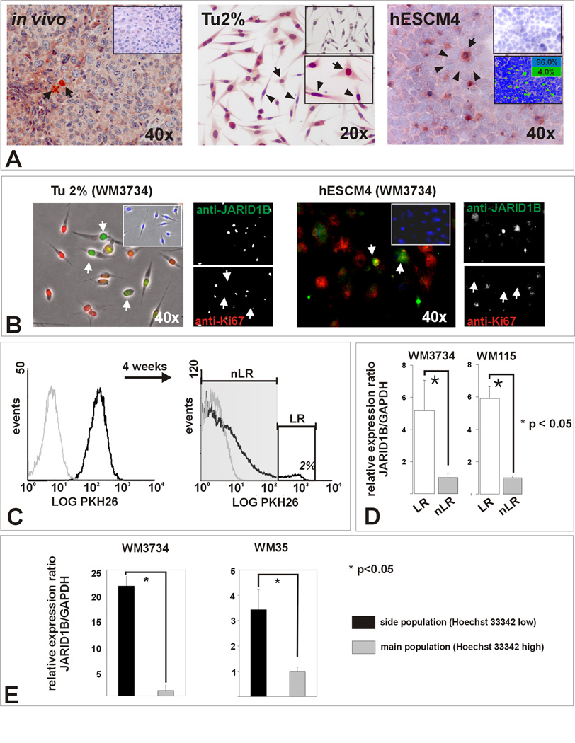
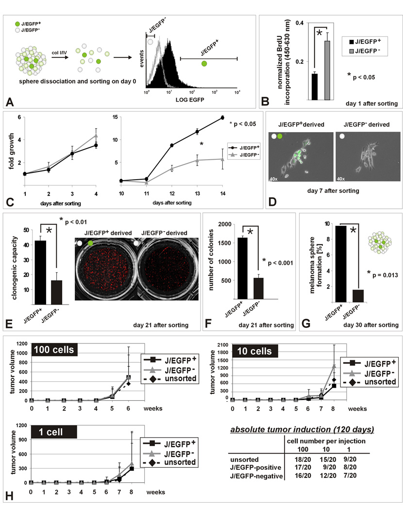
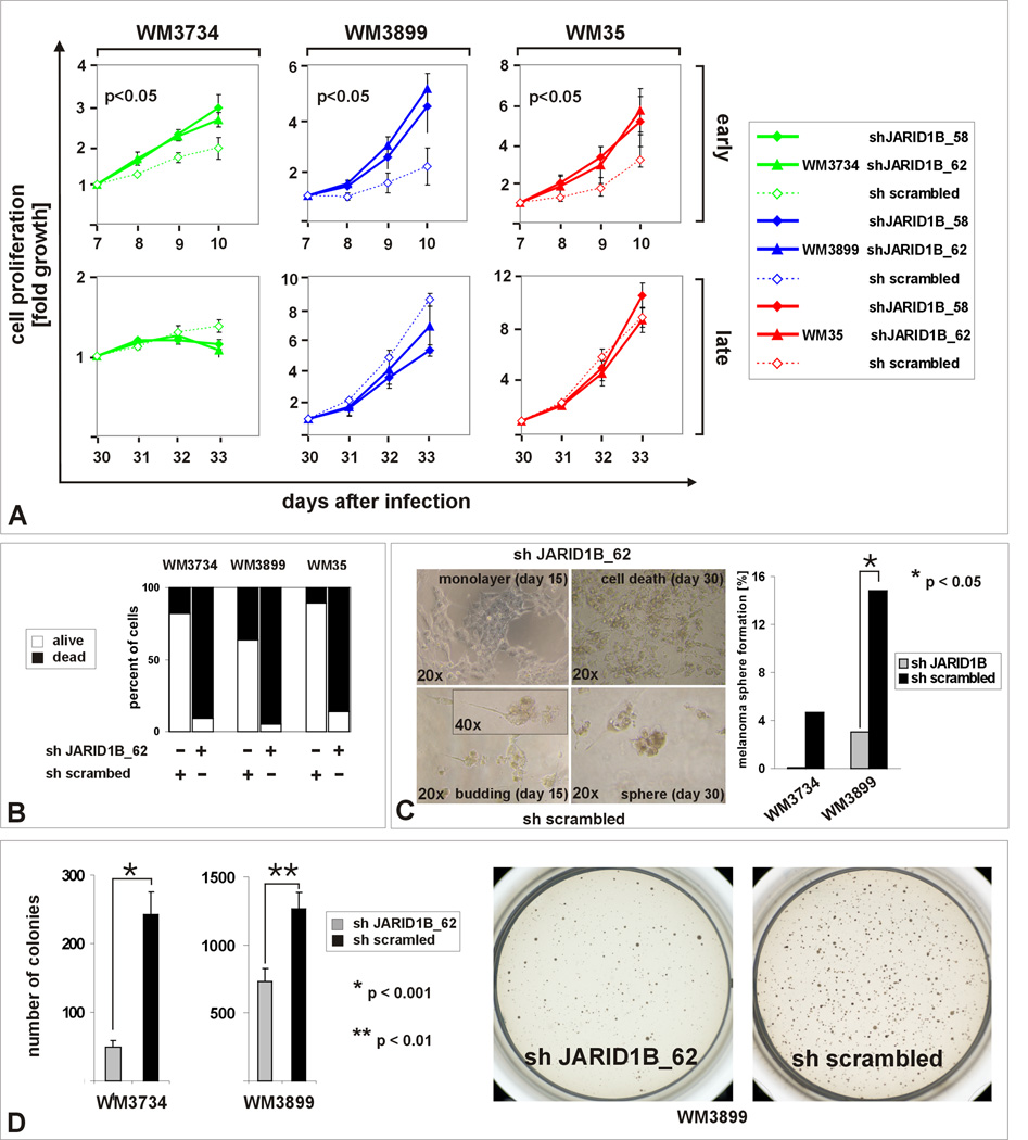
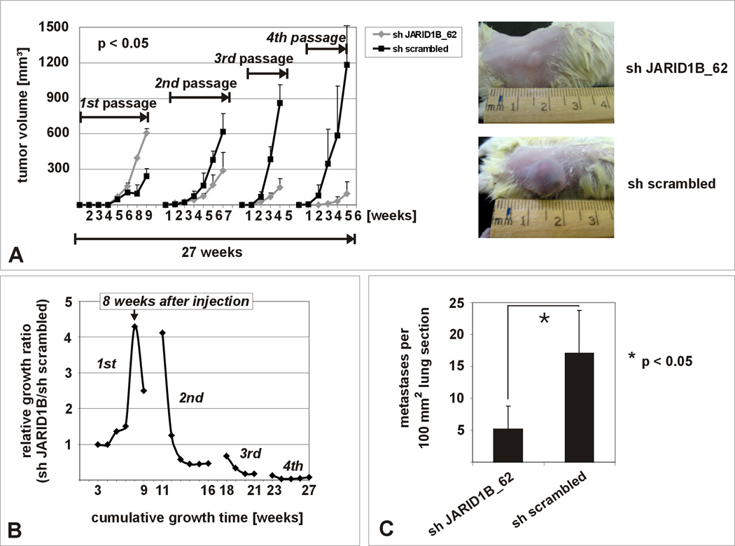
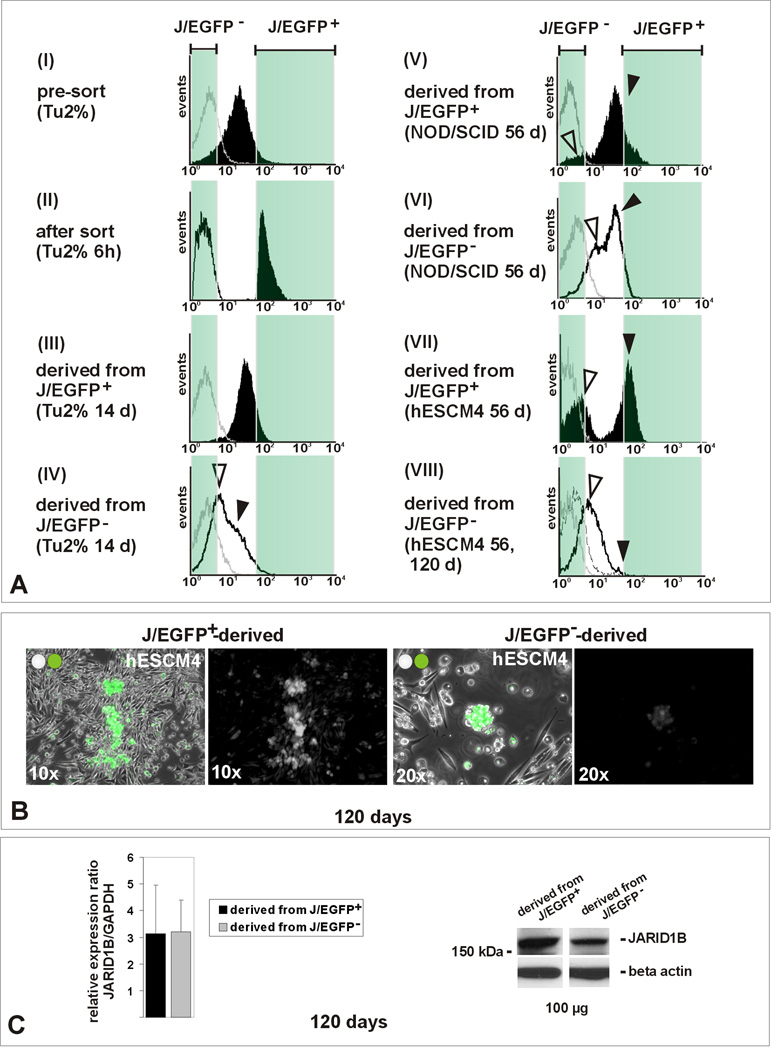
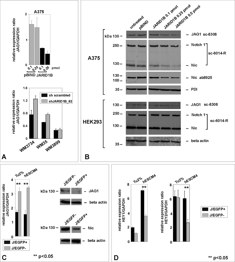
Melanomas are highly heterogeneous tumors, but the biological significance of their different subpopulations is not clear. Using the H3K4 demethylase JARID1B (KDM5B/PLU-1/RBP2-H1) as a biomarker, we have characterized a small subpopulation of slow-cycling melanoma cells that cycle with doubling times of >4 weeks within the rapidly proliferating main population. Isolated JARID1B-positive melanoma cells give rise to a highly proliferative progeny. Knockdown of JARID1B leads to an initial acceleration of tumor growth followed by exhaustion which suggests that the JARID1B-positive subpopulation is essential for continuous tumor growth. Expression of JARID1B is dynamically regulated and does not follow a hierarchical cancer stem cell model because JARID1B-negative cells can become positive and even single melanoma cells irrespective of selection are tumorigenic. These results suggest a new understanding of melanoma heterogeneity with tumor maintenance as a dynamic process mediated by a temporarily distinct subpopulation.


Figures

Figure 1
Figure 1. Melanomas contain a subpopulation of slow-cycling cells characterized by increased JARID1B expression
(A) Left panel: Representative example of a vertical growth phase melanoma after immunostaining for JARID1B. Single positive cells (arrows) with predominantly nuclear and minor cytoplasmic staining are surrounded by negative cells. Middle: Single adherent WM3734 melanoma cells show high JARID1B (arrows) while the bulk of cells shows median or low expression (arrowheads) when grown in conventional medium (Tu2%). Isotype control (top) and higher magnification (bottom) are shown as inserts. Right: More distinct JARID1B staining was seen in cryosections of WM3734 melanoma spheres (hESCM4). Digital quantitation after pseudocoloring revealed a relative frequency of 4% JARID1B-immunopositive (green) and 96% immunonegative cells (blue) for this section (see also Figure S1). (B) Anti- JARID1B-positive cells (Alexa Fluor® 488, green) are predominately negative for the proliferation marker Ki-67 (Alexa Fluor® 568, red). The result was reproduced in WM115 cells (not shown). (C) Flow cytometry analysis of WM3734 melanoma sphere cells after incubation with PKH26 (black line) compared to unlabeled control cells (grey line). After 4 weeks in hESCM4, 2% of the cells retained the maximum label (label-retaining ‘LR’ cells, white square). (D) LR cells displayed significantly enhanced JARID1B expression compared to non label-retaining (nLR) cells in QPCR. (E) QPCR screening of side population cells from WM3734 and WM35 melanoma sphere cells showing an upregulation of JARID1B compared to the main populations.
Figure 2
Figure 2. JARID1B is a biomarker for the label-retaining subpopulation in melanoma
(A) Lentiviral pLU-JARID1Bprom-EGFP-Blast. (B) Flow cytometric determination of the J/EGFP-positive subpopulation, exemplarily shown for WM3734JARID1Bprom-EGFP melanoma spheres grown in hESCM4 (see also Figure S2). QPCR and immunoblots of sorted populations showed a significant correlation between exogenous EGFP and endogenous JARID1B expression (p<0.05, t-test). The 5%-threshold was based on our in vitro and in vivo observations on endogenous JARID1B expression frequency (Figure 1). (C) PKH26 label-retention of WM3734JARID1Bprom-EGFP sphere cells in vitro. Left: LR cells grouped as a distinct J/EGFP-positive subpopulation (LR/JEGFP-positive, white square) and were enriched for small-sized cells. Right: LR-J/EGFP-positive cells (white bar) showed the highest capacity to re-form spheres. Depicted is one representative out of three independent experiments. (D) Left: Anti-BrdU-APC flow cytometry of dissociated tumor cells revealed that 6 weeks after BrdU incubation the majority of cells diluted out the BrdU (filled black). Unlabeled (grey line) and freshly BrdU-labeled cells (black line) served as controls. The threshold was set to the peak APC intensity of freshly BrdU-labeled cells. Right: In vivo BrdU-labeled cells grouped as a distinct J/EGFP-positive subpopulation (LR/JEGFP-positive, white square, 1.6% of total population).
Figure 3
Figure 3. In vitro self renewal capacity of the JARID1B-positive subpopulation
(A) Methodical scheme. (B) Decreased BrdU incorporation into J/EGFP-positive cells 24 hours after separation from spheres (hESCM4) as determined by ELISA after pulsed BrdU exposition. (C) MTS assays of sorted J/EGFP-positive and –negative cells in hESCM4 (see also Figure S3). After day 10, the progeny of J/EGFP-positive cells proliferated significantly faster and (D) microscopically consisted of J/EGFP-positive and an increasing number of J/EGFP-negative cells. (E) Enhanced clonogenicity of single J/EGFP-positive cells. (F) J/EGFP-positive cells had both a higher potential to form 3D-colonies in 0.35% hESCM4-soft agar and (G) to self-renew again into heterogeneous melanoma spheres in limited dilution assays (hESCM4). Shown are representative results from at least two independent experiments. (H) Xenotransplantation growth curves after subcutaneous injection of 100, 10 or 1 WM3734JARID1Bprom-EGFP melanoma cell into NOD/LtSscidIL2Rγnull mice. Growth curves were stopped when the first mouse of the respective series exceeded a tumor size >1000 mm3. Also during the subsequent observation of remaining mice, no differences were seen.
Figure 4
Figure 4. In vitro exhaustion after knockdown of JARID1B
(A) MTS proliferation assays between days 7–10 and days 30–33 after JARID1B knock down (clones 58 and 62, see also Figure S4). Experiments were done in three independent approaches in conventional medium (Tu2%). (B) Exposure of JARID1B knocked down cells (5×10⁴ cells) to hESCM4 resulted in a strong increase of the number of trypan blue-positive cells after 4–6 weeks. (C) Limited (single cell) dilution assays determined the reduction of melanoma sphere formation in JARID1B knockdown cells (right panel). The left panel shows the corresponding cell morphologies. (D) Reduced 3D colony formation after JARID1B knockdown in Tu2% softagar. Depicted are representative results from at least 3 independent experiments.
Figure 5
Figure 5. In vivo exhaustion after knockdown of JARID1B
(A) In vivo tumor growth is exhausted after 4 passages of serial xenotransplantation of JARID1B knockdown WM3734 cells in NOD/LtSscidIL2Rγnull mice (n=5 per sample) compared to the control (see also Figure S5). (B) Normalized growth ratio (tumor volumes of sh JARID1B tumors divided by volumes of sh scrambled tumors) over the cumulative growth phase of 27 weeks. (C) Significant decrease of pulmonary metastasis after subcutaneous injection of 5×10⁵ JARID1B knocked down WM3899 cells into NOD/LtSscidIL2Rγnull mice (n=5 per sample). Shown is one representative from two independent experiments.
Figure 6
Figure 6. The JARID1B phenotype is dynamic (see also Figure S6)
(A) WM3734JARID1Bprom-EGFP cells were sorted according to J/EGFP levels as described before using maximum and minimum thresholds set at 5%. Untreated parental cells were used as a background control (grey line). Panel I: typical histogram pre-sort in conventional medium (Tu2%). Panel II: isolated J/EGFP-positive (filled black) and J/EGFP-negative cells (black line) 6 hours after FACS. Panel III: during subsequent culturing in Tu2% medium, the J/EGFP-positive subpopulation re-established a heterogeneous progeny of J/EGFP-positive and – negative cells (filled black). Panel V: same result in vivo after injection into NOD/LtSscidIL2Rγnull mice (filled black). Panels IV and VI: also J/EGFP-negative cells gave rise to a heterogeneous progeny including J/EGFP-positive cells (black line). Panel VII: in hESCM4, the J/EGFP-positive-derived progeny maintained a high number of J/EGFP-positive cells (filled black). Panel VIII: also the reversion of J/EGFP-negative to -positive cells was considerably decelerated (50-day line dashed, 120-day line black). Black and white arrowheads indicate developing positive and negative subpopulations. Depicted tumor histograms resulted from injection of 100 J/EGFP-positive or –negative cells, respectively. Shown are representative histograms from at least 3 analyses. (B) Immunofluorescence of cultures originating from J/EGFP-positive (left) and J/EGFP-negative cells (right) after 120 days in hESCM4. (C) Similar overall JARID1B expression in J/EGFP-positive and -negative-derived progenies after long-term culturing as determined by QPCR and immunoblotting.
Figure 7
Figure 7. JARID1B affects Jagged 1/Notch 1 signaling
(A) QPCR of A375-SM melanoma cells transiently transfected with pBIND-JARID1B vs. empty vector control (upper panel) and of JARID1B-knocked down WM3734, WM35, and WM3899 cells vs. sh scrambled control (lower panel). (B) Immunoblotting of JARID1B-transfected A375-SM melanoma and HEK293 control cells with antibodies against JAG1, uncleaved and cleaved Notch 1. Notch cleavage was confirmed by a second antibody. (C) Differential JAG1 expression and Notch cleavage in FACS-isolated J/EGFP-positive and -negative cells. (D) Differential expression of HEY1 and 2 in J/EGFP-positive and -negative cells.

Comment in

  • Don't let sleeping cells lie.
    Villanueva T. Villanueva T. Nat Rev Cancer. 2010 Jul;10(7):452. doi: 10.1038/nrc2884. Nat Rev Cancer. 2010. PMID: 20589969 No abstract available.

References

    1. Adams JM, Strasser A. Is tumor growth sustained by rare cancer stem cells or dominant clones? Cancer Res. 2008;68:4018–4021. - PubMed
    1. Balint K, Xiao M, Pinnix CC, Soma A, Veres I, Juhasz I, Brown EJ, Capobianco AJ, Herlyn M, Liu ZJ. Activation of Notch1 signaling is required for beta-catenin-mediated human primary melanoma progression. J Clin Invest. 2005;115:3166–3176. - PMC - PubMed
    1. Blagosklonny MV. Why therapeutic response may not prolong the life of a cancer patient: selection for oncogenic resistance. Cell Cycle. 2005;4:1693–1698. - PubMed
    1. Bonnet D, Dick JE. Human acute myeloid leukemia is organized as a hierarchy that originates from a primitive hematopoietic cell. Nat Med. 1997;3:730–737. - PubMed
    1. Christensen J, Agger K, Cloos PA, Pasini D, Rose S, Sennels L, Rappsilber J, Hansen KH, Salcini AE, Helin K. RBP2 belongs to a family of demethylases, specific for tri-and dimethylated lysine 4 on histone 3. Cell. 2007;128:1063–1076. - PubMed

Publication types

MeSH terms