. 2010 May 14;141(4):704-16.
doi: 10.1016/j.cell.2010.03.035.

Mechanosensitive hair cell-like cells from embryonic and induced pluripotent stem cells

Affiliations

Mechanosensitive hair cell-like cells from embryonic and induced pluripotent stem cells

Kazuo Oshima et al. Cell. .

Abstract

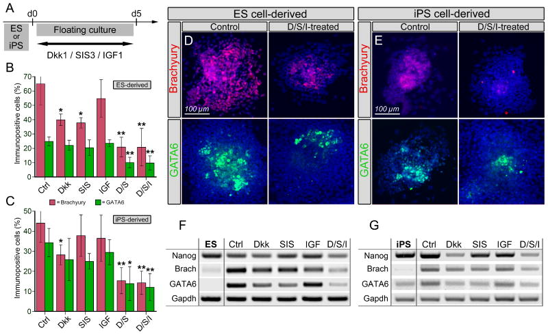
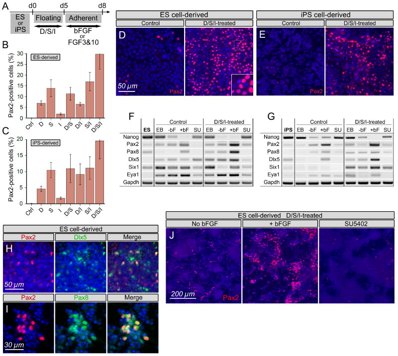
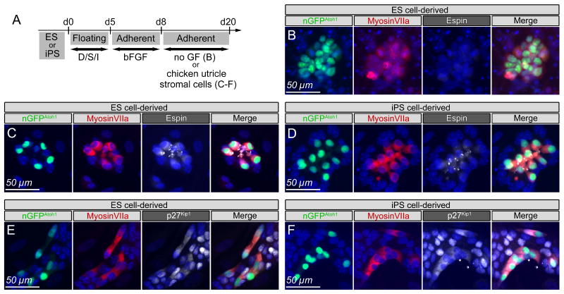
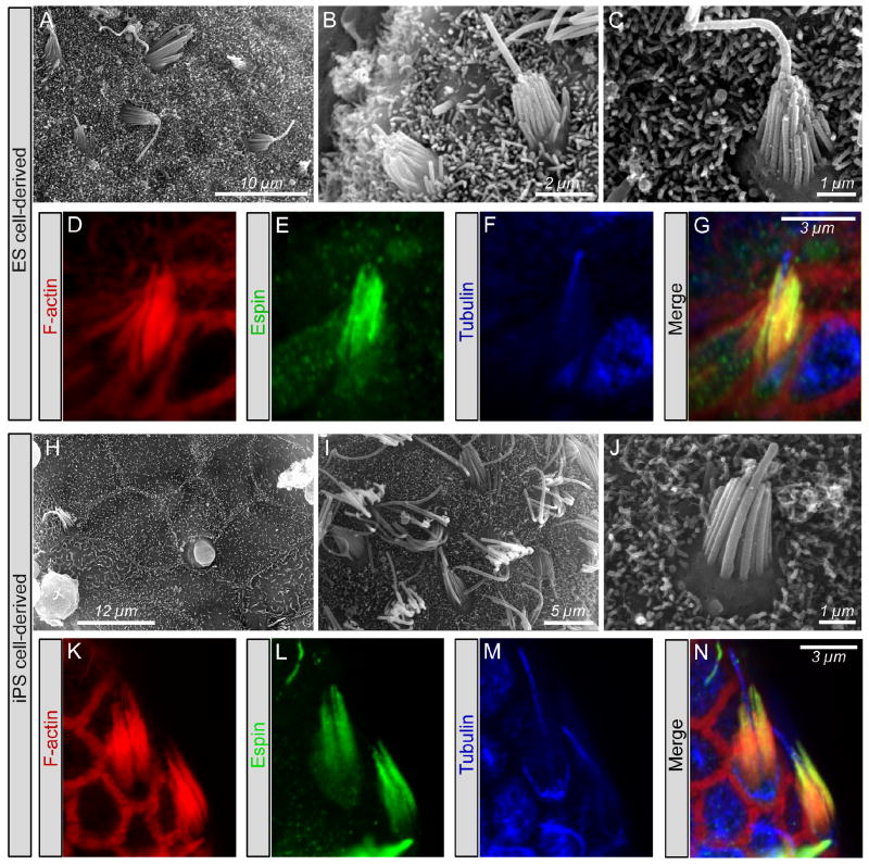
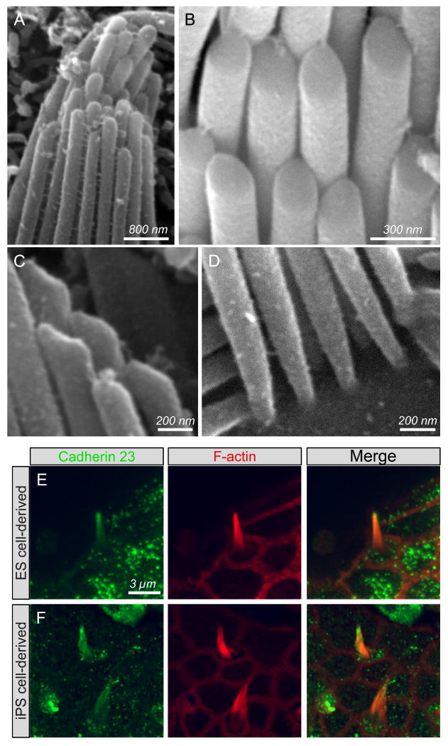
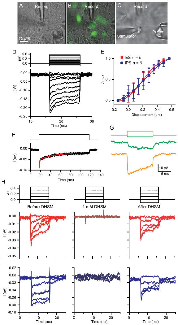
Mechanosensitive sensory hair cells are the linchpin of our senses of hearing and balance. The inability of the mammalian inner ear to regenerate lost hair cells is the major reason for the permanence of hearing loss and certain balance disorders. Here, we present a stepwise guidance protocol starting with mouse embryonic stem and induced pluripotent stem cells, which were directed toward becoming ectoderm capable of responding to otic-inducing growth factors. The resulting otic progenitor cells were subjected to varying differentiation conditions, one of which promoted the organization of the cells into epithelial clusters displaying hair cell-like cells with stereociliary bundles. Bundle-bearing cells in these clusters responded to mechanical stimulation with currents that were reminiscent of immature hair cell transduction currents.


Figures

Figure 1
Figure 1
Suppression of meso- and endodermal cell differentiation by interference with Wnt- and TGF-ß signaling. (A) C9 ES cells and No.25-5 iPS cells were dissociated into single cells and cultured in non-adhesive plates for five days to form embryoid bodies in presence of Dkk1, SIS3, and IGF-1, as indicated. (B,C) Embryoid bodies from ES (B) and iPS (C) cells were generated in presence of the factors indicated. Ctrl = no factors added, D/S = Dkk1 and SIS3, D/S/I = Dkk1, SIS3, and IGF-1. Error bars=S.D., n=5, * indicates p<0.05, ** indicates p<0.01, determined with paired, two-tailed t-tests. (D, E) Representative immunostainings of plated embryoid bodies from ES cells (D) and iPS cells (E). Treatment with D/S/I reduced the number of cells immunopositive for Brachyury and GATA6. (F, G) RT-PCR analyses show downregulation of transcripts for Brachyury and GATA6 in D/S/I-treated cultures of ES (F) and iPS (G) -derived populations. Expression of the pluripotent and ES cell marker Nanog is also reduced most noticeably after D/S/I treatment. Ctrl = embryoid bodies generated without factors added. ES = ES cells before differentiation; iPS = iPS cells before differentiation. Nuclear DAPI staining is shown in blue. See also Figures S1-S4.
Figure 2
Figure 2
Otic induction is most efficient in D/S/I-treated ES and iPS cells. (A) Embryoid bodies were generated for five days in presence of of Dkk1, SIS3, and IGF-1, as indicated; subsequent adherent culture was done for three days in fibronectin-coated plates in presence of otic inducers bFGF or FGF3&10. (B,C) ES- (B) and iPS- (C) cell derived cultures, exposed for three days to bFGF, were immunostained with antibodies to Pax2 and the percentage of Pax2-positive cells of the total cell population was determined. D/S/I treatment most efficiently increased the number of cells that responded with upregulation of Pax2 to bFGF-treatment. Ctrl = no added factors during embryoid body formation; error bars=S.D., n=3. (D, E) Representative immunocytochemical images showing upregulation of Pax2 in ES cell-derived (D) and iPS cell-derived (E) cultures in response to D/S/I treatment followed by bFGF exposure. Control = bFGF-treated cultures of embryoid bodies generated with no added factors. Inset = higher magnification view of the nuclear staining. (F, G) RT-PCR analyses for expression of Nanog and the early otic markers Pax2, Pax8, Dlx5, Six1, and Eya1 in cultures derived from ES (F) and iPS (G) cells. ES = ES cells before differentiation; iPS = iPS cells before differentiation. EB = embryoid bodies harvested at d5, -bF and +bF indicate absence and presence of bFGF between d5 and d8. SU = SU5402 treatment between d5 and d8. (H,I) Co-expression of otic markers Pax2 and Dlx5 (H) and of Pax2 and Pax8 (I) in the majority of ES cell-derived cultures after D/S/I and bFGF treatment. (J) Cultures of D/S/I-treated ES cell-derived embryoid body cells in absence and presence of bFGF as well as after treatment with SU5402, which virtually diminished Pax2 expression. Nuclear DAPI staining is shown in blue. See also Figure S5.
Figure 3
Figure 3
Differentiation into hair cell-like cells. (A) ES or iPS cells were cultured in non-adherent condition in presence of Dkk1, SIS3, and IGF-1 (D/S/I) and the resulting embryoid bodies were grown adherently in presence of bFGF. On day 8 (d8), the cells were replated and kept for 12 days without adding additional growth factors (no GF), or maintained on mitotically inactivated chicken utricle stromal cells. (B) When cultured without added growth factors, we observed differentiation into nGFPAtoh1 positive cells that were immunopositive for myosin VIIa, but did not display expression of espin or other hair bundle markers. (C,D) When the ES (C) and iPS (D) cell-derived progenitors were cultured on chicken utricle stromal cells, we found nGFPAtoh1 and myosinVIIa double-positive cells that coexpressed the hair bundle marker espin. (E,F) ES (E) and iPS (F) cell-derived progenitors that expressed nGFPAtoh1 displayed cytosolic immunoreactivity for p27Kip1 and were surrounded by nGFP-negative cells with nuclear p27Kip1 expression. Nuclear DAPI staining is shown in blue. See also Figure S6.
Figure 4
Figure 4
Hair bundle-like protrusions of ES and iPS cell-derived cells. (A-C, H-J) Scanning electron microscopic views of the surface of ES (A-C) and iPS (H-J) cell-derived cell clusters after 12 days differentiation on mitotically inactivated chicken utricle stromal cells. (D-G, K-N) Projections of confocal stacks of hair bundle-like protrusions of ES (D-G) and iPS (K-N) cell-derived cells. F-actin-filled membrane protrusions were visualized with TRITC-conjugated phalloidin (red). The actin-bundling stereociliary protein espin was visualized with FITC-conjugated secondary antibodies (green), and antibodies to acetylated tubulin were visualized with Cy5-conjugated secondary antibodies to visualize the kinocilium-like structures (blue). See also Figure S7.
Figure 5
Figure 5
Hair bundle-like protrusions and inter-stereociliary links revealed with scanning electron microscopy and expression of cadherin 23. (A) Many links between stereociliary-like protrusions on a ES cell-derived cell. (B) An iPS cell-derived cell shows stereociliary-like membrane protrusions that are connected at their tops with their taller neighbors. Also note the asymmetrical shape of the tops. (C) Asymmetrical tops and connections in an ES cell-derived cell. (D) Tapered bases of stereociliary-like protrusions in an ES cell-derived cell. (E,F) Hair bundle-like protrusions of ES (E) and iPS (F) cell-derived cells immunostained with antibodies to cadherin 23 (FITC, shown in green) and co-labeled with TRITC-conjugated phalloidin (shown in red).
Figure 6
Figure 6
Mechanical responses elicited from ES- and iPS-derived bundle-bearing cells. (A-C) Experimental setup showing transmitted light image (A) with recording electrode on a cell of choice, inset shows same cell with stimulus probe attached. The arrow points to the bundle. (B) Fluorescent overlay onto (A) showing that recorded cell was nGFPAtoh1-positive. (C) Typical recording arrangement showing placement of patch electrode, stimulating electrode and apical perfusion puffer. (D) Example of currents elicited from an iPS-derived cell in response to a series of mechanical deflections (shown above). Currents increased with stimulus intensity. (E) Normalized current displacement plots for ES- and iPS-derived cells showing no difference in either half activation or sensitivity (solid lines are fits with Boltzmann functions with r2 = 0.99 for both, details in text). (F) is the response to an intermediate displacement for an iPS-derived cell showing a time course for adaptation best fit by a double exponential, red line is fit with time constants of 0.89 and 16.7ms (r2 = 0.99). (G) is an example of the lack of directional sensitivity exhibited by many of the cells, here shown for an ES-derived cell. Mechanical deflections of opposite polarity, shown above, elicited inward currents. (H) ES-derived (red) and (I) iPS-derived (blue) cells with mechanically evoked currents that were reversibly blocked by 1mM dihydrostreptomycin (DHSM). Stimulus is shown above currents.
Figure 7
Figure 7
Green fluorescent cells derived from either ES or iPS cells were voltage clamped at -84mV and stepped between -120 and 50mV in 10mV increments. The resultant complex current responses are shown in (A) when K+ was in the internal solution and in (B) when Cs+ was the major monovalent ion. The percentages reflect the proportion of cells with this basic response type. Inward currents were also observed in about 30% of the cells. (C) Expanded view of the basic stimulus paradigm with Cs+ internally is shown to highlight the inward component of the complex current.

Similar articles

Cited by

References

    1. Alsina B, Abello G, Ulloa E, Henrique D, Pujades C, Giraldez F. FGF signaling is required for determination of otic neuroblasts in the chick embryo. Dev Biol. 2004;267:119–134. - PubMed
    1. Azuara V, Perry P, Sauer S, Spivakov M, Jorgensen HF, John RM, Gouti M, Casanova M, Warnes G, Merkenschlager M, Fisher AG. Chromatin signatures of pluripotent cell lines. Nat Cell Biol. 2006;8:532–538. - PubMed
    1. Beisel K, Hansen L, Soukup G, Fritzsch B. Regenerating cochlear hair cells: quo vadis stem cell. Cell Tissue Res. 2008;333:373–379. - PMC - PubMed
    1. Boeda B, El-Amraoui A, Bahloul A, Goodyear R, Daviet L, Blanchard S, Perfettini I, Fath KR, Shorte S, Reiners J, et al. Myosin VIIa, harmonin and cadherin 23, three Usher I gene products that cooperate to shape the sensory hair cell bundle. EMBO J. 2002;21:6689–6699. - PMC - PubMed
    1. Brigande JV, Heller S. Quo vadis, hair cell regeneration? Nat Neurosci. 2009;12:679–685. - PMC - PubMed

Publication types

MeSH terms

Substances