. 2010 Sep 1;345(1):1-11.
doi: 10.1016/j.ydbio.2010.05.008. Epub 2010 May 15.

Re-programming of C. elegans male epidermal precursor fates by Wnt, Hox, and LIN-12/Notch activities

Affiliations

Re-programming of C. elegans male epidermal precursor fates by Wnt, Hox, and LIN-12/Notch activities

Hui Yu et al. Dev Biol. .

Abstract

In Caenorhabditiselegans males, different subsets of ventral epidermal precursor (Pn.p) cells adopt distinct fates in a position-specific manner: three posterior cells, P(9-11).p, comprise the hook sensillum competence group (HCG) with three potential fates (1 degrees , 2 degrees , or 3 degrees ), while eight anterior cells, P(1-8).p, fuse with the hyp7 epidermal syncytium. Here we show that activation of the canonical BAR-1 beta-catenin pathway of Wnt signaling alters the competence of P(3-8).p and specifies ectopic HCG-like fates. This fate transformation requires the Hox gene mab-5. In addition, misexpression of mab-5 in P(1-8).p is sufficient to establish HCG competence among these cells, as well as to generate ectopic HCG fates in combination with LIN-12 or EGF signaling. While increased Wnt signaling induces predominantly 1 degrees HCG fates, increased LIN-12 or EGF signaling in combination with MAB-5 overexpression promotes 2 degrees HCG fates in anterior Pn.p cells, suggesting distinctive functions of Wnt, LIN-12, and EGF signaling in specification of HCG fates. Lastly, wild-type mab-5 function is necessary for normal P(9-11).p fate specification, indicating that regulation of ectopic HCG fate formation revealed in anterior Pn.p cells reflect mechanisms of pattern formation during normal hook development.


Figures

Figure 1
Figure 1
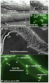
Formation of ectopic HCG fates in pry-1 mutant males. (A) Late L4. Two ventral invaginations with anchor-like shapes inside (arrowheads) were formed in the central region of a pry-1(mu38) male. (B) Overlay of Nomarski and fluorescence images of an adult pry-1(mu38) male. Two ventral protrusions (arrowheads) were observed in the middle body. Autofluorescence could be seen at the tip of the protrusions. (C–D) Nomarski (C) and fluorescence (D) images of an adult pry-1(mu38) male. An autofluorescent ventral protrusion (arrowhead) was associated with an ectopic 2° HOB hook neuron cell (arrow), as indicated by ceh-26::GFP expression. (E–F) Correlation of ectopic 1° PVV-like eat-4::GFP-expressing neurons (arrows) with ventral protrusions (arrowheads) in a pry-1(mu38) male (E, Nomarski; F, fluorescence). Scale bar, 20 μm. Left lateral views.
Figure 2
Figure 2
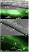
Alteration of male Pn.p fusion pattern by a lin-22 mutation. (A–D) Late L2. Nomarski (A) and fluorescence (B) images of a wild-type male. In the posterior, a line of AJM-1–GFP expression marked unfused P10.p and P11.p cells. P9.p and more anterior P(7–8).p were fused with hyp7 already. Nomarski (C) and fluorescence (D) images of a lin-22(n371) male in the same developmental stage. P9.p fused as indicated by the loss of AJM-1–GFP expression. However, P(7–8).p, as well as central P(3–6).p (not shown), remained unfused as do P(10–11).p. (E–H) Nomarski (E and F) and fluorescence (G and H) images of a lin-22 male in the middle L3 stage. The unfused central P(3–8).p and posterior P(10–11).p all divided once. Expression of AJM-1–GFP was observed in all those Pn.p descendants. The P11.pa cell was located a little above and thus the AJM-1–GFP expression was not shown in this picture. Scale bar, 20 μm. Left lateral views.
Figure 3
Figure 3
Preferential 2° HCG fate transformation in anterior Pn.p cells by activated Wnt and LIN-12 signaling. Nomarski (A) and fluorescence (B) images of an L4 pry-1; lin-12(gf)/lin-12(null) male. Three hook invaginations (arrowheads) were each coupled with a pair of osm-6::GFP-expressing hook neurons (arrows) among the three middle Pn.p cells (P4.p-P6.p). h-in, hook invagination. Scale bar, 20 μm. Left lateral views.
Figure 4
Figure 4
Induction of ectopic 2° HCG fates by the combined action of constitutive MAB-5 activity and Notch or EGF signaling. (A–D) 2°-like HCG fate transformation in anterior Pn.p cells by increased MAB-5 activity and activated LIN-12 signaling. An L4 mab-5(e1751gf) +/+ lin-12(gf) male had an nice hook invagination and a pair of HOA and HOB hook neurons (marked by osm-6::GFP) derived from the P1.p lineage (A, Nomarski; B, fluorescence). Another L4 mab-5(e1751gf) +/+ lin-12(gf) male generated hook invaginations at the central P5.p and P6.p (arrowheads) (C, Nomarski). Each invagination was associated with a pair of hook neurons (arrows) (D, fluorescence). (EG) Ectopic hook formation in P(1–2).p by extra MAB-5 activity and EGF signaling. (E) An L4 mab-5(e1751gf); lin-15(e1753) male formed two hook-like invaginations at P(1–2).p. Nomarski (F) and fluorescence (G) images of a P1.p hook in an adult mab-5(e1751gf); lin-15(e1753) male. h-in, hook invagination. Scale bar, 20 μm. Left lateral views.
Figure 5
Figure 5
2° HCG fate formation in male Pn.p cells caused by activated LIN-12 signaling in a lin-22 mutant background. (A–B) A L4 lin-12(gf)/lin-12(null); lin-22(n372) male. Nomarski (A) and fluorescence (B) images of two hook-like invaginations at P1.p and P2.p (arrowheads in A) were observed. osm-6::GFP is expressed in both 2° hook neurons. A single osm-6::GFP-expressing hook neuron was seen near the P1.p hook invagination, and the P2.p lineage made a complete hook sensillum with the presence of two osm-6::GFP-expressing cells (arrows in B). (C–D) Nomarski (C) and fluorescence (D) images of (P7-10).p in another L4 lin-12(gf)/lin-12(null); lin-22(n372) male. A P9.p hook sensillum was produced in addition to a P10.p wild-type hook sensillum. Furthermore, P(7–8).p were also induced to form hook invaginations. The P7.p hook invagination was associated with a pair of hook neurons, indicating a complete 2° HCG fate transformation. P8.p only generated a hook invagination, indicating a partial 2° fate transformation. One of the P9.p hook neurons was located in a slightly right focal plane and cannot be seen in the picture. h-in, hook invagination. Scale bar, 20 μm. Left lateral views.
Figure 6
Figure 6
Regulation of MAB-5–GFP expression in P(9–11).p by lin-17. (A–D) Early L3 wild-type male. Nomarski (A) and fluorescence (B) images of the HCG. P10.p and P11.p have moved slightly to the posterior. MAB-5–GFP was expressed in P11.p. Expression in P10.p was hardly detectable in this animal. (C-D) Early L3 lin-17(n671lf) male. MAB-5–GFP expression was absent in P11.p. However, this mutant male still retained some faint GFP expression in P10.p. Scale bar, 20 μm. Left lateral views.
Figure 7
Figure 7
Excess Wnt signaling or ectopic MAB-5 activity together with excess EGF or LIN-12/Notch signaling confers hook competence to male Pn.p cells anterior to the HCG. In wild-type males, only P(9–11).p possess HCG competence (blue). All anterior Pn.p cells (P(1–8).p) remain uninduced and fuse with hyp7 (grey). The most posterior cell P12.pa adopts a unique hypodermal fate (dark green). Increased Notch signaling due to a lin-12(gf) mutation or abnormal activation of EGF signaling by a lin-15(null) mutation causes the central Pn.p cells, P(3–8).p, to adopt a vulval-like fate (pink) but does not change the competence of the other Pn.p cells. Extra Wnt signaling caused by the pry-1(mu38) mutation confers HCG competence to P(3–8).p. Combined action of the pry-1(mu38) and lin-12(gf) mutations further extends the anterior boundary of the HCG to include P1.p and P2.p. A similar effect is also observed in conditions of excess MAB-5 activities combined with increased Notch (by a lin-12(gf) mutation) or EGF (by a lin-15(null) mutation) signaling whereby P(1–8).p is recruited into the HCG.
Figure 8
Figure 8
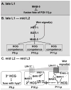
Model for development of the male HCG. (A) Regional MAB-5 activity preset during embryonic development keeps P(9–11).p unfused in the late L1 stage. (B) During the late L1 to mid-L2 stage, continuing presence of mab-5 activity in P(9–11).p, which is regulated by Wnt and probably other graded signals from the posterior tail region, makes these cells able to generate hook sensillum tissue (HCG competence). (C) During the mid-L2 to mid-L3 stage, a strong Wnt signal induces the 1° HCG fate in P11.p since it is closest to the posterior. The posterior signal(s) make P10.p assume an induced fate as well. With the presence of a 1° P11.p cell, the P10.p cell is locked into the 2° HCG fate by activated LIN-12 signaling. The P9.p cell, which has minimum mab-5 activity and receives the lowest level of inductive signals, adopts the uninduced 3° fate and finally fuses with hyp7.

Similar articles

Cited by

References

    1. Celniker SE, Dillon LA, Gerstein MB, Gunsalus KC, Henikoff S, Karpen GH, Kellis M, Lai EC, Lieb JD, MacAlpine DM, Micklem G, Piano F, Snyder M, Stein L, White KP, Waterston RH modENCODE Consortium. Unlocking the secrets of the genome. Nature. 2009;459:927–30. - PMC - PubMed
    1. Chamberlin HM, Sternberg PW. The lin-3/let-23 pathway mediates inductive signalling during male spicule development in Caenorhabditis elegans. Development. 1994;120:2713–2721. - PubMed
    1. Chamberlin HM, Sternberg PW. Mutations in the Caenorhabditis elegans gene vab-3 reveal distinct roles in fate specification and unequal cytokinesis in an asymmetric cell division. Dev Biol. 1995;170:679–689. - PubMed
    1. Clark SG, Chisholm AD, Horvitz HR. Control of cell fates in the central body region of C. elegans by the homeobox gene lin-39. Cell. 1993;74:43–55. - PubMed
    1. Clark SG, Lu WX, Horvitz HR. The Caenorhabditis elegans locus lin-15, a negative regulator of a tyrosine kinase signaling pathway, encodes two different proteins. Genetics. 1994;137:987–997. - PMC - PubMed

Publication types

MeSH terms