. 2010 Aug 1;344(1):259-71.
doi: 10.1016/j.ydbio.2010.05.007. Epub 2010 May 15.

A transitional extracellular matrix instructs cell behavior during muscle regeneration

Affiliations

A transitional extracellular matrix instructs cell behavior during muscle regeneration

Sarah Calve et al. Dev Biol. .

Abstract

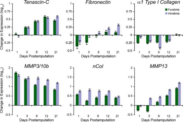
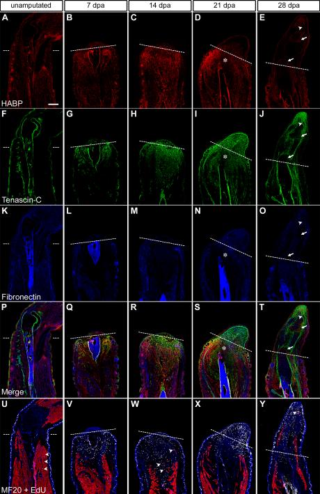
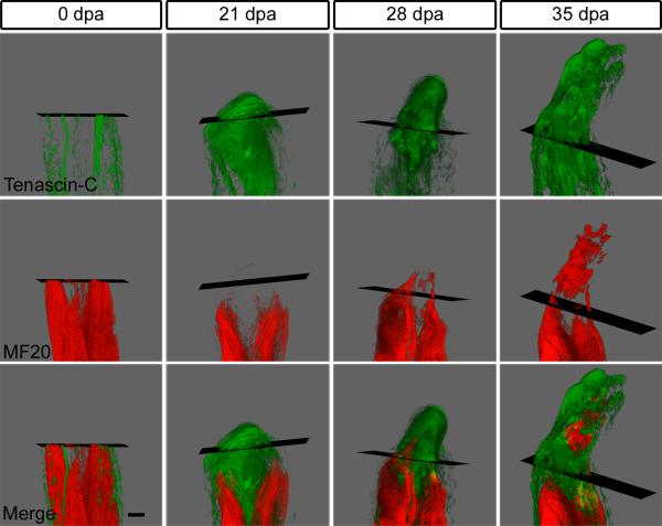
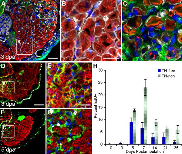
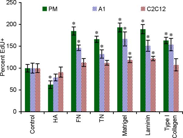
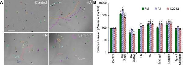
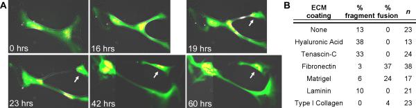
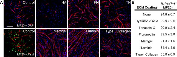
Urodele amphibians regenerate appendages through the recruitment of progenitor cells into a blastema that rebuilds the lost tissue. Blastemal formation is accompanied by extensive remodeling of the extracellular matrix. Although this remodeling process is important for appendage regeneration, it is not known whether the remodeled matrix directly influences the generation and behavior of blastemal progenitor cells. By integrating in vivo 3-dimensional spatiotemporal matrix maps with in vitro functional time-lapse imaging, we show that key components of this dynamic matrix, hyaluronic acid, tenascin-C and fibronectin, differentially direct cellular behaviors including DNA synthesis, migration, myotube fragmentation and myoblast fusion. These data indicate that both satellite cells and fragmenting myofibers contribute to the regeneration blastema and that the local extracellular environment provides instructive cues for the regenerative process. The fact that amphibian and mammalian myoblasts exhibit similar responses to various matrices suggests that the ability to sense and respond to regenerative signals is evolutionarily conserved.


Figures

Figure 1
Figure 1. Key ECM and MMPs are differentially expressed during appendage regeneration
Differential expression of key genes reveals dramatic matrix remodeling during newt forelimb and hindlimb regeneration. Each time point was normalized to intact (day 0) expression levels and log10 transformed. Geometric means and standard deviations are shown. dpa = days postamputation.
Figure 2
Figure 2. Limb amputation induces the deposition of a regeneration-specific transitional ECM
A - E: Hyaluronic acid (HA, red, indirectly labeled with hyaluronic acid binding protein, HABP), F – J: tenascin-C (TN, green), K – O: fibronectin (FN, blue) during forelimb regeneration at 7, 14, 21 and 28 days postamputation (dpa) in comparison to the normal, unamputated limb. HA was more highly expressed in the stump tissue and TN predominated the blastema; however, both were downregulated in areas of cartilage condensation (21 dpa, asterisk). Of note, HA was upregulated within the newly formed joints (28 dpa, arrows). U – Y: Adjacent sections were labeled to determine the distribution of the transitional ECM with respect to regenerating skeletal muscle (MF20 = red) and cells actively synthesizing DNA (EdU = white, DAPI = blue). In the unamputated limb, only a few EdU+ cells were detected outside of the epidermis (arrowheads). The number of EdU+ cells increased by 7 and 14 dpa and populated the same areas as the TN- and HA-rich matrix. Isolated fragments of MF20+ cells were present at 14 dpa (arrowheads). During the later stages, the majority of cells reentering the cell cycle were found distal to the amputation plane. TN and HA were downregulated within areas of skeletal muscle differentiation (28 dpa, arrowhead). Dashed line = amputation plane, dpa = days postamputation, bar = 400 μm. See also Figs. S1 and S2 for details on DNA synthesis and skeletal muscle regeneration.
Figure 3
Figure 3. The regeneration-specific ECM is absent from differentiating muscle and bone
A and A’: Adjacent sections of a 28 dpa forelimb were stained for either components of the transitional matrix (TN = green, HA = red, FN = blue) or skeletal muscle and cells that had reentered the cell cycle (MF20 = red, EdU = white, DAPI = blue). B and B’: TN and HA were downregulated in areas of differentiating skeletal muscle (m) whereas FN expression remained constant. C and C’: HA was expressed in a proximal to distal gradient in the regenerating ulna correlating with EdU+ chondrocytes (c). D and D’: EdU+ cells were co-localized with TN in the regenerating brachial nerve (n). Dashed line = amputation plane, dpa = days postamputation. A and A’: bar = 400 μm. B – D’: bar = 100 μm.
Figure 4
Figure 4. Regenerating skeletal muscle directly interacts with the transitional matrix
3D reconstruction of a regenerating forelimb at 0, 21, 28 and 35 dpa; 70 – 100 serial sections were imaged, assembled and rendered using ImageProPlus 6.1. See Videos 1 – 5. At 0 dpa, TN (green) was restricted to the tendons, periosteum, dermis and epidermis. A TN-rich matrix had infiltrated the stump at 21 dpa and skeletal muscle (MF20 = red) had regressed away from the amputation plane. By 28 dpa, the skeletal muscle regenerated through the amputation plane revealing both pockets of MF20 expression and extensions from stump musculature. Embedded within a TN-rich matrix, isolated MF20+ regions were found in the distal autopod. Black rectangle = amputation plane, dpa = days postamputation, bar = 400 μm.
Figure 5
Figure 5. The transitional matrix infiltrates the basement membrane of degenerating myofibers and specifies regions of proliferative activity
A: Cross-section of 3 dpa forelimbs shows that the humeroantebrachialis has both intact and degenerating myofibers (MF20 = red, type IV collagen = white, TN = green, DAPI = blue). B: Uninjured fibers were surrounded by an intact basement membrane, defined by type IV collagen expression, and nuclei were predominantly found in the periphery of the myofiber syncytium. C: The basement membrane of degrading myofibers was infiltrated by a TN-rich matrix and centrally located nuclei. D - G: Cross-sections of regenerating limbs revealed inhomogeneous distribution of both the transitional matrix (TN = green) and EdU+ (white) cells within the humeroantebrachialis muscle (MF20 = red, skeletal muscle; DAPI = blue). Notably, increased TN expression is apparent at 3 dpa (D and E) and precedes the induction of DNA synthesis (F and G). H: EdU incorporation within the mesodermal tissues dramatically increased after 3 dpa, reaching a peak around 7 dpa, with significantly more cells synthesizing DNA in the TN-rich transitional matrix. Two-way ANOVA for Interaction (p < 0.0001), Matrix (p < 0.0001) and Time (p < 0.0001). Bonferroni's post hoc test revealed statistically significant differences in EdU incorporation between TN-rich and TN-free regions (p < 0.001). dpa = days post amputation A: bar = 100 μm. B and C: bar = 40 μm. D and F: bar = 200 μm. E and G: bar = 50 μm. H: Error bars = S.D., n ≥ 5.
Figure 6
Figure 6. Matrix composition controls DNA synthesis
Primary newt myoblasts (PM), newt A1 and mouse C2C12 myoblast cell lines were cultured in vitro for 48 hours and the percentage of cells actively synthesizing DNA was determined by labeling for EdU incorporation. Data were normalized to uncoated control. Error bars = SD, n ≥ 4. One-way ANOVA, p < 0.0001 for each cell type. Tukey-Kramer post-hoc tests revealed statistically significant differences between EdU incorporation on specific matrices versus the uncoated control (* p < 0.05).
Figure 7
Figure 7. The ECM directs amphibian and murine myoblast migration
A: Myoblasts showed different migratory behavior on HA, TN, laminin and uncoated polystyrene. See Video 6. B: Migration of all cell lines was enhanced on TN and laminin, whereas cells plated on HA-coated substrates displayed a unique bimodal migration behavior with a subset of myoblasts migrating 10-fold farther than the control (note logarithmic scale of the y-axis). The rapidly migrating myoblasts eventually slowed down and took on the same spindle-shaped morphology displayed by the majority of cells plated on HA (arrowhead). Primary newt myoblasts (PM), newt A1 and mouse C2C12 cells were imaged for 19 hours and the total distance traveled was normalized to uncoated polystyrene. For all parameters n = 40 cells except HA (Fast) where n ≥ 8, error bars = SD. One-way ANOVA, p < 0.0001 for each cell type. Tukey-Kramer post-hoc tests revealed statistically significant differences between myoblast migration on specific matrices versus the uncoated control (* p < 0.05).
Figure 8
Figure 8. Components of the transitional matrix induce myotube fragmentation
A: Live cell imaging identified viable cells budding off newt multinuclear parent myotubes when cultured on HA-coated polystyrene (arrow). Cells were co-injected with plasmids to identify differentiated muscle (pXCarGFP3, green) and nuclei (pCMV-H2AmCherry, red). Adjacent images were tiled (42 and 60 hrs) to illustrate viable fragmentation (see Video 7). B: Primary newt myotubes cultured for five days preferentially fragmented on HA and TN, whereas FN and Matrigel inhibited fragmentation and induced fusion.
Figure 9
Figure 9. Basement membrane components enhance myoblast differentiation
A: Myoblast differentiation was defined by MF20+ staining (red) and undifferentiated muscle progenitors were identified by Pax7 expression (green). DAPI = blue, bar = 300 μm. B: Mononuclear undifferentiated cells (MF20-/Pax7+) on all substrates maintained their myogenic phenotype.

Similar articles

Cited by

References

    1. Alvarado AS. Regeneration in the metazoans: Why does it happen? BioEssays. 2000;22:578–590. - PubMed
    1. Ambrosio F, Ferrari RJ, Fitzgerald GK, Carvell G, Boninger ML, Huard J. Functional overloading of dystrophic mice enhances muscle-derived stem cell contribution to muscle contractile capacity. Arch. Phys. Med. Rehab. 2009;90:66–73. - PMC - PubMed
    1. Blau HM, Chiu CP, Webster C. Cytoplasmic activation of human nuclear genes in stable heterocaryons. Cell. 1983;32:1171–80. - PubMed
    1. Boonen KJM, Rosaria-Chak KY, Baaijens FPT, van der Schaft DWJ, Post MJ. Essential environmental cues from the satellite cell niche: Optimizing proliferation and differentiation. Am. J. Physiol.-Cell Ph. 2009;296:C1338–C1345. - PubMed
    1. Bouchentouf M, Benabdallah BF, Mills P, Tremblay JP. Exercise improves the success of myoblast transplantation in mdx mice. Neuromuscular Disord. 2006;16:518–529. - PubMed

Publication types

LinkOut - more resources