. 2010 Aug 1;344(1):293-303.
doi: 10.1016/j.ydbio.2010.05.010. Epub 2010 May 15.

AMPK supports growth in Drosophila by regulating muscle activity and nutrient uptake in the gut

Affiliations

AMPK supports growth in Drosophila by regulating muscle activity and nutrient uptake in the gut

Michelle L Bland et al. Dev Biol. .

Abstract

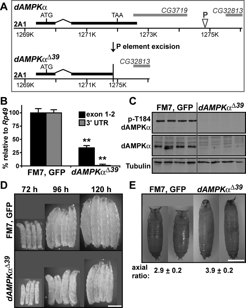
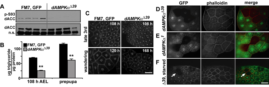
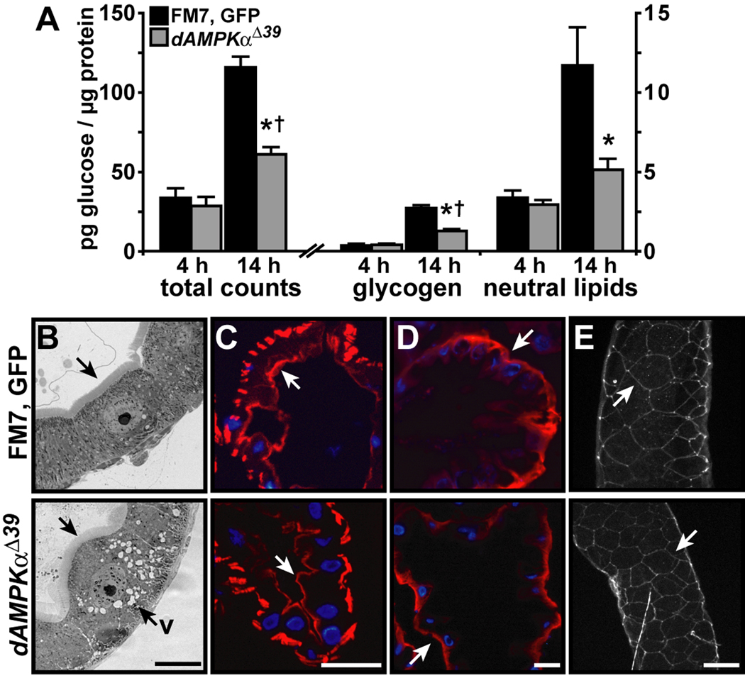
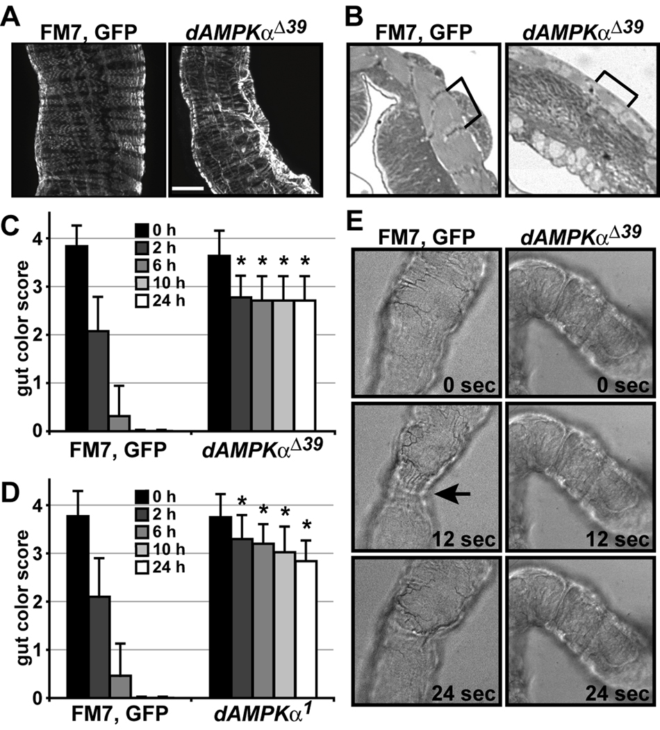
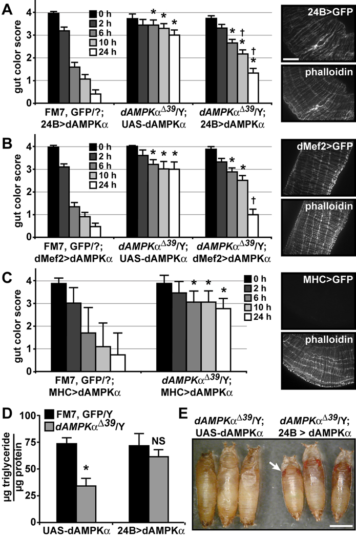
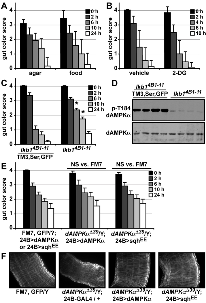
The larval phase of the Drosophila life cycle is characterized by constant food intake, resulting in a two hundred-fold increase in mass over four days. Here we show that the conserved energy sensor AMPK is essential for nutrient intake in Drosophila. Mutants lacking dAMPKalpha are small, with low triglyceride levels, small fat body cells and early pupal lethality. Using mosaic analysis, we find that dAMPKalpha functions as a nonautonomous regulator of cell growth. Nutrient absorption is impaired in dAMPKalpha mutants, and this defect stems not from altered gut epithelial cell polarity but from impaired peristaltic activity. Expression of a wild-type dAMPKalpha transgene or an activated version of the AMPK target myosin regulatory light chain (MRLC) in the dAMPKalpha mutant visceral musculature restores gut function and growth. These data suggest strongly that AMPK regulates visceral smooth muscle function through phosphorylation of MRLC. Furthermore, our data show that in Drosophila, AMPK performs an essential cell-nonautonomous function, serving the needs of the organism by promoting activity of the visceral musculature and, consequently, nutrient intake.


Figures

Figure 1
Figure 1. Generation of a dAMPKα mutant allele
A) Schematic of the dAMPKα locus (top) showing the dAMPKα gene (black), the P element (P) and the genes CG3719 and CG32813 (gray). Imprecise P element excision yielded the dAMPKαΔ39 allele (bottom). B) Q-PCR for exons 1 and 2 (black bars) and the 3’UTR (gray bars) of dAMPKα was performed on RNA isolated from wild type (FM7, GFP/Y) and dAMPKαΔ39/Y third instar larvae. dAMPKα transcript levels were normalized to Rp49 transcript levels. Data are presented as mean ± s.d., **p < 0.01 vs. FM7, GFP/Y. C) Western blot analyses of phosphorylated (p-Thr184, top) and total dAMPKα (middle) and tubulin (bottom) in lysates from FM7, GFP/Y and dAMPKαΔ39/Y third instar larvae. D) FM7, GFP/Y (top) and dAMPKαΔ39/Y larvae (bottom) at 72, 96 and 120 h AEL, scale bar = 1 mm. E) FM7, GFP/Y and dAMPKαΔ39/Y pupae, scale bar = 1 mm. The axial ratio (length/width, mean ± s.d.) is greater in dAMPKαΔ39 mutants compared with WT (p < 0.01).
Figure 2
Figure 2. Nutrient storage is decreased in dAMPKα mutant larvae
A) Western blot analyses of phosphorylated (p-Ser93, top) and total dACC (bottom) in lysates from FM7, GFP/Y and dAMPKαΔ39/Y third instar larvae, n.s., non-specific. B) Whole-body triglyceride levels were measured in FM7, GFP/Y (black bars) and dAMPKαΔ39/Y (gray bars) 108 h AEL larvae (n = 18/group) and white prepupae (n = 13/group) and normalized to protein. Data are presented as mean ± s.e.m., **p < 0.01 vs. FM7, GFP/Y. C) Fat bodies from 108 h AEL and wandering (FM7, GFP/Y: ~120 h AEL, dAMPKαΔ39/Y: ~168 h AEL) larvae were stained with phalloidin and DAPI to label plasma membranes and nuclei. Scale bar = 50 µm. D-F) Heat shock was used to induce clones of fat body cells homozygous for the dAMPKαΔ39 (D) or the dAMPKα1 (E) mutations (GFP negative cells, left panels). Cell membranes were labeled with phalloidin (center panels). Merged images are shown in the right panels, scale bar = 50 µm. F) 24 h starvation of larvae bearing clones had no additional effect on dAMPKαΔ39 homozygous cell size (arrows). Genotypes: D and F) UbiGFP, hsFLP, FRT19A/dAMPKαΔ39, FRT19A. E) UbiGFP, hsFLP, FRT19A/dAMPKα1, FRT19A.
Figure 3
Figure 3. Impaired nutrient absorption despite normal epithelial polarity in dAMPKα mutants
A) FM7, GFP/Y (black bars) and dAMPKαΔ39/Y larvae (gray bars) were fed radiolabeled glucose for 4 or 14 h, and incorporation of radioactivity into glycogen and neutral lipids was measured. Data are presented as mean ± s.d., *p < 0.05 vs. FM7, GFP/Y at the same time point, †p < 0.05 vs. dAMPKαΔ39/Y at 4 h. For FM7, GFP/Y, all 14 h values differ significantly from 4 h values, p < 0.05. B-E) Representative images of FM7, GFP/Y (top) and dAMPKαΔ39/Y (bottom) midguts. B) Toluidine blue-stained transverse sections of third instar midguts showing the brush border (arrows) in both genotypes and vacuoles (v) in the dAMPKαΔ39 midgut epithelium. C) Phalloidin labeling of the apical layer of filamentous actin (red, arrows) in sections of second instar guts. D, E) Immunocytochemistry for Na+/K+-ATPase (D, red, arrows), and Fasciclin III (E, white, arrows) in sections (D) or whole guts (E) from second instar larvae. Nuclei in C and D are stained with DAPI (blue), scale bars = 50 µm.
Figure 4
Figure 4. Visceral muscle function is impaired in dAMPKα mutants
A) Whole-mount immunocytochemistry for myosin heavy chain showing circular and longitudinal visceral muscle fibers in FM7, GFP/Y (left) and dAMPKαΔ39/Y (right) third instar midguts, scale bar = 50 µm. B) Toluidine blue staining of transverse sections of FM7, GFP/Y (left) and dAMPKαΔ39/Y (right) hindguts. Brackets indicate individual sarcomeres. C, D) Larvae were raised on food containing FD&C No. 1 Blue dye, transferred to Whatman paper at 96 h AEL and scored for the amount of food remaining in the gut over 24 h as described in the Materials and Methods. C) FM7, GFP/Y: n = 16, dAMPKαΔ39/Y: n = 13. D) FM7, GFP/Y: n = 12, dAMPKα1/Y: n = 11. Data are presented as mean ± s.d., **p < 0.01 vs. FM7, GFP/Y at the same time point. E) Still images of FM7, GFP/Y (left) and dAMPKαΔ39/Y (right) third instar guts imaged live in HL3 buffer. Each frame is separated from the next by 12 seconds. The arrow points to constriction of wild type gut muscles.
Figure 5
Figure 5. Muscle-specific expression of dAMPKα rescues dAMPKαΔ39 phenotypes
A–C) Visceral muscle function was tested using the blue food transit assay. Expression of wild type dAMPKα in muscle using 24B- (A) or dMef2-GAL4 (B) rescued the dAMPKαΔ39 phenotype (compare the center set of bars: dAMPKαΔ39 with UAS-dAMPKα alone, to the right set of bars: dAMPKαΔ39 with GAL4 > dAMPKα). C) MHC-GAL4 was unable to rescue the dAMPKαΔ39 gut transit phenotype. For A–C: FM7, GFP/Y, n = 23–24; dAMPKαΔ39/Y; UAS-dAMPKα, n = 5–7; and dAMPKαΔ39/Y; GAL4 > dAMPKα, n = 18–37. Data are presented as mean ± s.d., *p < 0.01 vs. FM7, GFP/Y, †p < 0.01 vs. dAMPKαΔ39/Y; UAS-dAMPKα. 24B-, dMef2-, and MHC-GAL4-driven GFP expression in visceral muscle (top panels) and phalloidin counter-staining (bottom panels) are shown to the right of each graph. D) Triglyceride levels in 120 h AEL FM7, GFP/Y (black bars) and dAMPKαΔ39/Y larvae (gray bars) with UAS-dAMPKα alone (FM7, GFP, n = 6, dAMPKαΔ39, n = 4) or 24B > dAMPKα (FM7, GFP, n = 8, dAMPKαΔ39, n = 11). Data are presented as mean ± s.d., *p < 0.01 and NS, not significant, vs. FM7, GFP/Y. E) dAMPKαΔ39 mutants (left) died after pupariation, while 24B-GAL4-rescued dAMPKαΔ39 mutants (right) underwent metamorphosis, as indicated by their red eyes (arrow), scale bar = 1 mm.
Figure 6
Figure 6. Expression of activated MRLC in muscle rescues the dAMPKαΔ39 gut phenotype
A, B) Visceral muscle function in 96 h AEL wild type larvae was tested using the blue food transit assay. Data are presented as mean ± s.d. A) Larvae were fed 1% agar for 22 h, followed by 4 h of blue-dyed 1% agar (n = 9) or cornmeal/molasses food (n = 14). B) Larvae were fed 10 mM glucose and 1% yeast extract in blue-dyed 1% agar with vehicle (n = 13) or 50 mM 2-DG (n = 11). C) Visceral muscle function in lkb14B1–11/TM3, Ser, GFP (n = 34) and lkb14B1–11 (n = 52) larvae. Data are presented as mean ± s.e.m., *p < 0.05 vs. lkb14B1–11/TM3, Ser, GFP. D) Western blot analysis of phosphorylated (p-Thr184, top) and total dAMPKα (bottom) in lysates from lkb14B1–11/TM3, Ser, GFP and lkb14B1–11 120 h AEL larvae. E) 24B-GAL4 driven expression of an activated MRLC transgene (sqhEE) provided a partial rescue of the dAMPKαΔ39 gut transit phenotype. Compare the middle set of bars: dAMPKαΔ39 with 24B > dAMPKα (n = 13), to the right set of bars: dAMPKαΔ39 with 24B > sqhEE (n = 15). Data are presented as mean ± s.e.m., NS, not significant, vs. FM7, GFP/Y. F) Whole-mount phalloidin labeling showing circular and longitudinal visceral muscle fibers in FM7, GFP/Y, dAMPKαΔ39/Y; 24B-GAL4 / +, dAMPKαΔ39/Y; 24B > dAMPKα and dAMPKαΔ39/Y; 24B > sqhEE third instar midguts.

Similar articles

Cited by

References

    1. Amin N, Khan A, St Johnston D, Tomlinson I, Martin S, Brenman J, McNeill H. LKB1 regulates polarity remodeling and adherens junction formation in the Drosophila eye. Proc Natl Acad Sci U S A. 2009;106:8941–8946. - PMC - PubMed
    1. Baumann O. Posterior midgut epithelial cells differ in their organization of the membrane skeleton from other drosophila epithelia. Exp Cell Res. 2001;270:176–187. - PubMed
    1. Bayliss WM, Starling EH. The movements and innervation of the small intestine. J Physiol. 1899;24:99–143. - PMC - PubMed
    1. Britton JS, Edgar BA. Environmental control of the cell cycle in Drosophila: nutrition activates mitotic and endoreplicative cells by distinct mechanisms. Development. 1998;125:2149–2158. - PubMed
    1. Britton JS, Lockwood WK, Li L, Cohen SM, Edgar BA. Drosophila's insulin/PI3-kinase pathway coordinates cellular metabolism with nutritional conditions. Dev Cell. 2002;2:239–249. - PubMed

Publication types

LinkOut - more resources