. 2010 Aug 15;344(2):578-92.
doi: 10.1016/j.ydbio.2010.05.012. Epub 2010 May 15.

Analysis of early human neural crest development

Affiliations

Analysis of early human neural crest development

Erin Betters et al. Dev Biol. .

Abstract

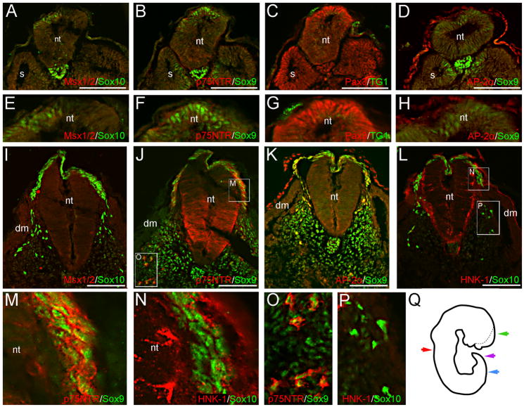
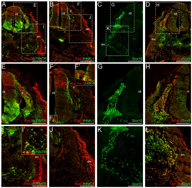
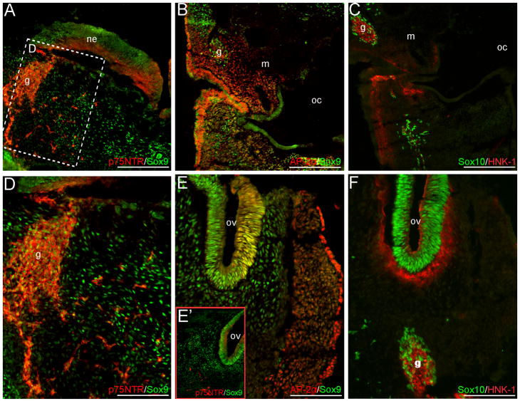
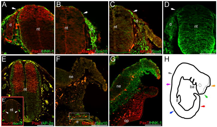
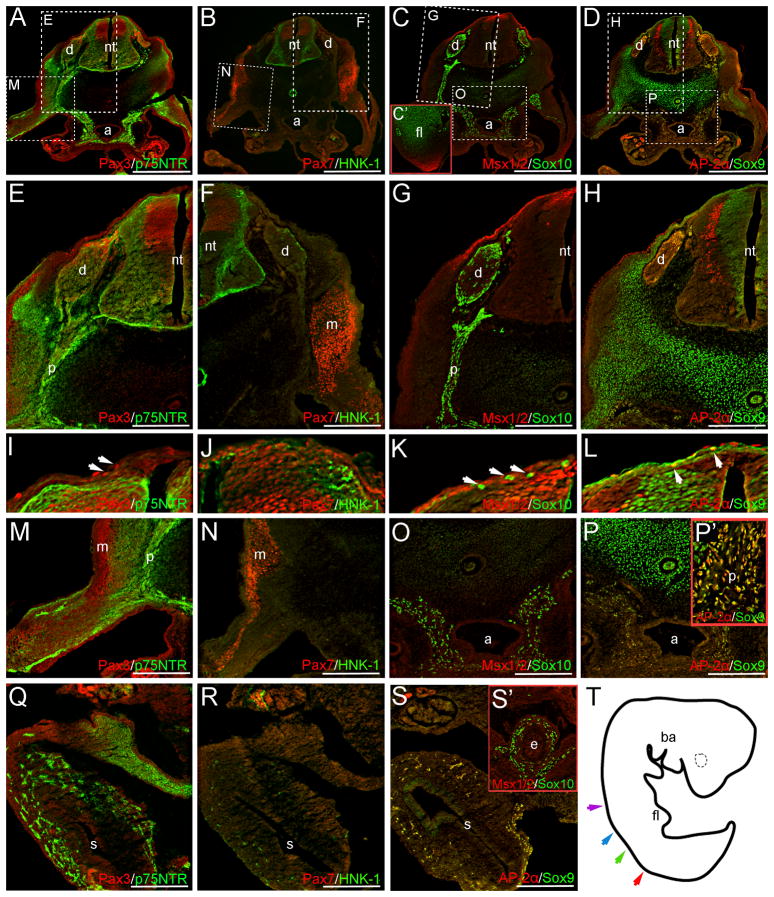
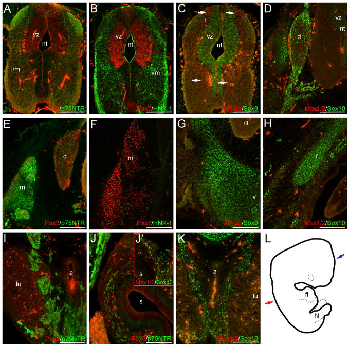
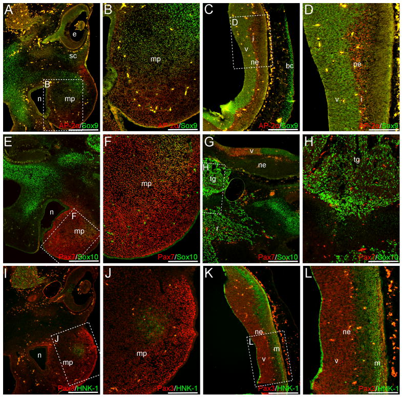
The outstanding migration and differentiation capacities of neural crest cells (NCCs) have fascinated scientists since Wilhelm His described this cell population in 1868. Today, after intense research using vertebrate model organisms, we have gained considerable knowledge regarding the origin, migration and differentiation of NCCs. However, our understanding of NCC development in human embryos remains largely uncharacterized, despite the role the neural crest plays in several human pathologies. Here, we report for the first time the expression of a battery of molecular markers before, during, or following NCC migration in human embryos from Carnegie Stages (CS) 12 to 18. Our work demonstrates the expression of Sox9, Sox10 and Pax3 transcription factors in premigratory NCCs, while actively migrating NCCs display the additional transcription factors Pax7 and AP-2alpha. Importantly, while HNK-1 labels few migrating NCCs, p75(NTR) labels a large proportion of this population. However, the broad expression of p75(NTR) - and other markers - beyond the neural crest stresses the need for the identification of additional markers to improve our capacity to investigate human NCC development, and to enable the generation of better diagnostic and therapeutic tools.


Figures

Fig. 1
Fig. 1
NCC markers are expressed in prospective premigratory and migratory NCCs at CS12. Sox10 (A), Sox9 (B, D) and Pax3 (C) signal occur in the dorsal neural tube of caudal trunk sections, prior to the appearance of the hindlimb. These cells likely represent premigratory NCCs. Images in (E–H) are magnifications of the dorsal neural tubes shown in (A–D). We do not observe definitive Msx1/2, p75NTR, or AP-2α signal in premigratory NCCs (A, B, D). The monoclonal antibody TG1 has previously been used to recognize other migratory cells and was thus here tried, but no signal can be associated with premigratory NCCs (C, G). In slightly rostral sections, Sox10 (I, L), Sox9 (J–K), p75NTR (J), and AP-2α (K) are expressed in migratory NCCs situated dorsal, or dorso-lateral to, the neural tube. The majority of AP-2α+ migratory NCCs appear to co-express Sox9 (K). Panels in (M–P) are magnifications of boxed regions shown in (J, L). Although robust p75NTR signal is co-expressed with Sox9 in migratory NCCs adjacent to the neural tube (M), or subadjacent to the dermomyotome (O), this cell population (when identified via Sox10 expression) does not express HNK-1 in similar sections (N, P); migratory NCCs generally appear HNK-1 negative. Image in (Q) is a tracing of the CS12 wholemount, prior to sectioning; the dotted line marks the contra-lateral side of the unfused neural tube. Color-coded arrows represent the axial levels of depicted sections as follows: purple (A–H), blue (I–P), red (Fig. 2), green (Fig. 3). Scale bars in all panels represent 100μm. Note: levels in (A–D) are modified separately from those in (I–L) to optimize signals in different embryonic regions. dm, dermomyotome; nt, neural tube; s, somite.
Fig. 2
Fig. 2
Pax3, p75NTR, Sox10, Sox9 and AP-2α are expressed in neural crest derivatives of CS12 rostral trunk sections. Images in (E–K) are magnifications of boxed regions in (A–D); image in (L) is from the contralateral side of the section shown in (D). Sox10, Sox9 and AP-2α expression appear in both the NCC-derived DRGs (C–D, G–H) and peripheral nerves (C, K, L). Pax3 and p75NTR signal additionally occur in the DRGs (A, E). In the ventral mesenchyme, NCCs that will contribute to either the sympathetic tissue or enteric nervous system are identified by their expression of p75NTR (A, I–I’), Sox10 (C, K), or Sox9 (I’). Respectively, images in (F’) and (I’) are magnifications of the boxed region in (F), and the aorta/mesonephric mesenchyme of a similar section. Axial level of sections in (A–D) is indicated by the red arrow in Fig. 1Q. Scale bars in (A–D) represent 200μm; those in (E–L) 100μm. Note: levels in (H) and (L) are modified separately to optimize green and red signals in different embryonic regions. d, dorsal root ganglia; dm, dermomyotome; nt, neural tube.
Fig. 3
Fig. 3
NCC markers are expressed in the mesenchyme or neural crest derivatives of cranial CS12 sections. Sox9 and AP-2α signal occur within the NCC-populated cranial mesenchyme or branchial arches (A–B, D–E’); panel in (D) is a higher magnification of the boxed region in (A). Additional Sox10+ and p75NTR+ cells are observed within this region (A, C–D, E’–F). NCC-derived cranial ganglia condensations or associated nerves express p75NTR, Sox9, Sox10, AP-2α and HNK-1 (A–D, F). Within the otic vesicles, Sox9, AP-2α, Sox10 and p75NTR signal occur (E–F). Axial level of sections is indicated by the green arrow in Fig. 1Q. Scale bars in (A–C) represent 200μm; those in (D–F) 100μm. g, ganglia condensation or associated nerve; m, mandibular/maxillay process; ne, neuroepithelium; oc, oral-pharyngeal cavity; ov, otic vesicle.
Fig. 4
Fig. 4
NCC markers are expressed within the caudal migratory neural crest of trunk sections, or within the cranial mesenchyme at CS13. Pax7 (A), Sox9 (C), Sox10 (B) and p75NTR (D) signal occur in migratory NCCs located directly dorsal, or dorso-lateral to (region marked by arrows), the neural tube within caudal trunk sections. Although HNK-1 is also expressed in this region, it does not appear to robustly co-localize with Pax7 (A). Pax7, Sox10, Sox9 and p75NTR are also expressed within regions of the forming DRGs (A–D). Definitive Msx1/2 or AP-2α signal are not observed in either migratory NCCs or the presumptive caudal DRGs (B, C). In cranial CS13 sections, Pax7 expression is observed in the mesenchyme adjacent to the presumptive olfactory placode, in addition to the distal-most tip of the unfused neuroepithelium (G). Punctate Sox10 expression occurs near the eye primordia of slightly caudal sections (F boxed region), and within the ganglia condensations adjacent to the neuroepithelium (E’ arrows); in similar sections, Pax7 appears in the dorsal portion of the neuroepithelium, with AP-2α signal occurring along the lateral margins (E). HNK-1 signal is also observed within the cranial mesenchyme of more rostral sections, but again appears largely independent of Pax7 expression (G). Image in (H) is a tracing of the CS13 embryo wholemount; the dotted line marks the contra-lateral side of the unfused neural tube. Color-coded arrows in (H) indicate axial levels of sections as follows: red (A–D); gray (E–E’); orange (F); green (G); blue (S1. A–C); purple (S1. D). Scale bars in (A–D) represent 100μm; those in (E–G) 200μm. Note: levels in (E’), (F) are modified separately to optimize green and red signals at different axial levels. ba, branchial arches; d, dorsal root ganglia; e, eye; fl, forelimb; ne, neuroepithelium; nt, neural tube; op, olfactory placode.
Fig. 5
Fig. 5
NCC markers are expressed in trunk neural crest derivatives at CS15. Panels in (E–P) are higher power images of boxed regions in (A–D). NCC-derived DRGs are positive for Pax3 (A, E), p75NTR (A, E), Sox10 (C, G), AP-2α (D, H), and Sox9 (D, H). Few HNK-1+ cells also occur in these structures (B, F). Neither Pax7 (B, F) or Msx1/2 (C, G) appear to be expressed in the DRGs at this stage. Sox10 (C, O, S’), AP-2α (D, P-P’, S), Sox9 (D, P-P’, S) and p75NTR (A, E, M, Q) staining are also observed within the mesenchyme surrounding the aorta, the presumptive peripheral nerves, and the wall of the gut; respectively, images in (C’, P’, S’) are magnifications of the forelimb bud, peripheral nerve, and esophagus in slightly rostral sections (see below). HNK-1+ cells also occur within the stomach wall at CS15 (R). Occasional Sox10+, Msx1/2+, AP-2α+ or Pax3+ cells are also observed directly dorsal to the neural tube (I, K, L arrows). Sox9 expressing cells are also suggested in similar regions (L). Outside of the neural crest or its derivatives, p75NTR (A, E, I), Pax3 (A, E, I), Pax7 (B, F, J), HNK-1 (B, F, J), Msx1/2 (C, G, K), AP-2α (D, H, L) and Sox9 (D, H, L) are all expressed in regions of the neuroepithelium. Additional Msx1/2 expression is observed in the distal forelimb mesenchyme (C’). Image in (T) depicts a tracing of the CS15 embryo wholemount prior to sectioning. Color-coded arrows indicate axial levels of sections as follows: red (A–D), green (P’, R–S), blue (Q) and purple (C’, S’). Scale bars in (A–D) represent 500μm, those in (E–S) represent 200μm. Note: levels in (C’), (E–P) and (Q–S’) are modified separately to optimize signals in different embryonic regions. a, aorta; ba, branchial arches; d, dorsal root ganglia; e, esophagus; fl, forelimb; nt, neural tube; m, presumptive musculature; p, peripheral nerve; s, stomach.
Fig. 6
Fig. 6
NCC markers are expressed within trunk neural crest and non-neural crest derivatives at CS18. Outside of neural crest-associated structures, Pax3 (A), Pax7 (B), Sox9 (C), AP-2α (C, arrows), Sox10 (D) and HNK-1 (B) occur in portions of the neuroepithelium. Sox10 and p75NTR signal also occur within the NCC-derived DRGs (D, E), sympathetic tissue (I, K) or presumptive enteric ganglia (J-J’); image in (J’) is a magnification of the gut wall within a similar section. Faint HNK-1 signal also occurs in the DRGs and associated nerve roots (F). Pax7 (F) and p75NTR (E) staining are additionally observed in the presumptive musculature of the back, and few Pax3+ cells occur in the dorso-medial margin of this region (E). Sox9 and Sox10 signal occur in the rib or vertebrae cartilage primordia (G, H). Image in (L) is a tracing of the CS18 embryo wholemount. Red and blue arrows represent axial levels of sections in (A–K) and Fig. 7, respectively. Scale bars in all panels indicate 200μm. Note: red levels in (B, F) are modified individually to optimize signal in the neural tube and musculature, respectively. Levels in (A, E) and (D, H) are modified separately from those in (I–K) to optimize signal in the sympathetic tissue and gut wall. a, aorta; d, dorsal root ganglia; fl, forelimb; hl, hindlimb; i, intermediate layer; i/m, intermediate/marginal layers; lu, lung; m, presumptive musculature; nt, neural tube; r, rib primordia; s, stomach; v, vertebrae primordia; vz, ventricular zone.
Fig. 7
Fig. 7
Neural crest markers are expressed in the maxillary process at CS 18. AP-2α (A, B), Sox9 (A, B), Sox10 (E, F), Pax7 (E, F), and Pax3 (I, J) signal are observed within the NCC-populated maxillary process. A circular patch of HNK-1+ cells also exists in this region (I, J). Additional Sox9 and Sox10 signal are observed throughout the facial mesenchyme or future skeletal elements of the skull (A, C, E, G). The cranial ganglia are positive for both Sox10 (G, H) and HNK-1 (I), with Sox10 signal also appearing in the associated nerve roots (G). Axial level of sections in (A, C, E, G, I, K) is indicated by blue arrow in Fig. 6L. Images in (B, D, F, H, J, L) are higher power magnifications of boxed regions in (A, C, E, G, I, K). Scale bars in (A, C, E, G, I, K) represent 500μm, those in (B, D, F, H, J, L) represent 200μm. Note: levels in (E, G), (F, H), (I, K) and (J, L) are modified individually to optimize red and green signals. bc, brain case primordia; e, eye; i, intermediate layer; m, marginal layer; mp, maxillary process; n, nasal cavity; ne, neuroepithelium; r, cranial ganglia-associated nerve root; sc, presumptive scleral cartilage; tg, trigeminal ganglia; v, ventricular zone.

References

    1. Abzhanov A, Tzahor E, Lassar AB, Tabin CJ. Dissimilar regulation of cell differentiation in mesencephalic (cranial) and sacral (trunk) neural crest cells in vitro. Development. 2003;130:4567–79. - PubMed
    1. Anderson RB, Stewart AL, Young HM. Phenotypes of neural-crest-derived cells in vagal and sacral pathways. Cell Tissue Res. 2006;323:11–25. - PubMed
    1. Auerbach R. Analysis of the developmental effects of a lethal mutation in the house mouse. Journal of Experimental Zoology. 1954;127:305–329.
    1. Aybar MJ, Mayor R. Early induction of neural crest cells: lessons learned from frog, fish and chick. Curr Opin Genet Dev. 2002;12:452–8. - PubMed
    1. Bajpai R, Chen DA, Rada-Iglesias A, Zhang J, Xiong Y, Helms J, Chang CP, Zhao Y, Swigut T, Wysocka J. CHD7 cooperates with PBAF to control multipotent neural crest formation. Nature. 463:958–62. - PMC - PubMed

Publication types

MeSH terms

Substances