. 2010 Sep;49(3):490-8.
doi: 10.1016/j.yjmcc.2010.05.003. Epub 2010 May 15.

Sonic hedgehog induces angiogenesis via Rho kinase-dependent signaling in endothelial cells

Affiliations

Sonic hedgehog induces angiogenesis via Rho kinase-dependent signaling in endothelial cells

Marie-Ange Renault et al. J Mol Cell Cardiol. 2010 Sep.

Abstract

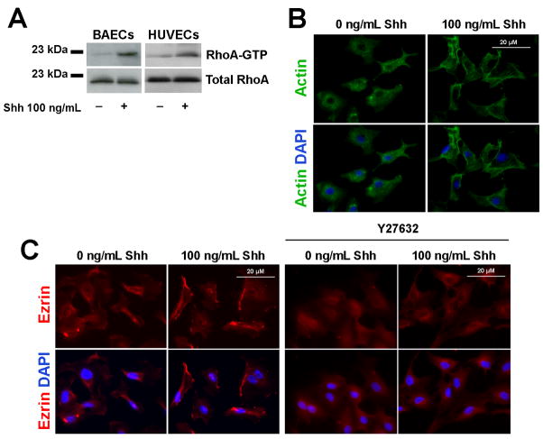
The morphogen Sonic hedgehog (Shh) promotes neovascularization in adults by inducing pro-angiogenic cytokine expression in fibroblasts; however, the direct effects of Shh on endothelial cell (EC) function during angiogenesis are unknown. Our findings indicate that Shh promotes capillary morphogenesis (tube length on Matrigel increased to 271+/-50% of the length in untreated cells, p=0.00003), induces EC migration (modified Boyden chamber assay, 191+/-35% of migration in untreated cells, p=0.00009), and increases EC expression of matrix metalloproteinase 9 (MMP-9) and osteopontin (OPN) mRNA (real-time RT-PCR), which are essential for Shh-induced angiogenesis both in vitro and in vivo. Shh activity in ECs is mediated by Rho, rather than through the "classic" Shh signaling pathway, which involves the Gli transcription factors. The Rho dependence of Shh-induced EC angiogenic activity was documented both in vitro, with dominant-negative RhoA and Rho kinase (ROCK) constructs, and in vivo, with the ROCK inhibitor Y27632 in the mouse corneal angiogenesis model. Finally, experiments performed in MMP-9- and OPN-knockout mice confirmed the roles of the ROCK downstream targets MMP-9 and OPN in Shh-induced angiogenesis. Collectively, our results identify a "nonclassical" pathway by which Shh directly modulates EC phenotype and angiogenic activity.


Figures

Figure 1
Figure 1. Shh significantly induces EC migration and tube formation
50×103 (A) serum-starved BAECs or (B) cultured HUVECs were seeded in the upper chamber of a modified Boyden chamber; the lower chamber contained 0–1000 ng/mL Shh. Migration was evaluated 6 hours later. (C) BAECs were suspended and cultured on Matrigel with or without 10 ng/mL Shh. Six hours later, tube formation was assessed under a phase-contrast microscope. Tube formation was quantified as the total length of tubes per high-power field. Results are presented as a percentage of measurements obtained in the absence of Shh. (D) Serum-starved BAECs and (E) HUVECs were treated with 0–1000 ng/mL Shh for 24 hours in the presence of 200 μM BrdU, then cells were stained for BrdU uptake with anti-BrdU antibodies (green); nuclei were stained with DAPI (blue). Proliferation was quantified as the number of BrdU+ cells. Three independent experiments, n=4 per condition for each experiment. ***p≤0.001 versus 0 ng/mL Shh, **p≤0.01 versus 0 ng/mL Shh; NS indicates not significant (p>0.05).
Figure 2
Figure 2. OPN and MMP-9 activity is essential for in vitro Shh-induced EC migration
(A–B) HUVECs were cultured with 0–1000 ng/mL Shh for 24 hours, then (A) VEGFA and (B) Ang1 mRNA expression was analyzed by real time RT-PCR and normalized to 18S rRNA expression. (C–D) HUVECs were cultured with 0–1000 ng/mL Shh for 24 hours or 48 hours, then (C) VEGFA and (D) Ang1 protein expression was analyzed by Western blot; α-tubulin expression was evaluated to control for loading. (E–F) HUVECs were cultured with 0 or 100 ng/mL Shh for 6 hours, then (E) MMP-9 and (F) OPN mRNA expression was analyzed by real-time RT-PCR and normalized to 18S rRNA expression. (G) HUVECs were transfected with GFP siRNA, MMP-9 siRNA, or OPN siRNA and cultured for 48 hours, then 50×103 cells were seeded in the upper chamber of a Boyden chamber; the lower chamber contained 0 or 100 ng/mL Shh. Migration was evaluated 6 hours later. Migration is presented as a percentage of the measurement obtained for GFP-siRNA–transfected cells in the absence of Shh. Panels A, B, E–G: 3 independent experiments, n=3 per condition for each experiment. Panels C–D: 3 independent experiments, n=1 per condition for each experiment. ***p≤0.001, **p≤0.01; NS indicates not significant (p>0.05).
Figure 3
Figure 3. Shh does not activate Gli-dependent transcription in ECs
(A) HUVECs, (B) BAECs, and (C) NIH3T3 fibroblasts were transiently co-transfected with GliBS luciferase or mutGli luciferase and RSV-β-galactosidase plasmids, cultured for 24 hours, then treated with 0–1000 ng/mL Shh for 6 or 24 hours. Cell lysates were assayed for luciferase and β-galactosidase activity, and luciferase activity was normalized to β-galactosidase activity to compensate for differences in transfection efficiency. (D) HUVECs were cultured with 0–1000 ng/mL Shh for 24 hours, then Ptch1 mRNA expression was analyzed by real time RT-PCR and normalized to 18S rRNA expression. Three independent experiments, n=3 per condition for each experiment. ***p≤0.001; NS indicates not significant (p>0.05).
Figure 4
Figure 4. Shh activates RhoA in ECs
(A) Serum-starved BAECs and HUVECs were treated with 0 or 100 ng/mL Shh for 5 minutes, then the level of RhoA-GTP was evaluated with the Rho pull down assay. (B) Serum-starved BAECs were treated with 0 or 100 ng/mL Shh for 10 minutes, then the actin cytoskeleton was stained with an anti-actin antibody (green), and nuclei were stained with DAPI (blue). (C) HUVECs cultured in 1% FBS-containing medium were treated with 0 or 100 ng/mL Shh in the presence or absence of 20 nM Y27632 for 10 minutes, then stained for Ezrin (red); nuclei were stained with DAPI (blue). Panel A: 3 independent experiments, n=1 per condition for each experiment; Panels B–C: 3 independent experiments, n=1 per condition for each experiment.
Figure 5
Figure 5. Shh-induced EC migration, tube formation, and gene expression are dependent on ROCK
(A) HUVECs and (B) BAECs were pretreated with 20 ng/mL Y27632, or control vehicle for 30 minutes then seeded in the upper chamber of a modified Boyden chamber; the lower chamber contained the indicated concentration of Shh. Migration was evaluated 6 hours later and expressed as the fold-change from measurements obtained in the absence of Shh. (C–D) BAECs were transfected with plasmids containing an empty vector (pcDNA3) or plasmids expressing RhoA N19 or ROCK RB/PH, then starved for 24 hours in serum-free medium. (C) 50×103 transfected BAECs were seeded in the upper chamber of a modified Boyden chamber; the lower chamber contained 0 or 100 ng/mL Shh. Migration was evaluated 6 hours later. (D) Transfected BAECs were suspended and cultured on Matrigel with or without 10 ng/mL Shh; tube formation was assessed 6 hours later, quantified as the total length of tubes per high-power field. (E) BAECs were transfected with plasmids containing an empty vector (pCB6) or dominant-negative Ezrin (Ez Nter), starved for 24 hours in serum-free medium, then 50×103 cells were seeded in the upper chamber of a modified Boyden chamber; the lower chamber contained 0 or 100 ng/mL Shh. Migration was evaluated 6 hours later. The results in panels C–E are presented as a percentage of the measurements obtained for pcDNA3- or pCB6-transfected cells in the absence of Shh. (F–G) HUVECs were treated with 0 or 100 ng/mL Shh in the presence of 20 ng/mL Y27632, or control vehicle for 6 hours. (F) OPN and (G) MMP-9 mRNA expression were analyzed by real time RT-PCR and normalized to 18S rRNA expression. Three independent experiments, n=3 per condition for each experiment. ***p≤0.001, **p≤0.01, *p≤0.05.
Figure 6
Figure 6. Shh-induced corneal angiogenesis is dependent on the activity of Smo and the Rho/ROCK pathway and on MMP-9 and OPN expression, but not on Gli expression
Pellets containing the indicated combinations of PBS, Shh, the Smo inhibitor cyclopamine (CYC), and the ROCK inhibitor Y27632 were implanted in the corneas of WT mice, MMP-9-knockout mice (MMP-9−/−) and their WT littermates, OPN-knockout mice (OPN−/−) and their WT littermates, Gli1-knockout (Gli1−/−) C57BL/6 mice, WT C57BL/6 mice, Gli3-haploinsufficient (Gli3+/−) C3HeB/FeJ mice, and WT C3HeB/FeJ mice; angiogenesis was evaluated 8 days later via in vivo fluorecein-BS-1 lectin injection. n≥4 mice (8 corneas) per group

Similar articles

Cited by

References

    1. Bitgood MJ, Shen L, McMahon AP. Sertoli cell signaling by Desert hedgehog regulates the male germline. Curr Biol. 1996;6:298–304. - PubMed
    1. Chiang C, Litingtung Y, Lee E, Young KE, Corden JL, Westphal H, et al. Cyclopia and defective axial patterning in mice lacking Sonic hedgehog gene function. Nature. 1996;383:407–13. - PubMed
    1. Johnson RL, Tabin CJ. Molecular models for vertebrate limb development. Cell. 1997;90:979–90. - PubMed
    1. Parmantier E, Lynn B, Lawson D, Turmaine M, Namini SS, Chakrabarti L, et al. Schwann cell-derived Desert hedgehog controls the development of peripheral nerve sheaths. Neuron. 1999;23:713–24. - PubMed
    1. Pepicelli CV, Lewis PM, McMahon AP. Sonic hedgehog regulates branching morphogenesis in the mammalian lung. Curr Biol. 1998;8:1083–6. - PubMed

Publication types

MeSH terms