Review
. 2010 Dec 30;62(15):1486-96.
doi: 10.1016/j.addr.2010.05.003. Epub 2010 May 15.

Cell penetrating elastin-like polypeptides for therapeutic peptide delivery

Affiliations
Review

Cell penetrating elastin-like polypeptides for therapeutic peptide delivery

Gene L Bidwell 3rd et al. Adv Drug Deliv Rev. .

Abstract

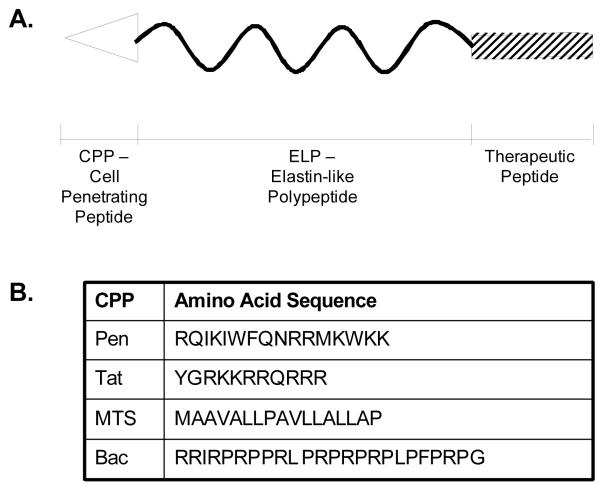
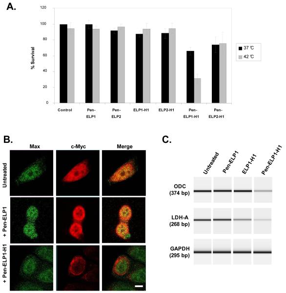
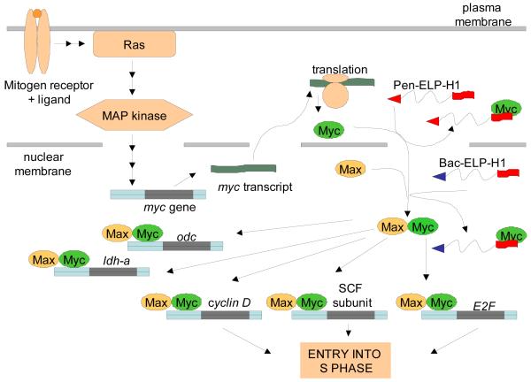
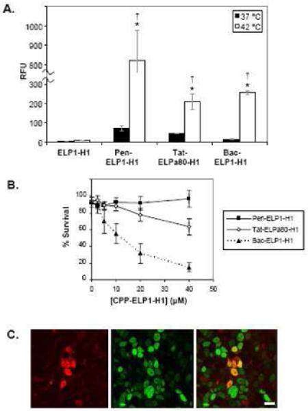
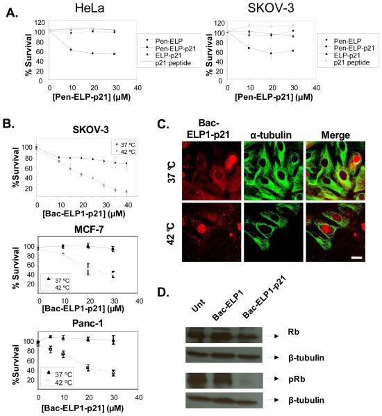
Current treatment of solid tumors is limited by side effects that result from the non-specific delivery of drugs to the tumor site. Alternative targeted therapeutic approaches for localized tumors would significantly reduce systemic toxicity. Peptide therapeutics are a promising new strategy for targeted cancer therapy because of the ease of peptide design and the specificity of peptides for their intracellular molecular targets. However, the utility of peptides is limited by their poor pharmacokinetic parameters and poor tissue and cellular membrane permeability in vivo. This review article summarizes the development of elastin-like polypeptide (ELP) as a potential carrier for thermally targeted delivery of therapeutic peptides (TP), and the use of cell penetrating peptides (CPP) to enhance the intracellular delivery of the ELP-fused TPs. CPP-fused ELPs have been used to deliver a peptide inhibitor of c-Myc function and a peptide mimetic of p21 in several cancer models in vitro, and both polypeptides are currently yielding promising results in in vivo models of breast and brain cancer.


Figures

Figure 1
Figure 1
Schematic representation of the ELP-based peptide delivery vector. A. The thermally responsive ELP polypeptide is fused at its N-terminus to a cell penetrating peptide (CPP) to mediate uptake of the macromolecule across the plasma membrane and dictate intracellular localization. At the C-terminus, a therapeutic peptide is added. B. Table of CPPs used to date for intracellular delivery of ELP.
Figure 2
Figure 2
A. Flow cytometry histograms showing relative fluorescence of HeLa cells after treatment with the indicated CPP-ELP-fluorescein (20μM) for 1 h at 37 °C. The results are representative of a typical experiment. B. Effect of CPP-ELP-fluorescein concentration on cellular uptake as expressed in relative fluorescence units (RFU) normalized to uptake of 5 μM ELP. Increasing concentrations of polypeptides were incubated with HeLa cells at 37 °C for 1 h. Results are represented as mean ± SEM of three independent experiments. C. Kinetics of internalization of CPP-ELPs. HeLa cells were incubated with 20 μM fluorescein labeled proteins for 1h. Cell fluorescence was measured by flow cytometry at 1, 2, 4 and 24 h after polypeptide exposure. The percentage internalized was calculated by dividing the trypan blue quenched (intracellular) fluorescence by the total unquenched fluorescence. The results represent the mean ± SEM of three independent experiments. Analysis of variance revealed that there is a difference in the initial internalization (1 and 2 hrs, p<0.05) between MTS-ELP and the other CPP-ELPs, but no CPP-ELPs are significantly different after 4 h and 24h incubation (p>0.17). D. Subcellular localization of CPP-ELPs. HeLa cells were treated with CPP-ELP-rhodamine (20 μM) for 1 h at 37 °C, and confocal images were taken 24 h later. Scale bar = 8 μm. Because of the considerable difference in fluorescence uptake between CPP-ELPs, the gain was adjusted individually during each image acquisition. Therefore, the fluorescence intensity of the images does not represent the relative amount of CPP-ELP in the cell.
Figure 3
Figure 3
Effect of inhibitors of endocytosis on CPP-ELP uptake. A. Effect of low temperature incubation on polypeptide uptake. HeLa cells were incubated with CPP-ELP-fluorescein (20 μM) for 1 h at 37 °C and 4 °C. B. Effect of ATP depletion on polypeptide uptake. The intracellular ATP pool was depleted by pre-incubation with sodium azide and deoxyglucose, followed by treatment of cells with CPP-ELP-fluorescein (20 μM) for 1 h at 37 °C. C. Effect of hyperosmolar sucrose on polypeptide uptake. Cells were incubated with 0.45 M sucrose for 1 h along with polypeptide treatment (20 μM). D. Effect of methyl-β-cyclodextrin on polypeptide uptake. Cells were preincubated with methyl-β-cyclodextrin (5 mM) for 30 min before polypeptide treatment. In all cases, non-internalized protein was quenched using trypan blue prior to flow cytometric analysis, and the cellular uptake is shown as relative fluorescence normalized to uptake of ELP. The results represent the mean ± SEM of three independent experiments. A Student’s t-test was used to determine statistical significance (*, p<0.01; +, p<0.05).
Figure 4
Figure 4
Intratumoral localization of the CPP-delivered ELP peptide carrier. Rhodamine-labeled Bac-ELP1-H1 (A.) or Tat-ELP1-H1 (B.) was injected IV, and one of two subcutaneous C6 tumors was heated with IR light for 60 min. 500 kDa FITC-dextran was injected 1 min prior to euthanasia in order to mark the perfused vessels, and tumors were frozen, sectioned, and stained with Hoechst 33342 to mark the cell nuclei. A representative section from multiple tumor sections from duplicate animals is shown. Scale bar = 30 μm.
Figure 5
Figure 5
A. Antiproliferative effect of Pen-ELP-H1. Proliferation of MCF-7 cells was determined 11 days after a single 1 h treatment with the indicated polypeptide (18 μM) at 37 °C or 42 °C. Cells were counted using the trypan blue dye exclusion assay. Results represent the mean ± SE of 3-5 experiments performed in duplicate. B. Effect of Pen-ELP1-H1 on c-Myc localization. The subcellular localization of c-Myc and Max was determined by confocal immunofluorescence microscopy in untreated cells (top row) and in cells treated with 18 μM Pen-ELP1 (middle row) or 18 μM Pen-ELP1-H1 (bottom row) for 1h. Images were taken 24 h after polypeptide treatment with a 100x oil immersion objective, scale bar = 8 μm. C. Effect of Pen-ELP1-H1 on transcriptional activation by c-Myc. The mRNA levels for the c-Myc responsive genes ODC (top panel) and LDH-A (middle panel) and a control gene GAPDH (bottom panel) were assayed by RT-PCR. MCF-7 cells were untreated (lane 1) or treated with 18 μM Pen-ELP1 (lane 2), ELP1-H1 (lane 3), or Pen-ELP1-H1 (lane 4) for 1h. RNA was purified 48 h after treatment. PCR products were analyzed by capillary electrophoresis using a Bioanalyzer Labchip with fluorescence detection. The fluorescence data was converted to a simulated gel using Agilent software. The experiment was repeated 2 times.
Figure 6
Figure 6
Proposed model for c-Myc inhibition by Pen-ELP-H1 and Bac-ELP-H1. Mitogen stimulation induces transcription of mRNA from the c-Myc gene. In the case of Pen-ELP-H1, newly translated c-Myc is bound by the polypeptide in the cytoplasm. Once bound, c-Myc can not be imported into the nucleus and interact with Max. In the case of Bac-ELP-H1, the polypeptide enters the nucleus and interferes directly with the c-Myc and Max interaction. Both situations result in the down-regulation of c-Myc-Max responsive genes and lead to inhibition of cell proliferation.
Figure 7
Figure 7
Optimization of ELP-H1 delivery with alternative CPPs. A. Cellular uptake of the CPP-ELP-H1 polypeptides. MCF-7 cells were treated for 1 h at 37 or 42 °C with fluorescein labeled polypeptides. Levels of each polypeptide were assessed using flow cytometry (n = 5,000 cells). Forward and side scatter gating were used to eliminate cell debris from the analysis, and fluorescence data was normalized to cellular autofluorescence and corrected for variations in labeling efficiency among the polypeptides. Data represent the average of 3 experiments; error bars, SEM.* Difference between 37 and 42 °C levels are statistically significant (ANOVA, p<0.01). † Difference is significant as compared to ELP at 42 °C. (ANOVA, p<0.01). B. MCF-7 proliferation after CPP-ELP-H1 treatment. MCF-7 cells were treated for 1 h at 37 or 42 °C with various concentrations of Pen-ELP1-H1, Tat-ELPa80-H1, or Bac-ELP1-H1, and the cell viability was determined after 7 days using the MTS assay. The data shown is an overlay of the 42 °C data for each CPP-ELP-H1. C. Subcellular localization of Bac-ELP1-H1. MCF-7 cells were treated for 1 h at 42 °C with rhodamine labeled Bac-ELP1-H1 (30 μM). 48 h after treatment, nuclei were stained with Sytox green and cells were imaged with a laser scanning confocal microscope. Scale bar = 20 μm.
Figure 8
Figure 8
CPP-ELP-p21 polypeptides. A. Inhibition of cell proliferation by Pen-ELP-p21. HeLa and SKOV-3 cells were exposed to the indicated concentration of Pen-ELP2-p21 at 37 °C for 1 h, and cell proliferation was determined 72 h later using the MTS assay. B. Inhibition of proliferation by Bac-ELP-p21. SKOV-3, MCF-7, and Panc-1 cells were exposed to the indicated concentration of Bac-ELP2-p21 at 37 °C or 42 °C for 1 h, and cell proliferation was determined 6 days later (SKOV-3) or 3 days later (MCF-7 and Panc-1) using the MTS assay. Data represent the mean ± SE of 3 independent experiments. C. Subcellular localization of rhodamine labeled Bac-ELP1-p21 in SKOV-3 cells as visualized by confocal microscopy. Cells were treated with 20 μM Bac-ELP1-p21 at 37°C or 42°C for 1 h. Confocal images were taken 24 h later. Tubulin was stained as a reference for cellular structure. Scale bar = 20 μm. The subcellular distribution of Bac-ELP1-p21 was also confirmed in live cells 24 h after a 1 h exposure at 37 or 42 °C (not shown). D. SDS-PAGE analysis of Rb protein in SKOV-3 cells following treatment with Bac-ELP1-p21. Cells were treated with the indicated polypeptide (30 μM) at 42°C for 1 h, harvested 24 h later, and lysed. Equal amounts of samples were loaded onto a 12% SDS gel and transferred to a blot which was probed with the indicated antibodies.

Similar articles

Cited by

References

    1. Greish K. Enhanced permeability and retention of macromolecular drugs in solid tumors: a royal gate for targeted anticancer nanomedicines. J Drug Target. 2007;15(7-8):457–464. - PubMed
    1. Iyer AK, Khaled G, Fang J, Maeda H. Exploiting the enhanced permeability and retention effect for tumor targeting. Drug Discov Today. 2006;11(17-18):812–818. - PubMed
    1. Maeda H, Bharate GY, Daruwalla J. Polymeric drugs for efficient tumor-targeted drug delivery based on EPR-effect. Eur J Pharm Biopharm. 2009;71(3):409–419. - PubMed
    1. Talelli M, Rijcken CJ, van Nostrum CF, Storm G, Hennink WE. Micelles based on HPMA copolymers. Adv Drug Deliv Rev. 2009 - PubMed
    1. Matsumura Y. Poly (amino acid) micelle nanocarriers in preclinical and clinical studies. Adv Drug Deliv Rev. 2008;60(8):899–914. - PubMed

Publication types

MeSH terms