. 2010 May 20;7(5):343-53.
doi: 10.1016/j.chom.2010.04.006.

Antiviral instruction of bone marrow leukocytes during respiratory viral infections

Affiliations

Antiviral instruction of bone marrow leukocytes during respiratory viral infections

Tamar Hermesh et al. Cell Host Microbe. .

Abstract

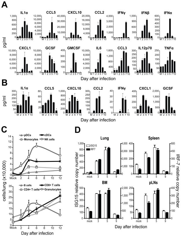
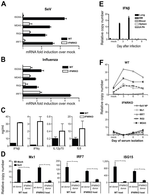
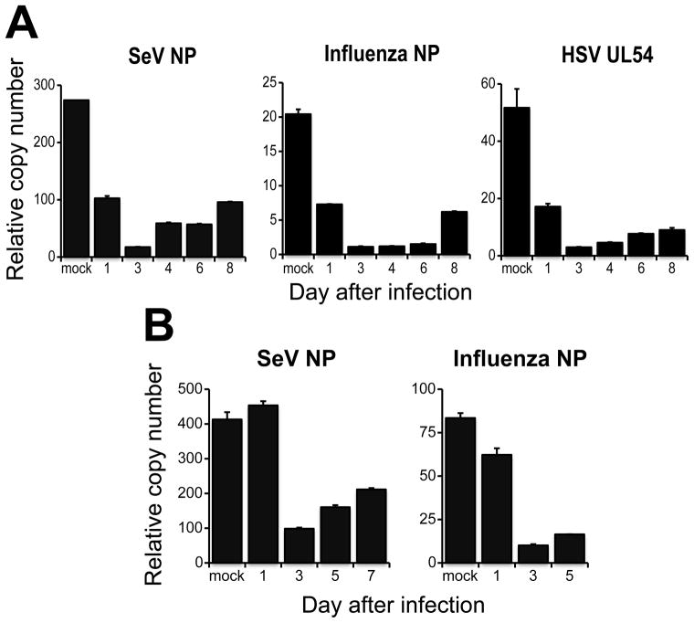
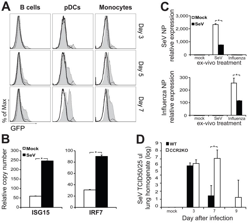
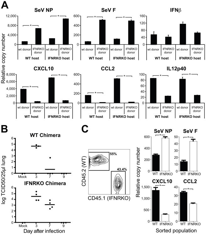
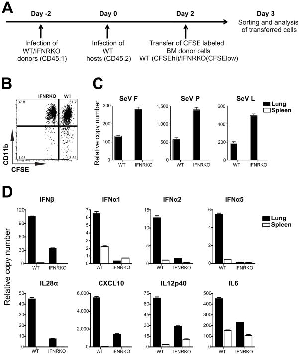
Respiratory viral infections trigger a robust inflammatory response in the lung, producing cytokines, chemokines, and growth factors that promote infiltration of effector leukocytes. Whereas the role of chemokines and infiltrating leukocytes in antiviral immunity is well studied, the effect that lung cytokines have on leukocytes in distal hematopoietic and lymphoid tissues and their role in antiviral immunity is unknown. We show that, during infection with influenza or Sendai virus, the lung communicates with the sterile bone marrow, the primary site of hematopoiesis, through type I interferons. While in the bone marrow, leukocytes exposed to type I interferons activate an antiviral transcriptional program and become resistant to infection with different viruses. The protected bone marrow leukocytes are capable of migrating to the infected lung and contribute to virus clearance. These findings show that appropriate instruction of cells during their development in the bone marrow is needed for effective control of infection.


Figures

Figure 1
Figure 1
Lung inflammation correlates with the systemic transcription of anti-viral genes. Cytokines from (A) fresh lung homogenates and (B) sera obtained from mice infected with SeV and sacrificed at different days post inoculation. Mock (M) infected animals were used as controls. (C) Lung cells count after tissue digestion. Cell numbers for specific cell population were extrapolated from flow cytometry analysis. The cells were pre-gated on live (PI) CD45+ cells. B cells (CD11bmPDCAB220+), pDCs (CD11bmPDCA+B220+), monocytes (Ly6c+Ly6gCD115+), Granulocytes (Ly6c+Ly6g+), NKs (NK1.1+CD11b+), cDCs (CD11c+MHCII+CD11b+), CD4+ T cells (CD3+CD4+CD8), CD8+ T cells (CD3+CD4CD8+). (D) ISG15 and IRF7 mRNA levels in the lung, pLNs, BM and spleen at different days after infection with SeV. mRNA was normalized to the house keeping genes α-tubulin, rps11 and β-actin of each sample analyzed. Error bars indicate the standard deviation of triplicate measurements in a representative experiment. See also Figure S1.
Figure 2
Figure 2
Transcription of anti-viral genes in the BM is induced by type I IFNs transported in the blood. (A–B) Gene expression in BM of wild type mice or type I IFN receptor KO mice infected with (A) SeV two days after infection or (B) influenza PR8 virus three days after infection. (C) Cytokines in lung homogenates obtained from WT and IFNRKO mice 3 days post-infection. (D) Gene expression in the BM of complete chimeric mice infected for five days with SeV (n=4 mice in each group; *p< 0.05). Error bars represent means±S.D. (E) IFNβ expression in the lung, BM, blood, peripheral lymph nodes (pLN) and spleen of wild type mice infected with SeV. See also figure S2. (F) Gene expression in naive BM cells isolated from WT (top panel) or IFNRKO (bottom panel) mice after a 6 h incubation with serum obtained from wild type mice at different times after infection with SeV. (n=3 mice in each group; *p< 0.05).
Figure 3
Figure 3
BM cells of mice undergoing a respiratory infection are protected from viral replication. Expression of viral genes in BM cells isolated at different days after infection with (A) SeV or (B) influenza virus strain X31 after 6 h of infection ex-vivo with SeV, influenza strain PR8 or HSV at a MOI of 2. n=4 mice in each group. Error bars represent means±S.D
Figure 4
Figure 4
Monocytes primed in the BM participate in the control of virus replication in the lung. (A) EGFP expression in BM B cells (PISSloB220+CD11bmPDCA), pDCs (PISSloB220+CD11bmPDCA+) and monocytes (PISSloCD115+CD11b+Ly6c+) from Mx-Cre x Rosa26-stopfloxEGFP mice infected for 3, 5 or 7 days with SeV (tinted), mock infected (black dashed line) or Mx-Cre(neg)Rosa26-stopfloxEGFP litter mates (black line). (B) Gene expression in BM monocytes (Ly6c+Ly6gCD115+) sorted from mock or SeV infected mice at day 4 post-infection (n=6 mice in each group; *P < 0.05). Error bars represent means±S.D. (C) Viral gene expression in BM monocytes isolated from mice four days post infection with SeV and after ex-vivo treatment with either SeV or influenza PR8. Mock infected animals were used as controls (n=6 mice in each group; *P < 0.05). Error bars represent means±S.D.(d) SeV titers in lung homogenates of wild type or CCR2KO mice after infection with SeV. 50% Tissue culture infectious dose (TCID50). n=3–4 mice in each group; *P < 0.05 Error bars represent means±S.D. See also figure S3
Figure 5
Figure 5
Type I IFNs arm hematopoietic cells against viral replication in vivo. (A) Gene expression on CD45+ cells isolated by magnetic beads from the lung of complete chimeric WT or IFNRKO mice after five days of infection with SeV. (B) SeV titers in lung homogenates of chimeric mice at different days after infection. (C) Viral gene expression analysis of CD45.2+ (WT) and CD45.1+ (IFNRKO) cells isolated from the lung of mixed chimeric mice after four days of infection with SeV. (n=3 mice in each group; *P < 0.05). Error bars represent means±S.D
Figure 6
Figure 6
Primed BM leukocytes are recruited to the lung and are protected from infection. (A) Experimental design for adoptive transfer of primed BM leukocytes. CD45.1+ WT or IFNRKO BM cells from mice infected with SeV for 4 days were labeled with 3.5μM or 0.5μM CFSE respectively, mixed at a 1/1 ratio and injected into WT CD45.2+ mice infected for 2 days with SeV. (B) Expression of CD11b by CD45.1+ CFSE high and low cells in the lungs of recipient mice. (C-D) qRT-PCR analysis of (C) viral mRNA and (D) cytokine mRNA in adoptively transferred BM WT and IFNRKO cells isolated from the lung and spleen.

Comment in

References

    1. Aldridge JR, Jr, Moseley CE, Boltz DA, Negovetich NJ, Reynolds C, Franks J, Brown SA, Doherty PC, Webster RG, Thomas PG. TNF/iNOS-producing dendritic cells are the necessary evil of lethal influenza virus infection. Proc Natl Acad Sci U S A. 2009;106:5306–5311. - PMC - PubMed
    1. Alexaki A, Wigdahl B. HIV-1 infection of bone marrow hematopoietic progenitor cells and their role in trafficking and viral dissemination. PLoS Pathog. 2008;4:e1000215. - PMC - PubMed
    1. Bernardini G, Sciume G, Bosisio D, Morrone S, Sozzani S, Santoni A. CCL3 and CXCL12 regulate trafficking of mouse bone marrow NK cell subsets. Blood. 2008;111:3626–3634. - PubMed
    1. Binder D, Fehr J, Hengartner H, Zinkernagel RM. Virus-induced Transient Bone Marrow Aplasia: Major Role of Interferon-alpha/beta during Acute Infection with the Noncytopathic Lymphocytic Choriomeningitis Virus. J Exp Med. 1997;185:517–530. - PMC - PubMed
    1. Biron CA, Nguyen KB, Pien GC, Cousens LP, Salazar-Mather TP. Natural killer cells in antiviral defense: function and regulation by innate cytokines. Annu Rev Immunol. 1999;17:189–220. - PubMed

Publication types