. 2010 May 20;7(5):376-87.
doi: 10.1016/j.chom.2010.04.009.

A Yersinia effector protein promotes virulence by preventing inflammasome recognition of the type III secretion system

Affiliations

A Yersinia effector protein promotes virulence by preventing inflammasome recognition of the type III secretion system

Igor E Brodsky et al. Cell Host Microbe. .

Abstract

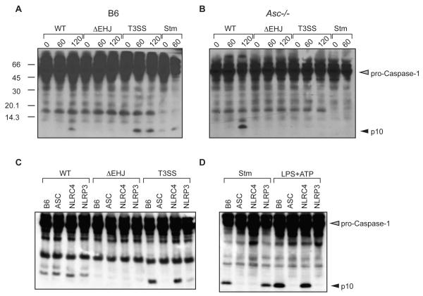
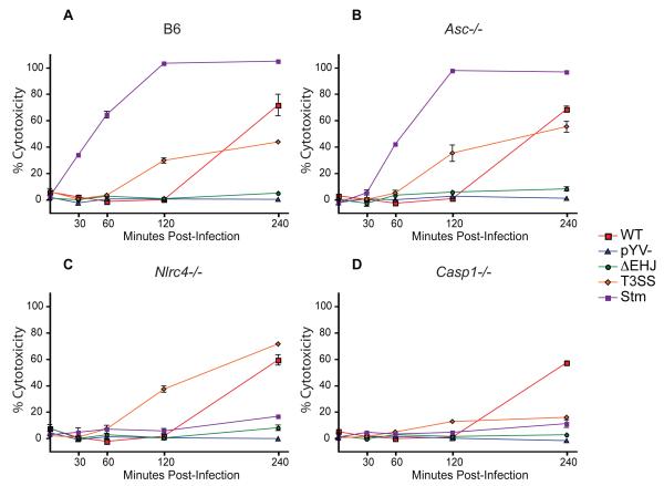
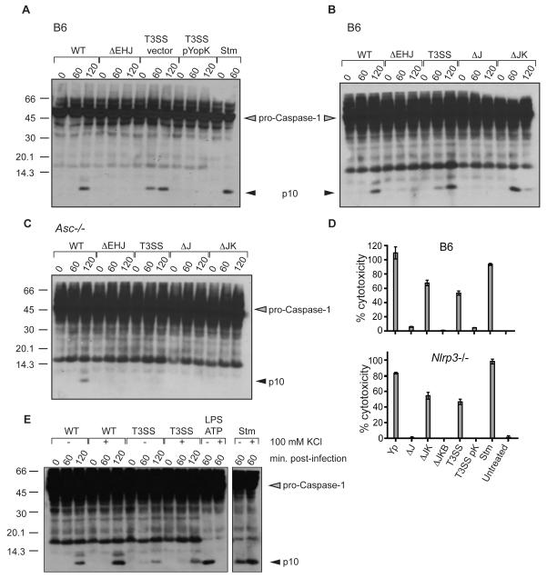
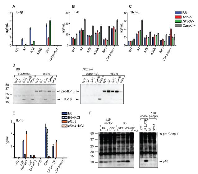
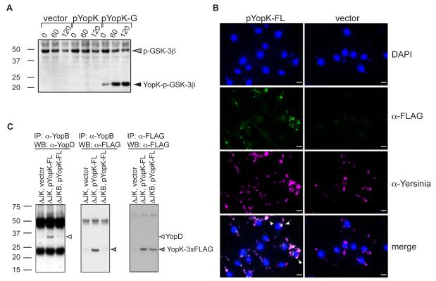
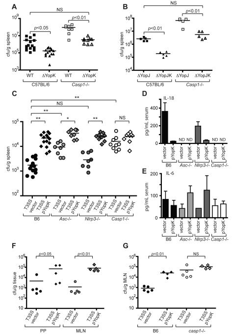
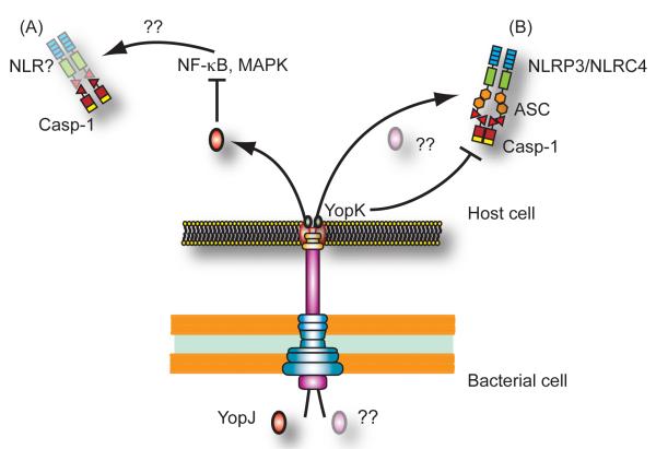
Bacterial pathogens utilize pore-forming toxins or specialized secretion systems to deliver virulence factors to modulate host cell physiology and promote bacterial replication. Detection of these secretion systems or toxins, or their activities, by nucleotide-binding oligomerization domain leucine-rich repeat proteins (NLRs) triggers the assembly of inflammasomes, multiprotein complexes necessary for caspase-1 activation and host defense. Here we demonstrate that caspase-1 activation in response to the Yersinia type III secretion system (T3SS) requires the adaptor ASC and involves both NLRP3 and NLRC4 inflammasomes. Further, we identify a Yersinia type III secreted effector protein, YopK, which interacts with the T3SS translocon to prevent cellular recognition of the T3SS and inflammasome activation. In the absence of YopK, inflammasome sensing of the T3SS promotes bacterial clearance from infected tissues in vivo. These data demonstrate that a class of bacterial proteins interferes with cellular recognition of bacterial secretion systems and contributes to bacterial survival within host tissues.


Figures

Figure 1
Figure 1. Macrophages require ASC for caspase-1 activation in response to the Yersinia T3SS
(A) BMMs derived from WT (B6), or (B) Asc-/- mice were infected with WT, ΔyopEHJ, or type III secretion system only (T3SS) strains of Y. pseudotuberculosis, as indicated, or with S. typhimurium (Stm) as control. Cell lysates were harvested at indicated times post-infection and probed with anti-caspase-1 antibodies. (C) BMMs from indicated mouse strains were harvested 120 minutes post-infection with WT, ΔyopEHJ, or T3SS Yersinia strains, or (D) 60 minutes post-infection with Salmonella enterica serovar Typhimurium (Stm). Cells were treated with LPS for 3 hours followed by 60 minutes ATP treatment (LPS+ATP). Data are representative of at least 3 independent experiments.
Figure 2
Figure 2. Caspase-1 is required for cell death triggered in response to the Yersinia T3SS but not YopJ
Percent cytotoxicity was measured by release of lactate dehydrogenase (LDH) in supernatants of (A) WT, (B) Asc-/-, (C) Nlrc4-/-, (D) Casp1-/- BMMs collected at indicated times following infection with indicated bacteria. pYV- - Y. pseudotuberculosis lacking Yersinia virulence plasmid. Data are representative of at least three independent experiments.
Figure 3
Figure 3. Caspase-1 activation and cell death triggered by macrophages in response to the Yersinia T3SS are inhibited by YopK
(A) WT (B6) BMMs were harvested at indicated times post-infection and assayed for caspase-1 activation by western blotting for cleaved p10 caspase-1 subunit. pYopK – YopK expression plasmid, Vector – vector control plasmid. (B) WT or (C) Asc-/- BMMs were infected with wild-type (WT), ΔJ – yopJ mutant, or ΔJK – yopJK mutant Y. pseudotuberculosis and assayed for caspase-1 activation by western blotting (D) Percent cytotoxicity in WT and isogenic Nlrp3-/- macrophages infected with indicated bacterial strains was assayed 4 hours post-infection (E) WT BMM lysates were harvested at indicated minutes post-infection and assayed for caspase-1 activation following infection with either WT or T3SS strains or S. typhimurium (Stm), or treated with LPS+ATP in the presence or absence of 100 mM KCl, as indicated. Data are representative of two to three independent experiments.
Figure 4
Figure 4. Processing and secretion of IL-1β in response to the Yersinia T3SS is inhibited by YopK and requires NLRP3 and ASC
(A, B, and C) BMMs were treated with LPS for 3 hours prior to infection, and supernatants harvested 2 hours post-infection and analyzed by ELISA for presence of (A) IL-1β (B) IL-6 or (C) TNF-α (D) WT or Nlrp3-/- BMMs were infected with indicated bacterial strains and supernatants and whole-cell lysates were immunoprecipitated with anti-IL-1β antibodies followed by western blotting for IL-1β (E) WT or Nlrc4-/- BMMs were treated with 100 mM KCl as indicated 15 minutes prior to infection and supernatants analyzed for IL-1β (F) BMMs were treated with 100 mM KCl prior to infection and cell lysates analyzed by western blotting with anti-caspase-1 antibodies.
Figure 5
Figure 5. YopK is translocated into cells and physically interacts with T3SS translocon
(A) BMMs were infected with T3SS expressing either empty vector control (vector), YopK expression plasmid (pYopK), or a GSK-3β tagged YopK expression vector (pYopK-G). Whole cell lysates were harvested and blotted for phospho-GSK-3β to determine the extent of translocation. (B) T3SS containing either pYopK-3XFLAG or vector control were used to infect BMMs on glass cover slides and stained with DAPI (top row), anti-FLAG (2nd row), or anti-total Yersinia (3rd row) antibodies. Bottom 2 panels are merged images. Arrows indicate colozalization of FLAG and Yersinia staining. Bar = 10 μM. (C) HeLa cells were infected with bacterial ΔyopJK bacteria containing vector control or YopK-3XFLAG plasmid, or infected with ΔyopJKB bacteria containing YopK-3XFLAG plasmid as additional control. Cell lysates were prepared, immunoprecipitated with anti-YopB antibodies (left and middle panels) or anti-FLAG M2 antibodies (right panel). Immunoprecipitated samples were run on SDS-PAGE and probed with antibodies against YopD (left panel) or YopK-FLAG (middle and right panels). Data are representative of two to three independent experiments.
Figure 6
Figure 6. YopK inhibits caspase-1 dependent bacterial clearance in vivo
(A) WT and Casp1-/- mice were infected intraperitoneally with WT or yopK mutant bacteria, or (B) yopJ or yopJK mutant bacteria, and spleen homogenates were plated 4 days post-infection to determine bacterial CFU per gram of tissue. (C) WT, Asc-/-, Nrlp3-/-, and Casp1-/- mice were infected as in (A) with either T3SS vector control or T3SS pYopK bacteria, and bacterial CFU per gram of tissue were determined. (D) Sera from mice in (C) were harvested and analyzed by ELISA for levels of circulating IL-18 or (E) IL-6. (F) WT mice were infected orally with indicated bacterial strains, and CFU per gram of Peyer’s patches and mesenteric lymph nodes was determined. (G) WT or Casp1-/- mice were infected as in (F) and bacterial load in mesenteric lymph nodes was determined. Statistical significance was analyzed by Student’s two-tailed t-test. * p<0.0002, ** p<0.0001. Data represent 2 to 3 independent pooled experiments (A, B, C) or at least three independent experiments for C57BL/6 mice and two independent experiments for knockout mice (F, G).
Figure 7
Figure 7. Yersinia infection triggers two distinct pathways of caspase-1 activation
(A) Inhibition of NF-κB and MAPK signaling by Y. pseudotuberculosis YopJ (red oval) activates caspase-1 independently of ASC, NLRP3 or NLRC4. (B) In contrast, the Yersinia T3SS triggers an ASC-dependent pathway of caspase-1 activation involving both NLRP3 and NLRC4. This pathway may be triggered by T3SS-induced pore formation, or by translocation of an unknown protein or small molecule (pink oval) into the cell. YopK (green ovals) interacts with the T3SS translocon and prevents inflammasome activation in response to detection of the T3SS by the host cell.

Comment in

  • Hiding from the inflammasome.
    David R. David R. Nat Rev Microbiol. 2010 Jul;8(7):466. doi: 10.1038/nrmicro2389. Nat Rev Microbiol. 2010. PMID: 21394955 No abstract available.

References

    1. Akira S, Takeda K, Kaisho T. Toll-like receptors: critical proteins linking innate and acquired immunity. Nat Immunol. 2001;2:675–680. - PubMed
    1. Auerbuch V, Golenbock DT, Isberg RR. Innate immune recognition of Yersinia pseudotuberculosis type III secretion. PLoS Pathog. 2009;5:e1000686. - PMC - PubMed
    1. Auerbuch V, Isberg RR. Growth of Yersinia pseudotuberculosis in mice occurs independently of Toll-like receptor 2 expression and induction of interleukin-10. Infect Immun. 2007;75:3561–3570. - PMC - PubMed
    1. Bartra SS, Jackson MW, Ross JA, Plano GV. Calcium-regulated type III secretion of Yop proteins by an Escherichia coli hha mutant carrying a Yersinia pestis pCD1 virulence plasmid. Infect Immun. 2006;74:1381–1386. - PMC - PubMed
    1. Bergman MA, Loomis WP, Mecsas J, Starnbach MN, Isberg RR. CD8(+) T cells restrict Yersinia pseudotuberculosis infection: bypass of anti-phagocytosis by targeting antigen-presenting cells. PLoS Pathog. 2009;5:e1000573. - PMC - PubMed

Publication types