. 2010 Jul 15;588(Pt 14):2533-44.
doi: 10.1113/jphysiol.2010.188409. Epub 2010 May 17.

Enteric glia modulate epithelial cell proliferation and differentiation through 15-deoxy-12,14-prostaglandin J2

Affiliations

Enteric glia modulate epithelial cell proliferation and differentiation through 15-deoxy-12,14-prostaglandin J2

Kalyane Bach-Ngohou et al. J Physiol. .

Abstract

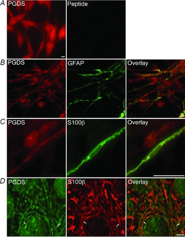
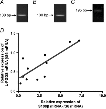
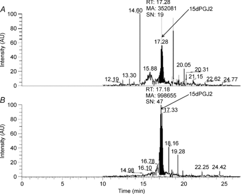
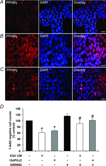
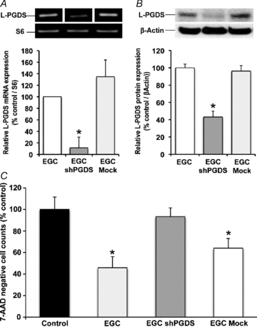
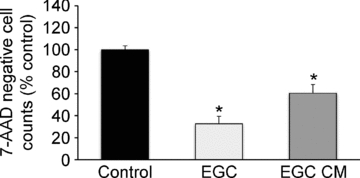
The enteric nervous system (ENS) and its major component, enteric glial cells (EGCs), have recently been identified as a major regulator of intestinal epithelial barrier functions. Indeed, EGCs inhibit intestinal epithelial cell (IEC) proliferation and increase barrier resistance and IEC adhesion via the release of EGC-derived soluble factors. Interestingly, EGC regulation of intestinal epithelial barrier functions is reminiscent of previously reported peroxisome proliferator-activated receptor gamma (PPARgamma)-dependent functional effects. In this context, the present study aimed at identifying whether EGC could synthesize and release the main PPARgamma ligand, 15-deoxy-(12,14)-prostaglandin J2 (15dPGJ2), and regulate IEC functions such as proliferation and differentiation via a PPARgamma dependent pathway. First, we demonstrated that the lipocalin but not the haematopoetic form for prostaglandin D synthase (PGDS), the enzyme responsible of 15dPGJ2 synthesis, was expressed in EGCs of the human submucosal plexus and of the subepithelium, as well as in rat primary culture of ENS and EGC lines. Next, 15dPGJ2 was identified in EGC supernatants of various EGC lines. 15dPGJ2 reproduced EGC inhibitory effects upon IEC proliferation, and inhibition of lipocalin PGDS expression by shRNA abrogated these effects. Furthermore, EGCs induced nuclear translocation of PPARgamma in IEC, and both EGC and 15dPGJ2 effects upon IEC proliferation were prevented by the PPARgamma antagonist GW9662. Finally, EGC induced differentiation-related gene expression in IEC through a PPARgamma-dependent pathway. Our results identified 15dPGJ2 as a novel glial-derived mediator involved in the control of IEC proliferation/differentiation through activation of PPARgamma. They also suggest that alterations of glial PGDS expression may modify intestinal epithelial barrier functions and be involved in the development of pathologies such as cancer or inflammatory bowel diseases.


Figures

Figure 1
Figure 1
Enteric glial cells express prostaglandin D synthase Immunofluorescence staining of prostaglandin D synthase (PGDS) in rat enteric glial cell lines without and with blocking peptides (A), in primary culture of rat enteric nervous system (ENS) (B), in human submucosal plexus (C) and in human subepithelial layer (D). In primary culture of rat ENS (B), glial fibrillary acidic protein (GFAP) immunoreactive cells were colocalized with PGDS. In human submucosal plexus (C) and subepithelial layer (D), some S100β-immunoreactive cells were colocalized with PGDS (white arrows). Each picture is representative of five experiments. Scales bars are 5 μm (A–C) and 100 μm (D).
Figure 2
Figure 2
Transcriptional expression of lipocalin-prostaglandin D synthase in enteric glial cells RT-PCR analysis of lipocalin-prostaglandin D synthase (L-PGDS) mRNA expression showed a 130 bp band in rat enteric glial cell lines (A) and in rat primary culture of enteric nervous system (B). In human colonic biopsies, a 195 bp band was detected (C). Quantitative PCR analysis of human colonic biopsies revealed a positive correlation between L-PGDS and S100β mRNA expression (D) (n = 11; r2 = 0.63; P = 0.01; linear regression).
Figure 3
Figure 3
Enteric glial cells secrete 15-deoxy-Δ12,14-prostaglandin J2 Gas chromatography associated with mass spectrometry analysis showed the presence of 15dPGJ2 in transformed enteric glial cell conditioned medium (representative pattern of three experiments) (A). A control pattern was generated with Dubelcco's modified Eagle medium supplemented with 15dPGJ2 (B).
Figure 6
Figure 6
Effect of enteric glial cells on peroxisome proliferator-activated receptor γ translocation in Caco-2 cells Immunofluorescence staining of peroxisome proliferator-activated receptor γ (PPARγ) in Caco-2 cells showed that it was uniformly distributed in the cytoplasm of cells cultured alone (A). In contrast, following coculture with enteric glial cells (B) or treatment with enteric glial cell conditioned medium (EGC CM) (C), PPARγ was mainly localized in the nucleus, identified by 4′,6′-diamidino-2-phenylindole (DAPI) staining. Data are representative of 3 independent experiments. Scale bars: 10 μm. D, fluorescence activated cell sorting analysis of 7-aminoactinomycin D (7-AAD) negative Caco-2 cells showed that EGC CM (n = 6) or 15dPGJ2 (5 μm; n = 3) induced a significant decrease in Caco-2 cell number as compared to control. PPARγ antagonist (GW9662, 10 μm) alone did not modify the cell number as compared to control. In the presence of GW9662, EGC CM (n = 6) or 15dPGJ2 (5 μm; n = 3) did not decrease Caco-2 cell number as compared to control. Values are expressed as means ± s.e.m. from 6 independents experiments (Kruskall–Wallis test followed by Dunn's post hoc test; P < 0.05 * as compared to non treated Caco-2; # as compared to Caco-2 treated with EGC CM or 15dPGJ2).
Figure 5
Figure 5
Transduction of EGC with L-prostaglandin D synthase shRNA abrogate their anti-proliferative effects RT-qPCR analysis showed that lipocalin-prostaglandin D synthase (L-PGDS) mRNA expression was significantly decreased in L-PGDS shRNA transduced enteric glial cells (EGC shPGDS) as compared to mock-transduced EGCs (EGC Mock) and non-transduced EGCs (EGC) (n = 5, Kruskall–Wallis test followed by Dunn's post hoc test; P < 0.05) (A). Western blot analysis showed that L-PGDS protein expression was significantly decreased in L-PGDS shRNA transduced enteric glial cells (EGC shPGDS) as compared to mock-transduced EGCs (EGC Mock) and non-transduced EGCs (EGC) (n = 7, Kruskall–Wallis test followed by Dunn's post hoc test; P < 0.05) (B). Fluorescence activated cell sorting analysis of 7-aminoactinomycin D (7-AAD) negative Caco-2 cells showed that coculture of Caco-2 cells with EGCs or EGC Mock but not EGC shPGDS induced a significant decrease in Caco-2 cell number as compared to control. Values are expressed as means ± s.e.m. (n = 4; Kruskall–Wallis test followed by Dunn's post hoc test; P < 0.05) (C).
Figure 4
Figure 4
Enteric glial cells inhibit Caco-2 cell proliferation Fluorescence activated cell sorting analysis of 7-aminoactinomycin D (7-AAD) negative Caco-2 cells showed that enteric glial cells (EGCs) or EGC conditioned medium (EGC CM) induced a significant decrease in Caco-2 cell number after 6 days of culture as compared to untreated (Control) Caco-2 monolayer.
Figure 7
Figure 7
Enteric glial cells induce mRNA expression of differentiation marker genes RT-qPCR analysis showed that mRNA expression of peroxisome proliferator-activated receptor γ (PPARγ) (A), intestinal alkaline phosphatase (hALPI) (B) and E-cadherin (C) was increased in Caco-2 cells cultured with enteric glial cell conditioned medium (EGC CM) as compared to Caco-2 cultured alone (n = 7). This effect was completely abrogated following culture with EGC CM in presence of GW9662 (n = 3). Values are expressed as means ± s.e.m. (Kruskall–Wallis test followed by Dunn's post hoc test; P < 0.05 * as compared to non treated Caco-2; # as compared to Caco-2 treated with EGC CM).

Similar articles

Cited by

References

    1. Beuckmann CT, Lazarus M, Gerashenko D, Mizoguchi A, Nomura S, Mohri I, Uesugi A, Kaneko T, Mizuno N, Hayaishi O, Urade Y. Cellular localization of lipocalin-type prostaglandin D synthase (beta-trace) in the central nervous sytem of the adult rat. J Comp Neurol. 2000;428:62–78. - PubMed
    1. Bull AW, Steffensen KR, Leers J, Rafter JJ. Activation of PPARγ in colon tumor cell lines by oxidized metabolites of linoleic acid, endogenous ligands for PPAR γ. Carcinogenesis. 2003;24:1717–1722. - PubMed
    1. Cabarrocas J, Savidge TC, Liblau RS. Role of enteric glial cells in inflammatory bowel disease. Glia. 2003;41:81–93. - PubMed
    1. Chen X, Whitney EM, Gao SY, Yang VW. Transcriptional profiling of Kruppel-like factor 4 reveals a function in cell cycle regulation and epithelial differentiation. J Mol Biol. 2003;326:665–677. - PMC - PubMed
    1. Chevalier J, Derkinderen P, Gomes P, Thinard R, Naveilhan P, Vanden Berghe P, Neunlist M. Activity-dependent regulation of tyrosine hydroxylase expression in the enteric nervous system. J Physiol. 2008;586:1963–1975. - PMC - PubMed

Publication types

MeSH terms

LinkOut - more resources