. 2010 Jul 1;588(Pt 13):2443-54.
doi: 10.1113/jphysiol.2010.189670. Epub 2010 May 17.

Postnatal development of mouse heart: formation of energetic microdomains

Affiliations

Postnatal development of mouse heart: formation of energetic microdomains

Jérôme Piquereau et al. J Physiol. .

Abstract

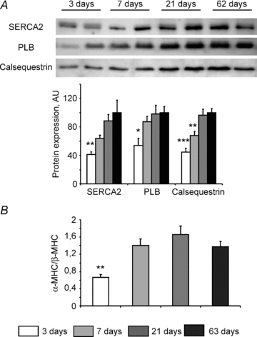
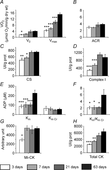
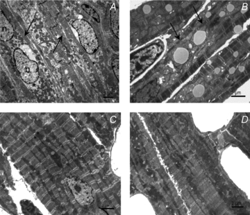
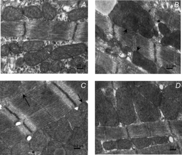
Cardiomyocyte contractile function requires tight control of the ATP/ADP ratio in the vicinity of the myosin-ATPase and sarcoplasmic reticulum ATPase (SERCA). In these cells, the main systems that provide energy are creatine kinase (CK), which catalyses phosphotransfer from phosphocreatine to ADP, and direct adenine nucleotide channelling (DANC) from mitochondria to ATPases. However, it is not known how and when these complex energetic systems are established during postnatal development. We therefore studied the maturation of the efficacy with which DANC and CK maintain ATP/ADP-dependent SR and myofibrillar function (SR Ca(2+) pumping and prevention of rigor tension), as well as the maturation of mitochondrial oxidative capacity. Experiments were performed on saponin-skinned fibres from left ventricles of 3-, 7-, 21-, 42- and 63-day-old mice. Cardiomyocyte and mitochondrial network morphology were characterized using electron microscopy. Our results show an early building-up of energetic microdomains in the developing mouse heart. CK efficacy for myosin-ATPase regulation was already maximal 3 days after birth, while for SERCA regulation it progressively increased until 21 days after birth. Seven days after birth, DANC for these two ATPases was as effective as in adult mice, despite a non-maximal mitochondrial respiration capacity. However, 3 days after birth, DANC between mitochondria and myosin-ATPase was not yet fully efficient. To prevent rigor tension in the presence of working mitochondria, the myosin-ATPase needed more intracellular MgATP in 3-day-old mice than in 7-day-old mice (pMgATP(50) 4.03 +/- 0.02 and 4.36 +/- 0.07, respectively, P < 0.05), whereas the intrinsic sensitivity of myofibrils to ATP (when mitochondria were inhibited) was similar at both ages. This may be due to the significant remodelling of the cytoarchitecture that occurs between these ages (cytosolic space reduction, formation of the mitochondrial network around the myofibrils). These results reveal a link between the maturation of intracellular energy pathways and cell architecture.


Figures

Figure 1
Figure 1. Pathways of energy supply for ATPases in the cardiomyocyte
1, creatine kinase (CK) bound to the myofilaments or the SR rephosphorylates locally produced ADP at the expense of PCr. 2, the juxtaposition of the mitochondrion and ATPase allows for direct adenine nucleotide channelling (DANC). 3, adenine nucleotides in the neighbourhood of ATPase exchange with those in the bulk of cytosol via diffusion.
Figure 3
Figure 3. Molecular characterization of the heart at different ages
A, Western blot analysis of SERCA2, phospholamban (PLB) and calsequestrin expression at different ages. Upper panel: representative original recording. Lower panel: mean values of protein content (n= 8 for each age). B, ratio of α-MHC to β-MHC mRNA expression measured by real-time quantitative RT-PCR. N= 8 animals for each age. *P < 0.05, **P < 0.01, ***P < 0.001 versus adult age (63 days).
Figure 2
Figure 2. SR calcium loading under different energetic conditions in permeabilized cardiac fibres at different ages
Calcium uptake at pCa 6.5 in the presence of different ATP sources: external (ATP), and endogenous, namely bound CK (+CK), mitochondria (+DANC) or both (+DANC+CK). By relating the subsequent contractile force elicited with caffeine to the calcium–force curve for each fibre, the [Ca2+]–time integral (SCa) was calculated as an index of SR calcium load. n= 7–10 fibres from 3–7 animals for each age. *P < 0.05, ***P < 0.001 versus adult age (63 days).
Figure 4
Figure 4. Sensitivity of rigor tension to MgATP from different sources in 3-, 7-, 42- and 63-day-old mice
pMgATP50 (pMgATP for half-maximal rigor tension) and role of creatine kinase and/or mitochondria in myofibrillar-ATPase energy supply. Rigor tension was recorded in solutions of decreasing MgATP concentration using different solutions that control ATP sources: external (ATP), and endogenous, namely bound CK (+CK), mitochondria (+DANC) or both (+DANC+CK). n= 6–11 fibres from 3–6 animals for each age. pMgATP50 : *P < 0.05 vs. 63 days.
Figure 5
Figure 5. Intrinsic energy metabolism parameters at different ages
A, oxidative capacities of ventricular fibres were measured in the presence of 2 mm ADP, at saturating concentration of substrates. (n= 15 fibre bundles from 5–9 animals for each age.) B, acceptor control ratio was calculated as the ratio between maximal respiration rate (Vmax) and basal respiration rate (V0). C, citrate synthase (CS) enzymatic activity. D, Complex I enzymatic activity. E, apparent Km of mitochondrial consumption for ADP without or with 12 mm creatine. F, ratio between Km without creatine and Km with creatine. G, Mi-CK expression. H, total CK activity. (N= 8 animals for each age.) *P < 0.05, **P < 0.01, ***P < 0.001 versus 63 days.
Figure 6
Figure 6. Electron micrographs of cardiomyocytes of papillary muscle
A, 3-day-old cardiomyocytes having a high content of free cytoplasm, myofibrils situated under the plasma membrane (arrows) and mitochondria clustered in the perinuclear regions (*). B, 7-day-old cardiomyocytes showing abundant mitochondrial clusters, and myofilaments of small diameters (arrows). C, 21-day-old cardiomyocytes showing regularly arranged myofilaments and mitochondria aligned in the longitudinal direction. D, 63-day-old cardiomyocytes demonstrating regular overall ultrastructure. Myofilaments and mitochondria are arranged in parallel along the longitudinal axis.
Figure 7
Figure 7. Spatial interaction between mitochondria and myofilaments in developing cardiomyocytes
A, 3-day-old cardiomyocytes with large cytoplasmic space (*) between myofilaments and mitochondria which are not in tight contact to each other. B, 7-day-old cardiomyocytes showing mitochondria in direct contact with myofilaments (short arrows) even though a thin layer of cytoplasm can still be found between mitochondria and myofibrils (*). C, 21-day-old cardiomyocytes with well developed longitudinal SR (arrow) and mitochondria in close contact with the myofibrils (short arrow). D, 63-day-old cardiomyocytes with mitochondria in direct contact with myofilaments.
Figure 8
Figure 8. Electron micrographs of cardiomyocytes showing spatial relationship of mitochondria and sarcoplasmic reticulum
A, mitochondrial cluster in 3-day-old mice cardiomyocytes. Profiles of rough endoplasmic reticulum (short arrow) and short stretches of sarcoplasmic reticulum (arrow) are present between mitochondria. B, 7-day-old cardiomyocytes show the presence of longitudinal sarcoplasmic reticulum between the myofilaments and the mitochondrial surface (arrow). Occasionally, short stretches of rough endoplasmic reticulum near the mitochondria are still observed (short arrow). C, 21-day-old cardiomyocytes with well developed longitudinal sarcoplasmic reticulum (arrows). The cisternae of SR of the dyads occur in the close vicinity of the mitochondrial surface (short arrow). D, 63-day-old cardiomyocytes display a rich network of sarcoplasmic reticulum (arrows) and cisternae of SR (short arrow) close to mitochondria.
Figure 9
Figure 9. Stereological measurements of cardiomyocytes of papillary muscles
Relative cell volume occupied by mitochondria (Mito), myofilaments (Myof), cytosol (Cyto) and lipids drops (LD), evaluated from 20 images taken from 2 hearts of animals for each age. **P < 0.01, ***P < 0.001 versus 63 days.

References

    1. Anmann T, Guzun R, Beraud N, Pelloux S, Kuznetsov AV, Kogerman L, Kaambre T, Sikk P, Paju K, Peet N, Seppet E, Ojeda C, Tourneur Y, Saks V. Different kinetics of the regulation of respiration in permeabilized cardiomyocytes and in HL-1 cardiac cells. Importance of cell structure/organization for respiration regulation. Biochim Biophys Acta. 2006;1757:1597–1606. - PubMed
    1. De Sousa E, Veksler V, Minajeva A, Kaasik A, Mateo P, Mayoux E, Hoerter J, Bigard X, Serrurier B, Ventura-Clapier R. Subcellular creatine kinase alterationsImplications in heart failure. Circ Res. 1999;85:68–76. - PubMed
    1. Estornell E, Fato R, Pallotti F, Lenaz G. Assay conditions for the mitochondrial NADH:coenzyme Q oxidoreductase. FEBS Lett. 1993;332:127–131. - PubMed
    1. Garnier A, Fortin D, Delomenie C, Momken I, Veksler V, Ventura-Clapier R. Depressed mitochondrial transcription factors and oxidative capacity in rat failing cardiac and skeletal muscles. J Physiol. 2003;551:491–501. - PMC - PubMed
    1. Hew KW, Keller KA. Postnatal anatomical and functional development of the heart: a species comparison. Birth Defects Res B Dev Reprod Toxicol. 2003;68:309–320. - PubMed

Publication types

MeSH terms