. 2010 Jul 23;285(30):22853-63.
doi: 10.1074/jbc.M110.111062. Epub 2010 May 17.

Molecular determinants of the CaVbeta-induced plasma membrane targeting of the CaV1.2 channel

Affiliations

Molecular determinants of the CaVbeta-induced plasma membrane targeting of the CaV1.2 channel

Benoîte Bourdin et al. J Biol Chem. .

Abstract

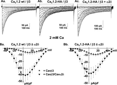
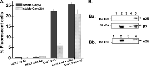
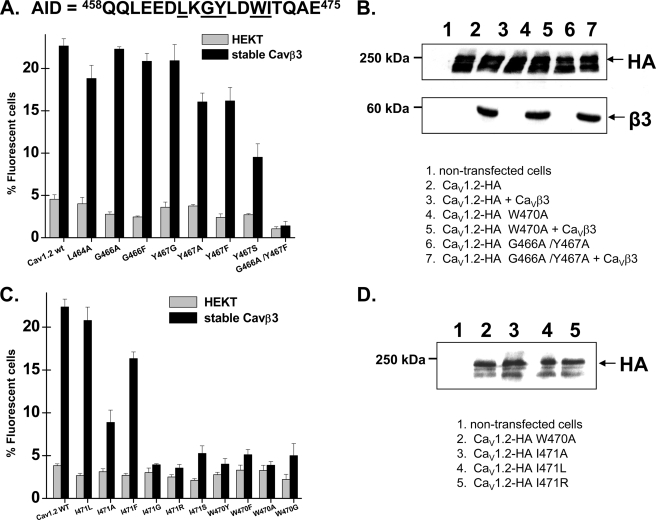
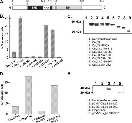
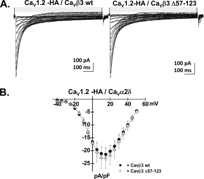
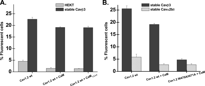
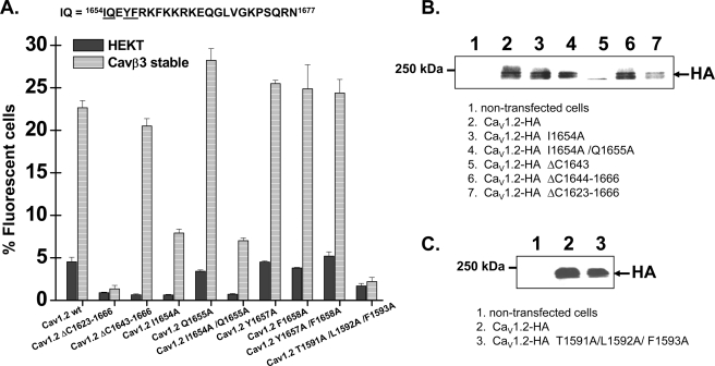
Ca(V)beta subunits modulate cell surface expression and voltage-dependent gating of high voltage-activated (HVA) Ca(V)1 and Ca(V)2 alpha1 subunits. High affinity Ca(V)beta binding onto the so-called alpha interaction domain of the I-II linker of the Ca(V)alpha1 subunit is required for Ca(V)beta modulation of HVA channel gating. It has been suggested, however, that Ca(V)beta-mediated plasma membrane targeting could be uncoupled from Ca(V)beta-mediated modulation of channel gating. In addition to Ca(V)beta, Ca(V)alpha2delta and calmodulin have been proposed to play important roles in HVA channel targeting. Indeed we show that co-expression of Ca(V)alpha2delta caused a 5-fold stimulation of the whole cell currents measured with Ca(V)1.2 and Ca(V)beta3. To gauge the synergetic role of auxiliary subunits in the steady-state plasma membrane expression of Ca(V)1.2, extracellularly tagged Ca(V)1.2 proteins were quantified using fluorescence-activated cell sorting analysis. Co-expression of Ca(V)1.2 with either Ca(V)alpha2delta, calmodulin wild type, or apocalmodulin (alone or in combination) failed to promote the detection of fluorescently labeled Ca(V)1.2 subunits. In contrast, co-expression with Ca(V)beta3 stimulated plasma membrane expression of Ca(V)1.2 by a 10-fold factor. Mutations within the alpha interaction domain of Ca(V)1.2 or within the nucleotide kinase domain of Ca(V)beta3 disrupted the Ca(V)beta3-induced plasma membrane targeting of Ca(V)1.2. Altogether, these data support a model where high affinity binding of Ca(V)beta to the I-II linker of Ca(V)alpha1 largely accounts for Ca(V)beta-induced plasma membrane targeting of Ca(V)1.2.


Figures

FIGURE 1.
FIGURE 1.
CaVα2bδ stimulated CaV1.2 whole cell currents. A, panel a, whole cell current traces recorded after the transient expression of the CaV1.2 wt channel in the stable CaVβ3 cell line. The charge carrier was 2 mm Ca2+. Panel b, whole cell current traces recorded after the transient expression of the CaV1.2-HA channel in the stable CaVβ3 cell line. Panel c, whole cell current traces recorded after the transient expression of the CaV1.2-HA channel and the CaVα2bδ in the stable CaVβ3 cell line. B, panel a, current-voltage relationships of CaV1.2 wt + CaVβ3 ± CaVα2bδ show a typical voltage-dependent activation with a mean current density of −7 ± 2 pA/pF (n = 6) for the wild type CaV1.2 channel in the stable CaVβ3 stable cell line as compared with a current density of −41 ± 9 pA/pF (n = 7) for the wild type CaV1.2 channel measured in the same cell line after transient transfection with CaVα2bδ subunit. The activation potential of 3 ± 3 mV for the CaV1.2 wt + CaVβ was shifted to −10 ± 2 mV in the presence of CaVα2bδ. Panel b, current-voltage relationships of CaV1.2-HA/CaVβ3 ± CaVα2bδ show a typical voltage-dependent activation with a mean current density of −9 ± 2 pA/pF (n = 5) for the CaV1.2-HA channel in the stable CaVβ3 stable cell line as compared with a current density of −36 ± 8 pA/pF (n = 7) for the CaV1.2-HA channel measured in the same cell line after transient transfection with CaVα2bδ subunit. The activation potential of 2 ± 3 mV for the CaV1.2-HA/CaVβ was shifted to −12 ± 2 mV in the presence of CaVα2bδ. Patch clamp experiments were carried out in the whole cell configuration in the presence of a 2 mm Ca2+ saline solution.
FIGURE 2.
FIGURE 2.
CaVβ stimulated CaV1.2 membrane expression in HEKT cells. A, HA-tagged CaV1.2 wt was co-expressed transiently either in the stable CaVβ3 or the stable CaVα2bδ cell line. Cell surface expression of CaV1.2 wt was determined in intact cells by flow cytometry using the anti-HA FITC conjugate antibody (Ab). The histogram shows the number of fluorescent cells as a function of the experimental conditions. Cell autofluorescence (HEKT no Ab) was <1% throughout, and the addition of the FITC did not significantly increase the level of fluorescence in HEKT cells not transfected with the HA-tagged CaV1.2 (HEKT with Ab). As seen, only co-expression of CaV1.2 with CaVβ3 significantly promoted membrane expression of CaV1.2 (p < 0.001). Co-expression of CaV1.2 with CaVα2bδ did not alter the number of CaV1.2 channels at the membrane (p > 0.1). Co-expression with both auxiliary subunits did not further improve the membrane expression of CaV1.2. The CaV1.2 + CaVβ3 + CaVα2bδ condition (either CaV1.2 + CaVβ3 expressed transiently in the stable CaVα2bδ cell line or CaV1.2 + CaVα2bδ expressed transiently in the CaVβ3 cell line) was not significantly different from the CaV1.2 + CaVβ3 condition (p > 0.1). Similar results were obtained with transient expression systems. The numerical values can be found in Table 1. B, Western blot analyses of HEKT cells transiently or stably transfected with CaVα2bδ or CaVβ3, using CaVα2δ-1 (1:200) and CaVβ3 (1:500) antibodies. Each lane was loaded with 50 μg of protein. Panel a, HEKT cells were transiently or stably transfected with CaVβ3. Lane 1, control nontransfected cells. Lane 2, transient transfection of CaVβ3. Lane 3, control nontransfected cells. Lane 4, stable CaVβ3 cell line. Lane 5, transient co-transfection of CaVβ3 and CaVα2bδ. Panel b, HEKT cells were transiently or stably transfected with CaVα2bδ. Lane 1, control nontransfected cells. Lane 2, transient transfection of CaVα2bδ. Lane 3, control nontransfected cells. Lane 4, stable CaVα2bδ cell line.
FIGURE 3.
FIGURE 3.
Point mutations within the C-terminal residues on the AID helix disrupted the CaVβ stimulation of CaV1.2 plasma membrane targeting. A, HA-tagged CaV1.2 wt and mutants were expressed transiently either in the HEKT cells or in the stable CaVβ3 cell line. Cell surface expression of the CaV1.2 protein was quantified as described for supplemental Fig. S2. The residues targeted in these experiments are underlined within the primary sequence of the AID region of CaV1.2. The number of fluorescent cells decreased in the order CaV1.2-HA wt ≈ L464A, G466A, G466F, Y467G > Y467A, Y467F, G466A/Y467A > Y467S, G466Y/Y467G ≫ G466A/Y467F. The numerical values can be found in supplemental Table SI. B, Western blot analyses of HEKT cells transiently transfected with CaV1.2 wt or mutants in stable CaVβ3 cells using HA (1:500) and CaVβ3 (1:500) antibodies. Lane 1, control nontransfected cells. Lane 2, CaV1.2-HA. Lane 3, CaV1.2-HA + CaVβ3. Lane 4, CaV1.2-HA W470A. Lane 5, CaV1.2-HA W470A + CaVβ3. Lane 6, CaV1.2-HA G466A/Y467A. Lane 7, CaV1.2-HA G466A/Y467A + CaVβ3. Western blot analyses confirmed that the W470A mutant was expressed in total cell lysates and recognized by the anti-HA (1:500). Each lane was loaded with 50 μg of protein. C, HA-tagged CaV1.2 wt and mutants were expressed transiently either in the HEKT cells or in the stable CaVβ3 cell line. Cell surface expression of the CaV1.2 protein was quantified as described for supplemental Fig. S2. The residues targeted in these experiments are underlined within the primary sequence of the AID region of CaV1.2 shown in A. The number of fluorescent cells decreased in the order CaV1.2-HA wt ≈ I471L > I471F > I471A > I471S > I471R ≫ I471G, W470A, W470F, W470G, W470Y. The numerical values can be found in supplemental Table SI. D, Western blot analyses confirmed that the CaV1.2 W470A, I471A, I471L, and I471R mutants expressed with the expected molecular weight in total cell lysates and were recognized by the anti-HA (1:500). Each lane was loaded with 50 μg of protein. Lane 1, control nontransfected cells. Lane 2, CaV1.2-HA W470A. Lane 3, CaV1.2-HA I471A. Lane 4, CaV1.2-HA I471L. Lane 5, CaV1.2-HA I471R.
FIGURE 4.
FIGURE 4.
The NK domain of CaVβ3 is critical for the plasma membrane targeting of CaV1.2. A, schematic diagram of the domain organization of the CaVβ3 subunit based on the crystal structure and adapted from (21). B, HA-tagged CaV1.2 wt and CaVβ3 mutants were expressed transiently in the HEKT cells. Cell surface expression of the CaV1.2 protein was quantified as described for supplemental Fig. S2. The residues targeted in these experiments are identified in the primary sequence of CaVβ3. The Δ57–123 deletion removed the SH3 domain; the Δ170–175 removed the PYDVVP sequence; and the Δ195–200 removed the MMQKAL sequence, also termed the ABP domain. The numerical values are provided in supplemental Table SIII. C, Western blot analyses of HEKT cell lysates transiently transfected with CaVβ3 constructs using anti-CaVβ3 (1:500). Each lane was loaded with 10 μg of protein except for CaVβ3 Δ54–362 loaded with 50 μg of protein. Lane 1, control nontransfected cells. Lane 2, CaVβ3 wt. Lane 3, CaVβ3 M196A. Lane 4, CaVβ3 Δ170–175. Lane 5, CaVβ3 Δ195–200. Lane 6, CaVβ3 Δ57–123. Lane 7, CaVβ3 Δ57–180. Lane 8, CaVβ3 Δ180–364. Lane 9, CaVβ3 Δ54–362. Western blot analyses confirmed that the CaVβ3 deleted proteins were detected in total cell lysates with the expected molecular weight. D, HA-tagged CaV1.2 wt and CaVβ3 fragments were expressed transiently in the HEKT cells. Cell surface expression of the CaV1.2 protein was quantified as described for supplemental Fig. S2. The residues targeted in these experiments are identified in the primary sequence of the CaVβ3. The 181–362 fragment is equivalent to the NK domain. The numerical values are provided in supplemental Table SIII. E, Western blot analyses of HEKT cell lysates transiently transfected with the CaVβ3 fragments using anti-c-Myc (1:500). Each lane was loaded with 50 μg of protein. Lane 1, control nontransfected cells. Lane 2, CaVβ3 58–120. Lane 3, CaVβ3 58–362. Lane 4, empty lane. Lane 5, CaVβ3 181–362. The CaVβ3 58–120 fragment formed a 7.4-kDa protein that cannot be seen in this figure. Western blot analyses confirmed that the CaVβ3 fragments were detected in total cell lysates with the expected molecular weight.
FIGURE 5.
FIGURE 5.
CaVβ3 Δ57–123 stimulated CaV1.2 whole cell currents. A, whole cell current traces recorded after the transient expression of the CaV1.2-HA channel with CaVα2bδ and CaVβ3 wt (left panel) or with CaVα2bδ and CaVβ3 Δ57–123 (right panel) in HEKT cells. All of the subunits were transiently expressed. B, current-voltage relationships of CaV1.2-HA + CaVα2bδ + CaVβ3 wt (filled circles) and CaV1.2-HA + CaVα2bδ + CaVβ3 Δ57–123 (open triangles) show a typical voltage-dependent activation with a mean current density of −21 ± 2 pA/pF (n = 5) for CaV1.2-HA + CaVα2bδ + CaVβ3 wt as compared with a current density of −23 ± 4 pA/pF (n = 4) for CaV1.2-HA + CaVα2bδ + CaVβ3 Δ57–123 measured under the same conditions. Patch clamp experiments were carried out in the whole cell configuration in the presence of a 2 mm Ca2+ saline solution.
FIGURE 6.
FIGURE 6.
Overexpressing CaM wt and CaM1,2,3,4 did not alter the pattern of CaVβ stimulation of CaV1.2 plasma membrane targeting. A, HA-tagged CaV1.2 wt was expressed transiently either in the HEKT cells or in the stable CaVβ3 cell line. Cell surface expression of the CaV1.2 protein was quantified as described for supplemental Fig. S2. The number of fluorescent cells was not significantly influenced by overexpressing the wild type CaM or the dominant negative mutant CaM1,2,3,4. The numerical values can be found in Table 1 and supplemental Table SIV. B, HA-tagged CaV1.2 wt and the double W470A/I471A mutant were expressed transiently either in the stable CaVβ3 or in the stable CaVα2bδ cell line. Cell surface expression of the CaV1.2 protein was quantified as described for supplemental Fig. S2. The number of fluorescent cells was not significantly increased by overexpressing CaM wt in any cell line as compared with the control CaV1.2 cells (p > 0.05). Similar data were obtained with the W470A mutant that abrogated CaVβ3 binding and stimulation of CaV1.2 plasma membrane targeting. The numerical values can be found in Table 1 and supplemental Table SIV.
FIGURE 7.
FIGURE 7.
Mutations within the high affinity CaM binding motif did not alter the CaVβ stimulation of CaV1.2 plasma membrane targeting. A, flow cytometry data. HA-tagged CaV1.2 wt and mutants were expressed transiently either in HEKT cells or in the stable CaVβ3 cell line. Cell surface expression of the CaV1.2 protein was quantified as described for supplemental Fig. S2. The number of fluorescent cells decreased significantly for the mutants ΔC1623–1666, I1654A, I1654A/Q1655A, and T1591A/L1592A/F1593A (p < 0.01) as compared with the CaV1.2-HA wt protein under the same conditions. From left to right, the channels were CaV1.2-HA wt, ΔC1623–1666, ΔC1643–1666, I1654A, Q1655A, I1654A/Q1655A, Y1657A, F1658A, Y1657A/F1658A, and T1591A/L1592A/F1593A. The numerical values can be found in supplemental Table SIV. B, Western blot analyses confirmed that the CaV1.2 mutant proteins were detected in total cell lysates by the anti-HA (1:500) with the expected molecular weight. Lane 1, nontransfected cells. Lane 2, CaV1.2-HA. Lane 3, CaV1.2-HA I1654A. Lane 4, CaV1.2-HA I1654A/Q1655A. Lane 5, CaV1.2-HA ΔC1643. Lane 6, CaV1.2-HA ΔC1644–1666. Lane 7, CaV1.2-HA ΔC1623–1666. Each lane was loaded with 50 μg of protein. C, Western blot analyses confirmed that the CaV1.2 mutant proteins were detected in total cell lysates by the anti-HA (1:500) with the expected molecular weight. Lane 1, nontransfected cells. Lane 2, CaV1.2-HA wt. Lane 3, CaV1.2-HA T1591A/L1592A/F1593A. Each lane was loaded with 50 μg of protein.

Similar articles

Cited by

References

    1. Snutch T. P., Reiner P. B. (1992) Curr. Opin. Neurobiol. 2, 247–253 - PubMed
    1. Zhang J. F., Randall A. D., Ellinor P. T., Horne W. A., Sather W. A., Tanabe T., Schwarz T. L., Tsien R. W. (1993) Neuropharmacology 32, 1075–1088 - PubMed
    1. Birnbaumer L., Campbell K. P., Catterall W. A., Harpold M. M., Hofmann F., Horne W. A., Mori Y., Schwartz A., Snutch T. P., Tanabe T. (1994) Neuron 13, 505–506 - PubMed
    1. Perez-Reyes E., Cribbs L. L., Daud A., Lacerda A. E., Barclay J., Williamson M. P., Fox M., Rees M., Lee J. H. (1998) Nature. 391, 896–900 - PubMed
    1. Cribbs L. L., Lee J. H., Yang J., Satin J., Zhang Y., Daud A., Barclay J., Williamson M. P., Fox M., Rees M., Perez-Reyes E. (1998) Circ. Res. 83, 103–109 - PubMed

Publication types

LinkOut - more resources