. 2010 Jun;135(6):619-28.
doi: 10.1085/jgp.200910328. Epub 2010 May 17.

A malignant hyperthermia-inducing mutation in RYR1 (R163C): alterations in Ca2+ entry, release, and retrograde signaling to the DHPR

Affiliations

A malignant hyperthermia-inducing mutation in RYR1 (R163C): alterations in Ca2+ entry, release, and retrograde signaling to the DHPR

Eric Estève et al. J Gen Physiol. 2010 Jun.

Abstract

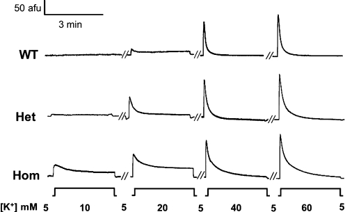
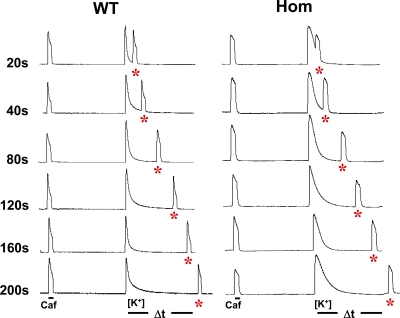
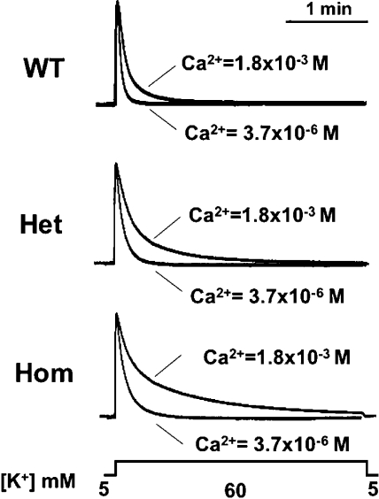
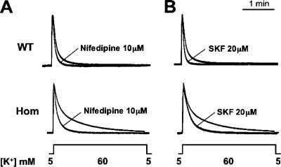
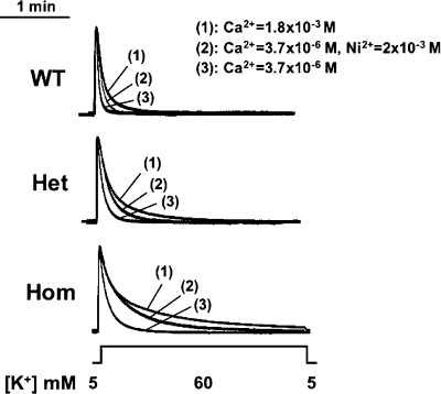
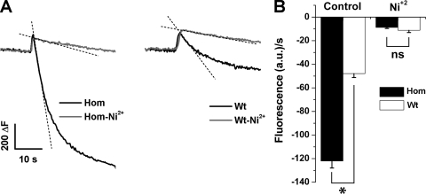
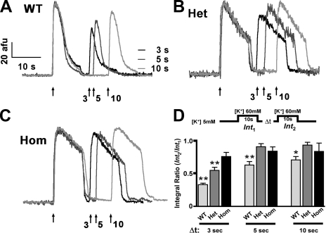
Bidirectional signaling between the sarcolemmal L-type Ca(2+) channel (1,4-dihydropyridine receptor [DHPR]) and the sarcoplasmic reticulum (SR) Ca(2+) release channel (type 1 ryanodine receptor [RYR1]) of skeletal muscle is essential for excitation-contraction coupling (ECC) and is a well-understood prototype of conformational coupling. Mutations in either channel alter coupling fidelity and with an added pharmacologic stimulus or stress can trigger malignant hyperthermia (MH). In this study, we measured the response of wild-type (WT), heterozygous (Het), or homozygous (Hom) RYR1-R163C knock-in mouse myotubes to maintained K(+) depolarization. The new findings are: (a) For all three genotypes, Ca(2+) transients decay during prolonged depolarization, and this decay is not a consequence of SR depletion or RYR1 inactivation. (b) The R163C mutation retards the decay rate with a rank order WT > Het > Hom. (c) The removal of external Ca(2+) or the addition of Ca(2+) entry blockers (nifedipine, SKF96365, and Ni(2+)) enhanced the rate of decay in all genotypes. (d) When Ca(2+) entry is blocked, the decay rates are slower for Hom and Het than WT, indicating that the rate of inactivation of ECC is affected by the R163C mutation and is genotype dependent (WT > Het > Hom). (e) Reduced ECC inactivation in Het and Hom myotubes was shown directly using two identical K(+) depolarizations separated by varying time intervals. These data suggest that conformational changes induced by the R163C MH mutation alter the retrograde signal that is sent from RYR1 to the DHPR, delaying the inactivation of the DHPR voltage sensor.


Figures

Figure 1.
Figure 1.
K+ dose response for WT and R163C Het and Hom myotubes. Representative fluorescence imaging records of WT and R163C (Het and Hom) myotubes loaded with Fluo-4 AM exposed to 10, 20, 40, and 60 mM [K+]e for 180 s to observe the entire time course of the Ca2+ transients. The peak of the Ca2+ transients increased and the duration shortened as [K+]e was elevated from 10 to 60 mM in all three genotypes. Afu, arbitrary fluorescence units.
Figure 2.
Figure 2.
Decay of the Ca2+ transient is not due to store depletion or to RYR1 inactivation. Representative fluorescence records of WT and Hom R163C myotubes loaded with Fluo-4 AM in response to a 10-s exposure to 20 mM caffeine (Caf) and a 180-s exposure to 60 mM [K+]e, followed by a second 10-s application of 20 mM caffeine (*) applied at different intervals (20–200 s) after the Ca2+ transient was elicited by 60 mM [K+]e. The decay of the Ca2+ transient during K+ depolarization does not appear to be related to SR Ca2+ depletion, nor is it due to RYR1 inactivation based on the amplitude of the second caffeine response (*).
Figure 3.
Figure 3.
Low [Ca2+]e increases the rate of decay of the Ca2+ transient in response to 60 mM [K+]e. The average transients obtained from WT and R163C Het and Hom myotubes exposed to 60 mM [K+]e for 3 min in normal [Ca2+]e and in low [Ca2+]e solution (no added Ca2+ and no EGTA). Data were normalized by making the peak transient of each response equal to 1.
Figure 4.
Figure 4.
The effect of nifedipine and SKF-96365 on the decay of the K+-induced Ca2+ transients. The average normalized traces (n = 8–10) of Ca2+ transients from WT and Hom R163C myotubes in response to 60 mM [K+]e for 180 s before and after exposure to 10 µM nifedipine (A) or 20 µM SKF96365 (B). The cells were exposed to nifedipine or SKF96365 for 10 s before and throughout the K+ depolarization.
Figure 5.
Figure 5.
Ni2+ partially reverses the effect of low [Ca2+]e on the Ca2+ transient. The average normalized Ca2+ transients from WT (n = 14), Het (n = 12), and Hom (n = 18) R163C myotubes, which were depolarized for 180 s by 60 mM [K+]e in the presence of 1.8 × 10⁻³ M Ca2+, followed by a 5-min rest in normal Ringer’s solution, depolarized for 180 s again in low Ca2+ solution (3.7 × 10⁻⁶ M), followed by a second 5-min rest, and then depolarized for a third time in low Ca2+ solution supplemented with 2 × 10⁻³ M Ni2+ (see Table II for the rates of Ca2+ transient decay for each condition).
Figure 6.
Figure 6.
Mn2+ quench in WT and Hom R163C myotubes in the absence and presence of 2 mM Ni2+. (A) Representative Fura-2 emission traces from WT and R163C Hom myotubes before and after stimulation with 60 mM KCl in the presence and absence of Ni2+. (B) The average rates ± SEM of Mn2+ quench during the linear phase of quench after KCl exposure with (left) and without (right) the addition of Ni2+ to the bath solution. *, P < 0.01.
Figure 7.
Figure 7.
Inactivation of the DHPR voltage sensor is genotype dependent. Representative Ca2+ transients for WT (A), Het (B), and Hom (C) myotubes induced by the following protocol: 60 mM [K+]e (Int1) for 10 s; 2.5 mM [K+]e for 3, 5, or 10 s; 60 mM [K+]e (Int2). (D) The integrals of the second Ca2+ transient were expressed as a percentage of the first fluorescence signal (**, P < 0.01; Tukey’s test for multiple comparisons). The amplitude of the second Ca2+ transient was used as an index of DHPR inactivation at the time the second stimulus was applied.

Similar articles

Cited by

References

    1. Almers W., Palade P.T. 1981. Slow calcium and potassium currents across frog muscle membrane: measurements with a vaseline-gap technique. J. Physiol. 312:159–176 - PMC - PubMed
    1. Araya R., Liberona J.L., Cárdenas J.C., Riveros N., Estrada M., Powell J.A., Carrasco M.A., Jaimovich E. 2003. Dihydropyridine receptors as voltage sensors for a depolarization-evoked, IP3R-mediated, slow calcium signal in skeletal muscle cells. J. Gen. Physiol. 121:3–16 10.1085/jgp.20028671 - DOI - PMC - PubMed
    1. Armstrong C.M., Bezanilla F.M., Horowicz P. 1972. Twitches in the presence of ethylene glycol bis(-aminoethyl ether)-N,N’-tetracetic acid. Biochim. Biophys. Acta. 267:605–608 10.1016/0005-2728(72)90194-6 - DOI - PubMed
    1. Bannister R.A., Beam K.G. 2009. The cardiac alpha(1C) subunit can support excitation-triggered Ca2+ entry in dysgenic and dyspedic myotubes. Channels (Austin). 3:268–273 - PMC - PubMed
    1. Bannister R.A., Pessah I.N., Beam K.G. 2009. The skeletal L-type Ca2+ current is a major contributor to excitation-coupled Ca2+ entry. J. Gen. Physiol. 133:79–91 10.1085/jgp.200810105 - DOI - PMC - PubMed

Publication types

MeSH terms

Substances