. 2010 Jun 7;207(6):1273-81.
doi: 10.1084/jem.20100348. Epub 2010 May 17.

Superior antigen cross-presentation and XCR1 expression define human CD11c+CD141+ cells as homologues of mouse CD8+ dendritic cells

Affiliations

Superior antigen cross-presentation and XCR1 expression define human CD11c+CD141+ cells as homologues of mouse CD8+ dendritic cells

Annabell Bachem et al. J Exp Med. .

Abstract

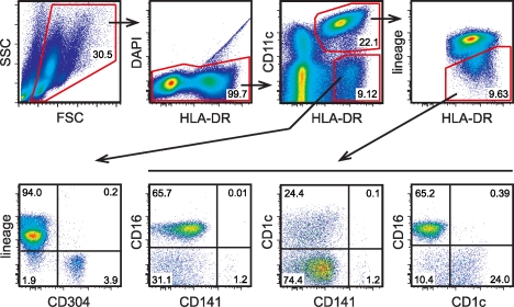
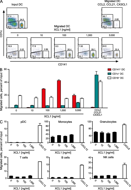
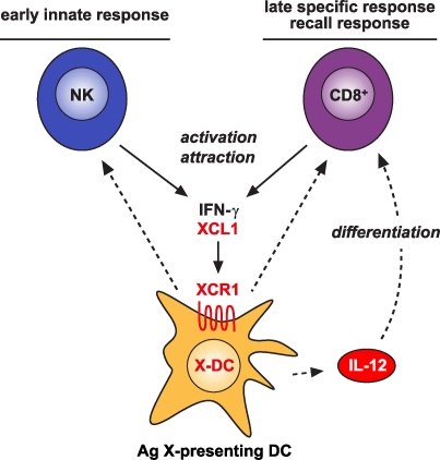
In recent years, human dendritic cells (DCs) could be subdivided into CD304+ plasmacytoid DCs (pDCs) and conventional DCs (cDCs), the latter encompassing the CD1c+, CD16+, and CD141+ DC subsets. To date, the low frequency of these DCs in human blood has essentially prevented functional studies defining their specific contribution to antigen presentation. We have established a protocol for an effective isolation of pDC and cDC subsets to high purity. Using this approach, we show that CD141+ DCs are the only cells in human blood that express the chemokine receptor XCR1 and respond to the specific ligand XCL1 by Ca2+ mobilization and potent chemotaxis. More importantly, we demonstrate that CD141+ DCs excel in cross-presentation of soluble or cell-associated antigen to CD8+ T cells when directly compared with CD1c+ DCs, CD16+ DCs, and pDCs from the same donors. Both in their functional XCR1 expression and their effective processing and presentation of exogenous antigen in the context of major histocompatibility complex class I, human CD141+ DCs correspond to mouse CD8+ DCs, a subset known for superior antigen cross-presentation in vivo. These data define CD141+ DCs as professional antigen cross-presenting DCs in the human.


Figures

Figure 1.
Figure 1.
Strategy for defining human DC subsets in the blood. PBMCs from human blood were enriched by density gradient centrifugation and stained for the indicated cell-surface markers (for the mAb used for staining see Materials and methods). Flow sorting (after magnetic cell enrichment) was performed on the principle of the gating strategy shown. This approach allowed us to isolate CD304+ pDCs, CD16+ DCs, CD1c+ DCs, and CD141+ DCs to very high purities. The inset numbers represent the percentage of the gated cells in the respective gating step. FSC, forward scatter; SSC, side scatter.
Figure 2.
Figure 2.
The chemokine receptor XCR1 is selectively expressed in CD141+ DCs. (A) Organization of the human XCR1 gene (E1, exon 1; E2, exon 2; and E3, exon 3); the coding region is shown in black, and the primer–probe sets used for expression analyses are indicated. (B) qRT-PCR of total RNA from T cells, B cells, NK cells, granulocytes, monocytes, pDCs, CD1c+ DCs, CD16+ DCs, and CD141+ DCs isolated to a very high purity (>97.5%) from PBMCs and tested with primer–probe set 1 (F1, R1, and probe P1). Cultured MoDCs were also tested. Identical results were obtained using primer–probe set 2 (F2, R2, and P2; not depicted). The test systems used allowed the detection of ≥200 copies of XCR1 in cDNA reverse transcribed from 100 ng of total RNA. All PCR analyses were performed with cell subsets from at least two donors. Error bars represent means ± SEM.
Figure 3.
Figure 3.
XCL1 induces a [Ca2+]i signal in CD141+ DCs. CD141+ DCs were flow sorted to a purity >98.7%, immobilized on poly–l-lysine–coated glass coverslips, and loaded with 2 µM fura-2/AM. Cells were imaged in a monochromator-assisted digital video imaging system and challenged with 1 µg/ml XCL1 as indicated (left arrow). Subsequently, the same cells were challenged again with a mixture of 100 ng/ml CCL2, 200 ng/ml CCL21, 200 ng/ml CXCL9, and 1 ng/ml CX3CL1 used as a positive control (right arrow). The data shown represent [Ca2+]i concentrations of 300 single cells (gray lines) measured in two independent experiments. The mean [Ca2+]i signal averaged over all cells responding to XCL1 is indicated (black line).
Figure 4.
Figure 4.
XCL1 selectively induces chemotaxis in CD141+ DCs. (A) A mixture of highly purified, flow-sorted DC subtypes (20% CD141+, 40% CD16+, and 40% CD1c+ DCs; Input DC) was tested for migration in response to medium alone or to serial dilutions of XCL1 (10–5,000 ng/ml) in a Transwell system. A combination of the chemokines CCL2, CCL21, and CX3CL1 was used as a positive control for the DC subsets (Migrated DC). The absolute numbers of CD141+, CD1c+, and CD16+ DCs in input and migrated cell populations are truly represented in the dot plots, because all cells within a defined volume were included in the analysis in each instance. (B) Proportion of migrated CD1c+, CD16+, and CD141+ DCs in the experiment shown in A. (C) Proportion of migrated pDCs, monocytes, granulocytes, T cells, B cells, and NK cells in response to XCL1 (10–1,000 ng/ml) or the chemokines CXCL12 and CXCL8, which were used as positive controls. For migration assays of B cells, NK cells, and monocytes, PBMCs were magnetically depleted of T cells, and for T cell migration, PBMCs were used directly. For migration assays of granulocytes, whole blood cells were used after erythrocyte lysis with ACK buffer, and pDCs were magnetically enriched from PBMCs with the Plasmacytoid Dendritic Cell Isolation Kit (Miltenyi Biotec). All experiments with DCs were performed three times; all other populations were assayed twice. Error bars represent means ± SEM.
Figure 5.
Figure 5.
Capacity of CD141+, CD1c+, and CD16+ DCs to cross-present soluble and cell-associated HCMV pp65 antigen. (A) CD8+ T cell clone 10, specific for the HLA-A*0201–restricted HCMV pp65 peptide NLVPMVATV (pp65495–503), was co-cultured with CD141+ DCs, CD1c+ DCs, CD16+ DCs, or pDCs obtained from the buffy coat of one HLA-A*0201+ blood donation, with 3 µg/ml of recombinant soluble HCMV pp65 added for the entire culture period. The activation of the T cell clone was determined by measuring the concentration of IFN-γ in the supernatants at the termination of culture after 20 h. Negative controls included addition of irrelevant protein OVA (3 µg/ml) and cultures with only the T cell clone or DCs; addition of 1 µg/ml of pp65495–503 peptide to the co-cultures served as a positive control. Shown is one representative experiment out of nine; each experiment was performed with cells from a different donor (all experiments included all cDCs subsets; four of them also included pDCs). (B) CD141+, CD1c+, or CD16+ DCs isolated from leukapheresis PBMCs of one HLA-A*0201+ donor were co-cultured with CD8+ T cell clone 61 at variable ratios (from 1:1 to 1:16) with cell-associated pp65 antigen added for the entire culture period, and IFN-γ was determined in the supernatant after 24 h. Negative controls included irrelevant MART-127–35 peptide, and positive controls included HCMV pp65495–503 peptide (both at 1 µg/ml). Shown are results representative of three experiments with different donors. Error bars represent means ± SEM.
Figure 6.
Figure 6.
Involvement of the XCL1–XCR1 communication axis in the innate and adaptive cytotoxic responses to cross-presented microbial and tumor antigens. Secretion of the chemokine XCL1 by activated NK cells specifically attracts XCR1-expressing DCs capable of antigen cross-presentation. This ensures an effective communication between these cells in the innate phase of the immune response. In the adaptive phase, secretion of XCL1 by activated CD8+ T cells optimizes the communication with antigen cross-presenting DCs and facilitates the differentiation of CD8+ T cells to cytotoxic cells.

References

    1. Andrews D.M., Maraskovsky E., Smyth M.J. 2008. Cancer vaccines for established cancer: how to make them better? Immunol. Rev. 222:242–255 10.1111/j.1600-065X.2008.00612.x - DOI - PubMed
    1. Bevan M.J. 2006. Cross-priming. Nat. Immunol. 7:363–365 10.1038/ni0406-363 - DOI - PubMed
    1. Caminschi I., Proietto A.I., Ahmet F., Kitsoulis S., Shin Teh J., Lo J.C., Rizzitelli A., Wu L., Vremec D., van Dommelen S.L., et al. 2008. The dendritic cell subtype-restricted C-type lectin Clec9A is a target for vaccine enhancement. Blood. 112:3264–3273 10.1182/blood-2008-05-155176 - DOI - PMC - PubMed
    1. Caminschi I., Lahoud M.H., Shortman K. 2009. Enhancing immune responses by targeting antigen to DC. Eur. J. Immunol. 39:931–938 10.1002/eji.200839035 - DOI - PubMed
    1. Colonna M., Trinchieri G., Liu Y.J. 2004. Plasmacytoid dendritic cells in immunity. Nat. Immunol. 5:1219–1226 10.1038/ni1141 - DOI - PubMed

Publication types

MeSH terms