. 2010 Jun 7;207(6):1261-71.
doi: 10.1084/jem.20092618. Epub 2010 May 17.

Characterization of human DNGR-1+ BDCA3+ leukocytes as putative equivalents of mouse CD8alpha+ dendritic cells

Affiliations

Characterization of human DNGR-1+ BDCA3+ leukocytes as putative equivalents of mouse CD8alpha+ dendritic cells

Lionel Franz Poulin et al. J Exp Med. .

Abstract

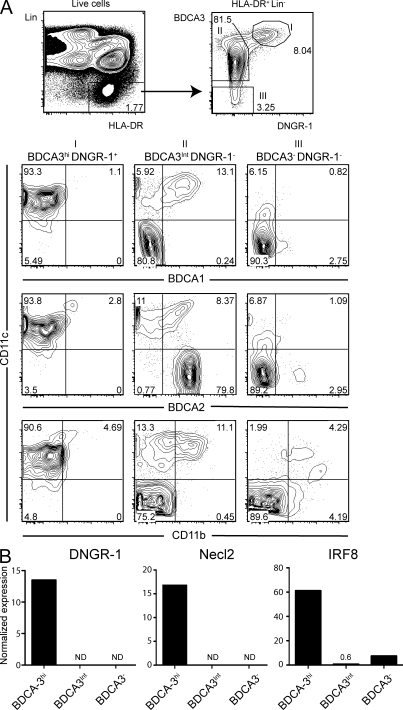
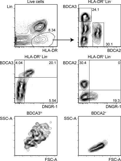
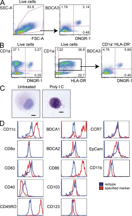
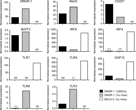
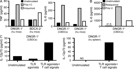
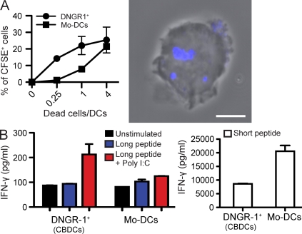
In mouse, a subset of dendritic cells (DCs) known as CD8alpha+ DCs has emerged as an important player in the regulation of T cell responses and a promising target in vaccination strategies. However, translation into clinical protocols has been hampered by the failure to identify CD8alpha+ DCs in humans. Here, we characterize a population of human DCs that expresses DNGR-1 (CLEC9A) and high levels of BDCA3 and resembles mouse CD8alpha+ DCs in phenotype and function. We describe the presence of such cells in the spleens of humans and humanized mice and report on a protocol to generate them in vitro. Like mouse CD8alpha+ DCs, human DNGR-1+ BDCA3hi DCs express Necl2, CD207, BATF3, IRF8, and TLR3, but not CD11b, IRF4, TLR7, or (unlike CD8alpha+ DCs) TLR9. DNGR-1+ BDCA3hi DCs respond to poly I:C and agonists of TLR8, but not of TLR7, and produce interleukin (IL)-12 when given innate and T cell-derived signals. Notably, DNGR-1+ BDCA3+ DCs from in vitro cultures efficiently internalize material from dead cells and can cross-present exogenous antigens to CD8+ T cells upon treatment with poly I:C. The characterization of human DNGR-1+ BDCA3hi DCs and the ability to grow them in vitro opens the door for exploiting this subset in immunotherapy.


Figures

Figure 1.
Figure 1.
DNGR-1+ BDCA3hi DCs are found in the spleens of humans. (A) Flow cytometry analysis of human spleen cell suspensions from cadaveric donors. Live HLA-DR+ Lin cells were gated as shown (top left) and analyzed for expression of BDCA3 versus DNGR-1 (top right). Three populations were defined (gate I: BDCA3hi, DNGR-1+; gate II: BDCA3int, DNGR-1; gate III: BDCA3, DNGR-1) and analyzed for the expression of CD11c versus BDCA1, BDCA2 or CD11b (bottom). Numbers indicate percentage of cells in each of the indicated gates or quadrants. Arrows show gating strategy. (B) Normalized expression of DNGR-1, Necl2, and IRF8 mRNA on sorted HLA-DR+ Lin BDCA2 BDCA1 DCs, from human spleen, expressing high (BDCA3hi), intermediate (BDCA3int), or low levels of BDCA3 (BDCA3). ND, not detectable. Data in A and B are representative of at least three cadaveric donors. Additional data analysis is shown in Fig. S1 and a summary of DC population frequency across three donors is presented in Table S1.
Figure 2.
Figure 2.
DNGR-1+ BDCA3hi DCs are found in the spleens of humanized mice. HLA-DR+ Lin live spleen cells from humanized mice (NOD/SCID/γc) were analyzed as in Fig. 1 for the expression of BDCA3 versus BDCA2 (top right), BDCA3 versus DNGR-1 (middle left), and BDCA2 versus DNGR-1 (middle right). The scatter profile of BDCA3+ and BDCA2+ cells is also shown (bottom). Numbers indicate percentage of cells in each of the indicated gates or quadrants. Arrows show gating strategy. Data are representative of multiple mice from three independent cohorts of mice engrafted with human HSCs/HPCs.
Figure 3.
Figure 3.
Phenotype of in vitro–generated DNGR-1+ BDCA3+ CBDCs. CBDCs were generated as described in the Materials and methods. Live cells were analyzed for the expression of BDCA3 versus DNGR-1 (A), CD1a versus DNGR-1 (B, left), and CD1a versus HLA-DR (B, middle). Gated CD1a+ HLA-DR+ cells were analyzed for the expression of BDCA3 versus DNGR-1 (B, right). Numbers indicate percentage of cells in each of the indicated gates or quadrants. Arrows show gating strategy. (C) CBDCs were treated with or without poly I:C (10 µg/ml) overnight and subsequently sorted into live HLA-DR+ Lin DNGR-1+ cells. Cytospins were prepared and morphology was assessed by hematoxylin and eosin staining. Bar, 5 µm. (D) Live DNGR-1+ DCs were analyzed for the expression (red) of CD11c, BDCA1, CD8α, BDCA2, CD83, CD86, CD40, CD103, CD45RO, CD123, CCR7, EpCam, and CD11b. Isotype-matched control mAb staining is shown in the blue histograms. Data in A–D are representative of multiple BDC cultures with two independent pools of CB-derived HSCs/HPCs.
Figure 4.
Figure 4.
DNGR-1+ BDCA3+ DCs display a gene expression profile characteristic of mouse CD8α+ DCs. Normalized expression of DNGR-1, Necl2, CD207, BATF3, IRF8, IRF4, TLR7, TLR9, DAP12, TLR8, and TLR3 mRNA in BDCA3+ DNGR-1+ DCs (DNGR-1+) purified either from CBDCs or from pooled spleens of two to five humanized mice (hu mice). Expression was compared with that of BDCA2+ pDCs purified from the same humanized mouse spleens. Data are representative of two independent experiments. ND, not detectable.
Figure 5.
Figure 5.
DNGR-1+ BDCA3+ DCs respond to poly I:C and to TLR8, but not TLR7 agonists. (A) Purified DNGR-1+ BDCA3+ DCs from CBDCs (DNGR-1+ CBDCs) or from humanized mice (DNGR-1+ hu mice), or BDCA2+ pDCs from the same humanized mice (BDCA2+ hu mice), were cultured with poly I:C (10 µg/ml), imiquimod (10 µg/ml), or medium alone. After overnight incubation, culture supernatant was tested for TNF and IL-6 content. (B) DNGR-1+ CBDCs as in (A) were cultured with resiquimod (10 µg/ml), poly U (10 µg/ml) or medium alone. Supernatant was tested for IL-6 after overnight culture. (C) DNGR-1+ BDCA3+ DCs purified from CBDCs (DNGR-1+ CBDCs) or human spleen (DNGR-1+ hu spleen) were cultured with a mix of TLR agonists and cytokines with or without antigen-specific T cells and antigen, as described in the Materials and methods. Supernatant was tested for IL-12 p70 after overnight culture. Data in A–C are representative of at least two independent experiments with independent sources of cells. ND, not detectable.
Figure 6.
Figure 6.
DNGR-1+ BDCA3+ DCs internalize dead cell debris and cross-present exogenous antigens. (A) CBDCs or Mo-DCs were incubated with labeled dead melanoma cells at the indicated ratios, as described in the Materials and methods. Uptake of dead cell material by DNGR-1+ DCs or Mo-DCs was quantified by flow cytometry (left) and confirmed by confocal microscopy (right; blue shows dead cell material). Data are mean ± SEM of three biological replicates from one experiment representative of two independent experiments. Bar, 5 µm. (B) 104 purified BDCA3+ DNGR-1+ CBDCs or Mo-DCs, as indicated, were pulsed for 2–3 h with or without 1 µM MelanA/MART-1 long peptide as antigen source for cross-presentation or the same concentration of short peptide as a processing-independent control, in the presence or absence of poly I:C. Cells were subsequently washed twice and co-cultured with a MelanA-specific CD8+ T cell clone at a 5:1 T:DCs ratio. IFN-γ accumulation in culture supernatants was assessed after 40 h. Data are the mean value of duplicate wells ± range and are representative of two independent experiments.

References

    1. Albert M.L., Sauter B., Bhardwaj N. 1998. Dendritic cells acquire antigen from apoptotic cells and induce class I-restricted CTLs. Nature. 392:86–89 10.1038/32183 - DOI - PubMed
    1. Ban Y.-L., Kong B.-H., Qu X., Yang Q.-F., Ma Y.-Y. 2008. BDCA-1+, BDCA-2+ and BDCA-3+ dendritic cells in early human pregnancy decidua. Clin. Exp. Immunol. 151:399–406 - PMC - PubMed
    1. Bedoui S., Whitney P.G., Waithman J., Eidsmo L., Wakim L., Caminschi I., Allan R.S., Wojtasiak M., Shortman K., Carbone F.R., et al. 2009. Cross-presentation of viral and self antigens by skin-derived CD103+ dendritic cells. Nat. Immunol. 10:488–495 10.1038/ni.1724 - DOI - PubMed
    1. Bursch L.S., Wang L., Igyarto B., Kissenpfennig A., Malissen B., Kaplan D.H., Hogquist K.A. 2007. Identification of a novel population of Langerin+ dendritic cells. J. Exp. Med. 204:3147–3156 10.1084/jem.20071966 - DOI - PMC - PubMed
    1. Caminschi I., Proietto A.I., Ahmet F., Kitsoulis S., Shin Teh J., Lo J.C., Rizzitelli A., Wu L., Vremec D., van Dommelen S.L., et al. 2008. The dendritic cell subtype-restricted C-type lectin Clec9A is a target for vaccine enhancement. Blood. 112:3264–3273 10.1182/blood-2008-05-155176 - DOI - PMC - PubMed

Publication types

MeSH terms