. 2010 Oct;24(10):3706-19.
doi: 10.1096/fj.09-151159. Epub 2010 May 17.

Expansion of CUG RNA repeats causes stress and inhibition of translation in myotonic dystrophy 1 (DM1) cells

Affiliations

Expansion of CUG RNA repeats causes stress and inhibition of translation in myotonic dystrophy 1 (DM1) cells

Claudia Huichalaf et al. FASEB J. 2010 Oct.

Abstract

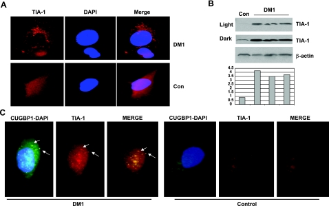
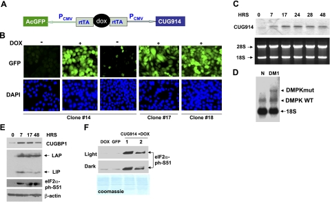
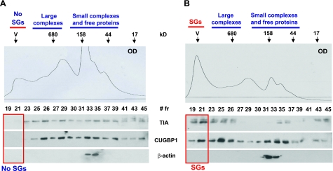
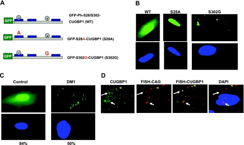
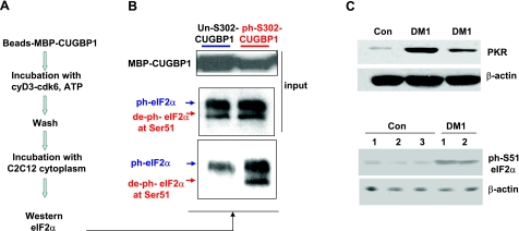
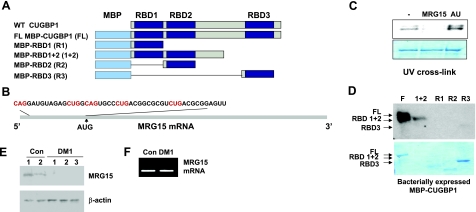
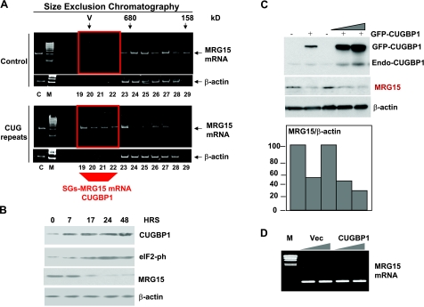
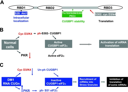
The purpose of this study was to investigate the role of the mutant CUGn RNA in the induction of stress in type 1 myotonic dystrophy (DM1) cells and in the stress-mediated inhibition of protein translation in DM1. To achieve our goals, we performed HPLC-based purification of stress granules (SGs), immunoanalysis of SGs with stress markers TIA-1, CUGBP1, and ph-eIF2, site-specific mutagenesis, and examinations of RNA-protein and protein-protein interactions in myoblasts from control and DM1 patients. The cause-and-effect relationships were addressed in stable cells expressing mutant CUG repeats. We found that the mutant CUGn RNA induces formation of SGs through the increase of the double-stranded RNA-dependent protein kinase (PKR) and following inactivation of eIF2α, one of the substrates of PKR. We show that SGs trap mRNA coding for the DNA repair and remodeling factor MRG15 (MORF4L1), translation of which is regulated by CUGBP1. As the result of the trapping, the levels of MRG15 are reduced in DM1 cells and in CUG-expressing cells. These data show that CUG repeats cause stress in DM1 through the PKR-ph-eIF2α pathway inhibiting translation of certain mRNAs, such as MRG15 mRNA. The repression of protein translation by stress might contribute to the progressive muscle loss in DM1.


Figures

Figure 1.
Figure 1.
The increase of TIA-1-containing SGs in DM1 myoblasts. A) IF images with antibodies to TIA-1 of control and DM1 myoblasts were taken under identical conditions of brightness and time of exposure to allow comparative quantification of the signal. View is ×100. Nuclei were stained with DAPI. B) Examination of TIA-1 levels in DM1 myoblasts by Western blotting assay. Cytoplasmic protein extracts from control and from 3 DM1 myoblast lines were examined by Western blotting with antibodies to TIA-1. The membrane was reprobed with β-actin. Light and dark exposures are shown for TIA-1. Bar graphs show levels of TIA-1 as ratios to β-actin (calculated from the membrane with dark exposure). C) CUGBP1 is a component of the TIA-1-containing SGs in DM1 myoblasts. CUGBP1 was detected in DM1 and control myoblasts by immunostaining with monoclonal antibodies (3B1) and with secondary antibodies labeled by FITC. The same cells were stained with rabbit polyclonal antibodies to TIA-1 and with secondary antibodies labeled with TR. Arrows indicate examples of the cytoplasmic CUGBP1-TIA-1 aggregates in DM1 myoblasts.
Figure 2.
Figure 2.
Expression of the mutant RNA CUG914 in CHO double-stable cells causes DM1-like molecular abnormalities. A) Structure of the inducible TRE-GFP-CUG914 construct that was integrated into CHO cells. B) Fluorescence images of inducible CHO TRE-GFP-CUG914 clones expressing GFP and CUG914 are shown. Three clones were induced with Dox, and the expression of GFP was monitored by IF analysis. Images of the uninduced clone 14 following Dox addition and withdrawal are shown. Images of cells from clones 17 and 18 after induction with Dox are also shown. Nuclei were stained with DAPI (blue). C) Northern blot analysis of RNA CUG914 in the inducible CHO cells. RNA was extracted from cells at different time points (0, 7, 17, 24, 28, and 48 h) after Dox addition. RNA (5 μg) was separated by gel-electrophoresis, transferred on membrane, and hybridized with CAG10 probe. Gel was stained with ethidium bromide before transfer to verify RNA loading. Positions of 28S and 18S rRNAs are shown on the left. D) Northern blot analysis of the expression of endogenous DMPK mRNA in myoblasts from control and DM1 patient. Total RNA (5 μg) was used for hybridization with the CAG10 probe under conditions described above. Positions WT (5 CUG repeats) and mutant (320 CUG repeats) DMPK mRNAs are shown. Probe interacts with 18S rRNA, which serves as an internal control. E) Expression of RNA CUG914 causes alterations in protein expression that are identical to those observed in DM1 patients. Cytoplasmic and nuclear extracts were prepared from inducible CUG914-expressing cells at different time points and examined by Western blotting with antibodies to CUGBP1 (cytoplasm), C/EBPβ (nucleus), ph-S51-eIF2α (cytoplasm), and β-actin (cytoplasm). F) Phosphorylation of eIF2α at S51 is caused by CUG repeats. Western blotting was performed with cytoplasmic extracts from unmodified, regular CHO cells treated with Dox as control, from a stable clone expressing GFP only and from CUG914 stable clone treated with Dox (2 protein preparations). Dark and light exposures are shown. Membrane was stained with Coomassie blue to verify protein loading.
Figure 3.
Figure 3.
Ectopic expression of the mutant CUG914 RNA causes formation of SGs containing CUGBP1 and TIA-1. Cytoplasm from uninduced (A) and induced (B) CHO TRE-GFP-CUG914 cells was subjected to size exclusion chromatography (SEC400 column, HPLC, Bio-Rad) to separate free TIA-1 protein from TIA-1 bound to SGs. Top panels: optical density (OD, A280) profiles of chromatography fractions. Arrows indicate positions of molecular weight markers used for calibration of column. Blue lines indicate chromatography fractions, typically containing small and large complexes and free RNA-binding proteins. Fractions 19–21 contain SGs (marked with red boxes in bottom panels). Note that the OD profile shows that the fractions 19–21 in inducible cells (B) contain large peak in the position of SGs. Bottom panels: expression of CUG914 RNA shifts TIA-1 and CUGBP1 to the region containing SGs (indicated by red box). Chromatography fractions were sequentially analyzed by Western blotting with Abs to TIA-1, CUGBP1, and β-actin. Reprobe of the filters with Abs to β-actin shows that expanded CUG repeats do not change localization of β-actin.
Figure 4.
Figure 4.
CUGBP1-S302G mutant accumulates in cytoplasmic aggregates. A) Structure of wild-type and GFP-CUGBP1 mutants Ser-28A and Ser-302G. Green box indicates GFP fusion part; gray circles indicate sites of phosphorylation; blue boxes indicate RNA binding domains within CUGBP1. B) Intracellular localization of the CUGBP1 molecule with mutant phosphorylation sites. Wild-type, S28A, and S302G mutants of GFP-CUGBP1 were transfected into control human myoblasts, and their intracellular localization was determined by fluorescence analysis. Typical pictures are shown. Green, GFP-CUGBP1; blue, nuclei stained with DAPI. View is ×100. C) Transiently transfected GFP-CUGBP1 localizes in cytoplasmic aggregates of DM1 myoblasts. Control and DM1 myoblasts were transfected with GFP-CUGBP1. Transfected cells were examined by fluorescence analysis. Average percentages of cells containing GFP-CUGBP1-positive aggregates were calculated based on 3 experiments, counting transfected cells as 100%. D) A portion of CUGBP1-positive aggregates in DM1 myoblasts contains DMPK mRNA. Left to right: IF with antibodies to CUGBP1 (green), FISH with CAG probe (red), merge of CUG and CUGBP1 signals, merge of CUG signal and DAPI. White arrows indicate examples of CUGBP1 aggregates containing DMPK mRNA; yellow arrowheads indicate CUGBP1 signals that are not colocalized with DMPK mRNA.
Figure 5.
Figure 5.
CUGBP1 unphosphorylated at S302 interacts preferentially with inactive form of eIF2α. A) Diagram of experimental procedure. Purified CUGBP1-MBP bound to amylose beads was phosphorylated with the cyclin D3/cdk6, and its interaction with active and inactive forms of eIF2α was tested by Western blotting assay. B) Top panel: input of CUGBPI. Middle panel: input of eIF2α. Bottom panel: Western blotting of the pulldown samples of unphosphorylated CUGBP1 and CUGBP1 phosphorylated by cyclin D3/cdk6 with antibodies to total eIF2α. Arrows indicate positions of ph-S51-eIF2α (inactive protein) and deph-S51-eIF2α. C) Protein levels of PKR are increased in DM1 myoblasts. Top panel: cytoplasmic extracts from control myoblasts and myoblasts from 2 DM1 patients were analyzed by Western blotting with Abs to PKR. Membrane was reprobed with antibodies to β-actin to verify protein loading. Bottom panel: levels of ph-S51-eIF2α are increased in DM1 myoblasts. Cytoplasmic protein extracts from control and DM1 myoblasts were examined by Western blotting with antibodies to ph-S51-eIF2α. Membrane was reprobed with antibodies to β-actin.
Figure 6.
Figure 6.
CUGBP1 interacts with the 5′ region of MRG15 mRNA. A) Structure of the fusion MBP-CUGBP1 proteins used in these studies. B) Nucleotide sequence of the 5′ region of MRG15 mRNA. CUG/CAG repeats are in red. C) Purified MBP-CUGBP1 binds to the 5′ region of MRG15 mRNA. UV cross-link was performed with MRG15 RNA probe and full-length MBP-CUGBP1. Competition with specific RNA (unlabeled MRG15) and nonspecific (AU-rich) is shown. D) CUGBP1 binds to MRG15 RNA mainly through RBD1 + 2 domains. Full-length (FL) and truncated MBP-CUGBP1 proteins (see diagram in A) were used in UV cross-link with MRG15 RNA. E) MRG15 protein, but not mRNA, is reduced in DM1 myoblasts. Western blotting analysis of the protein isolated from control and DM1 myoblasts with MRG15 antibodies. Membrane was reprobed with antibodies to β-actin to verify protein loading. F) Real time RT-PCR of MRG15 mRNA shows the same levels of MRG15 mRNA in control and DM1 myoblasts using GAPDH as a standard.
Figure 7.
Figure 7.
CUGBP1 recruits MRG15 mRNA into SGs and inhibits its translation. MRG15 RNA is observed in SGs of CHO cells expressing the mutant CUG914 RNA. A) Cytoplasmic samples were fractionated as shown in Fig. 3. Total RNA was isolated from the region of the chromatographic profile (fractions 19–29), including fractions containing SGs from uninduced and induced cells, and RNA was examined by real time RT-PCR with primers specific to MRG15 mRNA and to β-actin mRNA. Fractions 19–22 (indicated by red box) in CUG-expressing cells contain MRG15, while the same fractions in cells without CUG repeats do not have MRG15 mRNA. B) Induction of CUG914 increases CUGBP1 and ph-S51-eIF2α isoform and reduces MRG15 protein. Western blotting was performed with protein extracts from induced cells using antibodies to MRG15, CUGBP1, ph-eIF2α, and β-actin. C) Transiently expressed GFP-CUGBP1 inhibits translation of MRG15. GFP-CUGBP1 was transfected into HeLa cells, and the levels of CUGBP1 and MRG15 were determined by Western blot with antibodies to CUGBP1 and MRG15, respectively. Note that expression of GFP-CUGBP1 also increases levels of endogenous CUGBP1 migrating in the position of 51 kDa. Bar graphs show levels of MRG15 as ratios to β-actin that are 2.3- to 3.0-fold reduced by GFP-CUGBP1. D) Ectopic expression of CUGBP1 does not change MRG15 mRNA levels. Total RNA extracted from cells transfected with an empty vector or with CUGBP1 was examined by real time RT-PCR with primers specific for MRG15. GAPDH levels were used as control.
Figure 8.
Figure 8.
Phosphorylation controls biological functions of CUGBP1. A) Positions of phosphorylation sites and their role in the CUGBP1 functions. B) Hypothetical model for phosphorylation-dependent regulation of CUGBP1 translational activity in normal cells. C) Hypothetical mechanism by which RNA CUG repeats inhibit translation of certain mRNAs via increase of stress in DM1 cells. CUG repeats increase levels of inactive eIF2α and translational inactive CUGBP1, which are recruited into cytoplasmic SGs and form inactive CUGBP1-eIF2α-S51-ph complexes. As a result, translation of mRNAs associated with these inactive complexes within SGs is inhibited.

Similar articles

Cited by

References

    1. Harper P. S. (2001) Myotonic Dystrophy, W. B. Saunders, London
    1. Aslanidis C., Jansen G., Amemiya C., Shutler G., Mahadevan M., Tsilfidis C., Clen C., Alleman J., Wormskamp N. G., Vooijs M., Buxton J., Johnson K., Sweets H. J. M., Lennon G. G., Carrano A. V. R. G., Korneluk R. G., Wieringa B., deJong P. J. (1992) Cloning of essential myotonic dystrophy region and mapping of the putative defect. Nature 355, 548–551 - PubMed
    1. Brook J. D., McCurrah M. E., Harley H. G., Buckler A. J., Church D., Aburatani H., Hunter K., Stanton V. P., Thirion J.-P., Hudson T., Sohn R., Zemelman B., Snell R. G., Rundle S. A., Crow S., Davies J., Shelbourne P., Buxton J., Jones C., Juvonen V., Johnson K., Harper P. S., Shaw D. J., Housman D. E. (1992) Molecular basis of myotonic dystrophy: expansion of a trinucleotide (CTG) repeats at the 3′ end of a transcript encoding a protein kinase family member. Cell 68, 799–808 - PubMed
    1. Buxton J., Shelbourne P., Davies J., Jones C., Van Tongeren T., Aslanidis C., de Jong P., Jansen G., Avret M., Riley B., Williamson R., Johnson K. (1992) Detection of an unstable fragment of DNA specific to individuals with myotonic dystrophy. Nature 355, 547–548 - PubMed
    1. Fu Y. H., Pizzuti A., Fenwick R. G., Jr., King J., Rajnarayan S., Dunne P. W., Dubel J., Nasser G. A., Ashizawa T., de Jong P., Wieringa B., Korneluk R., Perryman M. B., Epstein H. F., Caskey C. T. (1992) An unstable triplet repeat in a gene related to myotonic muscular dystrophy. Science 255, 1256–1258 - PubMed

Publication types