. 2010 Jul;30(14):3722-31.
doi: 10.1128/MCB.01507-09. Epub 2010 May 17.

Molecular symbiosis of CHOP and C/EBP beta isoform LIP contributes to endoplasmic reticulum stress-induced apoptosis

Affiliations

Molecular symbiosis of CHOP and C/EBP beta isoform LIP contributes to endoplasmic reticulum stress-induced apoptosis

Calin-Bogdan Chiribau et al. Mol Cell Biol. 2010 Jul.

Abstract

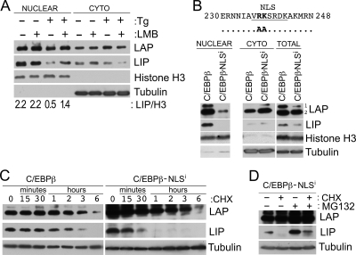
Induction of the transcription factor CHOP (CCAAT-binding homologous protein; GADD 153) is a critical cellular response for the transcriptional control of endoplasmic reticulum (ER) stress-induced apoptosis. Upon nuclear translocation, CHOP upregulates the transcription of proapoptotic factors and downregulates antiapoptotic genes. Transcriptional activation by CHOP involves heterodimerization with other members of the basic leucine zipper transcription factor (bZIP) family. We show that the bZIP protein C/EBP beta isoform LIP is required for nuclear translocation of CHOP during ER stress. In early ER stress, LIP undergoes proteasomal degradation in the cytoplasmic compartment. During later ER stress, LIP binds CHOP in both cytoplasmic and nuclear compartments and contributes to its nuclear import. By using CHOP-deficient cells and transfections of LIP-expressing vectors in C/EBP beta(-/-) mouse embryonic fibroblasts (MEFs), we show that the LIP-CHOP interaction has a stabilizing role for LIP. At the same time, CHOP uses LIP as a vehicle for nuclear import. LIP-expressing C/EBP beta(-/-) MEFs showed enhanced ER stress-induced apoptosis compared to C/EBP beta-null cells, a finding in agreement with the decreased levels of Bcl-2, a known transcriptional control target of CHOP. In view of the positive effect of CHOP-LIP interaction in mediating their proapoptotic functions, we propose this functional cooperativity as molecular symbiosis between proteins.


Figures

FIG. 1.
FIG. 1.
LIP is degraded by cytoplasmically localized proteasomes during ER stress. (A) C6 cells were pretreated or not with LMB (10 ng/ml) for 1 h before the induction of ER stress (Tg, 3 h) and then fractionated, and extracts were probed (20 μg [nuclear] and 40 μg [cytoplasmic]) for the expression of C/EBPβ isoforms. Tubulin and histone H3 were used as loading controls for the cytoplasmic and nuclear fractions, respectively. (B) The upper portion shows a schematic representation of the NLS mutation for C/EBPβ. The NLSi construct contains the RK-to-AA mutation within the NLS. The lower panel shows C/EBPβ−/− MEFs, stably transfected with the C/EBPβ wild type or C/EBPβ-NLSi expressing plasmids were fractionated, and the expression of the indicated proteins was detected by Western blot analysis. (C) C/EBPβ-WT and C/EBPβ-NLSi cell lines were treated with 10 μg of CHX/ml. The C/EBPβ LIP isoform half-lives in the two cell lines were determined by quantifying the intensity of the bands from the Western blots using the ImageJ software. Tubulin was used for loading control and normalization of the quantified bands. (D) The C/EBPβ-NLSi cell line was treated or not with 10 μg of CHX/ml with or without 10 μM MG132 for 1 h, and total cell lysates were probed for the expression of C/EBPβ isoforms. Tubulin was used for the loading control.
FIG. 2.
FIG. 2.
Characterization of the subcellular association of CHOP and C/EBPβ-LIP during ER stress. (A) CHOP−/− and CHOP+/+ MEFs were treated with Tg for the indicated times. Total and nuclear (right side) cell extracts were prepared and analyzed by Western blotting with the indicated antibodies. (B) C/EBPβ−/− MEFs, expressing the LIP isoform only were treated with Tg for the indicated times or 6 h for the first two lanes and used for the preparation of total cell lysates. Cytoplasmic (CYTO) and nuclear extracts were prepared from 6 h-treated LIP-expressing cells. The total lysate and the subcellular fractions were either analyzed directly (lysate) or immunoprecipitated with C/EBPβ antibodies (+) or with rabbit nonspecific IgG (−). The immunoprecipitates were analyzed by Western blotting with the indicated antibodies. (C) C/EBPβ−/− MEFs, expressing the NLS-inactivated LIP isoform only (LIP-NLSi), were treated with Tg for 6 h. Total, cytoplasmic (CYTO), and nuclear (NUCL) cell extracts were either analyzed directly (lysate) or immunoprecipitated with C/EBPβ antibodies (+) or with rabbit nonspecific IgG (−). The immunoprecipitates were analyzed by Western blotting with the indicated antibodies.
FIG. 3.
FIG. 3.
Nuclear accumulation of CHOP in early ER stress is dependent on the presence of the C/EBPβ isoform LIP. (A) C/EBPβ−/− MEFs stably transfected with the indicated plasmid constructs were stressed with Tg for the indicated times, fractionated, and probed for the expression of CHOP and C/EBPβ (LAP-1, LAP-2, and LIP) in nuclear extracts (20 μg/gel lane). The right side shows the same experiment, with the cytoplasmic samples (40 μg/gel lane). Tubulin and histone H3 were used as loading controls and as cytoplasmic and nuclear markers, respectively. (B) Total CHOP mRNA levels were detected by qPCR as described in Materials and Methods in cells stably transfected with the indicated plasmids and stressed for 0, 3, and 6 h with Tg. (C) Cellular localization of CHOP was assessed by immunocytochemistry in C/EBPβ−/− MEFs stably transfected with the indicated plasmids and stressed with Tg for 5 h. DAPI was used for specific staining of the nuclei.
FIG. 4.
FIG. 4.
Interaction of LIP with CHOP provides heterodimers with a functional NLS. (A) Amino acid sequence of the mouse CHOP and mutagenesis strategy. The putative NLS of CHOP is underlined in the sequence. (B) CHOP−/− MEFs were transiently cotransfected with the indicated plasmid constructs (pCDNA3.1A-CHOP/pEGFP-N1 ratio of 5:1), fractionated into nuclear extracts and cytoplasmic lysates, and probed for the expression of CHOP. In the pCDNA3.1A-CHOP construct, the expression of CHOP is under the control of CMV promoter. Histone H3 was used as a nuclear marker and loading control; green fluorescent protein (GFP) was used as a cytoplasmic marker and transfection efficiency control. (C) C/EBPβ−/− MEFs, stably expressing LIP and the LIP-NLSi mutant, were stressed with Tg, fractionated, and probed for the expression of CHOP and LIP. (D) LIP and LIP-NLSi MEFs were stressed with Tg, and the localization of CHOP was assessed in early ER stress by immunocytochemistry, as described in Materials and Methods. Wild-type (+/+) and CHOP-deficient (−/−) MEFs were assessed in the experimental settings described above in order to test the specificity of the CHOP antibody. DAPI was used for specific nuclear staining.
FIG. 5.
FIG. 5.
The expression of C/EBPβ isoform LIP-only is sufficient for the efficient induction of ER stress-caused apoptosis. (A) C/EBPβ−/− MEFs stably transfected with the indicated plasmid constructs were treated with Tg. Phase-contrast pictures were taken after 18 h of treatment. C/EBPβ−/− MEFs (KO), C/EBPβ−/− MEFs expressing the LIP isoform only (LIP), and C/EBPβ−/− MEFs expressing all three isoforms (C/EBPβ) are depicted. (B) Apoptosis was measured using FACS analysis as described in Materials and Methods. White columns indicate the sub-G1-phase number of cells in the untreated (−Tg) conditions, and gray columns indicate the sub-G1-phase number of cells in Tg-treated cells for 18 h. (C) C/EBPβ−/− (KO) and C/EBPβ−/− MEFs expressing the LIP isoform only (LIP) were treated with Tg for the indicated times. Total cell lysates were probed for the expression of LIP, caspase 3, and PARP. Tubulin was used as a loading control. (D) C/EBPβ−/− (KO) and C/EBPβ−/− MEFs expressing the LIP isoform only (LIP) were treated with Tg for the indicated times. Total cell lysates (upper) and total mRNA (lower) were probed for the indicated proteins and mRNA levels (Bcl-2 [transcript 1] and 18S rRNA), respectively.
FIG. 6.
FIG. 6.
Schematic representation of the regulatory functions of the C/EBPβ isoform LIP in mediating induction of apoptosis during ER stress. During early ER stress, degradation of LIP occurs in the cytoplasm following nuclear export. During late ER stress, increased accumulation of CHOP in the cytoplasm leads to CHOP-LIP heterodimerization and nuclear translocation. Nuclear localization of CHOP at late ER stress induces a transcriptional program for apoptotic cell death.

References

    1. Akira, S., H. Isshiki, T. Sugita, O. Tanabe, S. Kinoshita, Y. Nishio, T. Nakajima, T. Hirano, and T. Kishimoto. 1990. A nuclear factor for IL-6 expression (NF-IL6) is a member of a C/EBP family. EMBO J. 9:1897-1906. - PMC - PubMed
    1. Buck, M., L. Zhang, N. A. Halasz, T. Hunter, and M. Chojkier. 2001. Nuclear export of phosphorylated C/EBPβ mediates the inhibition of albumin expression by TNFα. EMBO J. 20:6712-6723. - PMC - PubMed
    1. Buck, M., V. Poli, T. Hunter, and M. Chojkier. 2001. C/EBPβ phosphorylation by RSK creates a functional XEXD caspase inhibitory box critical for cell survival. Mol. Cell 8:807-816. - PubMed
    1. Cao, Z., R. M. Umek, and S. L. McKnight. 1991. Regulated expression of three C/EBP isoforms during adipose conversion of 3T3-L1 cells. Genes Dev. 5:1538-1552. - PubMed
    1. Descombes, P., and U. Schibler. 1991. A liver-enriched transcriptional activator protein, LAP, and a transcriptional inhibitory protein, LIP, are translated from the same mRNA. Cell 67:569-579. - PubMed

Publication types

MeSH terms

LinkOut - more resources