. 2010 Jun 1;107(22):10103-8.
doi: 10.1073/pnas.1004824107. Epub 2010 May 17.

Information processing at the foxa node of the sea urchin endomesoderm specification network

Affiliations

Information processing at the foxa node of the sea urchin endomesoderm specification network

Smadar Ben-Tabou de-Leon et al. Proc Natl Acad Sci U S A. .

Abstract

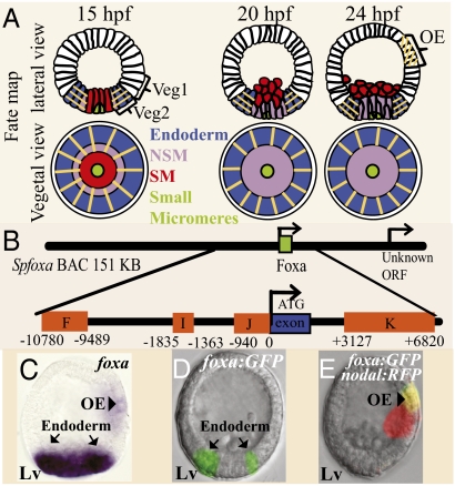
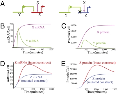
The foxa regulatory gene is of central importance for endoderm specification across Bilateria, and this gene lies at an essential node of the well-characterized sea urchin endomesoderm gene regulatory network (GRN). Here we experimentally dissect the cis-regulatory system that controls the complex pattern of foxa expression in these embryos. Four separate cis-regulatory modules (CRMs) cooperate to control foxa expression in different spatial domains of the endomesoderm, and at different times. A detailed mutational analysis revealed the inputs to each of these cis-regulatory modules. The complex and dynamic expression of foxa is regulated by a combination of repressors, a permissive switch, and multiple activators. A mathematical kinetic model was applied to study the dynamic response of foxa cis-regulatory modules to transient inputs. This study shed light on the mesoderm-endoderm fate decision and provides a functional explanation, in terms of the genomic regulatory code, for the spatial and temporal expression of a key developmental control gene.


Conflict of interest statement

The authors declare no conflict of interest.

Figures

Fig. 1.
Fig. 1.
Expression and regulatory structure of the foxA gene. (A) Lineage fate map showing lateral and vegetal views at 15, 20, and 24 hpf. The orange lines indicate the domains where foxa is expressed. Green, small micromeres; red, SM; purple, veg2 NSM; blue, veg2 endoderm; white, veg1 and the ectoderm; OA, oral ectoderm. (B) Diagram of foxA CRMs (orange boxes) in SpfoxA BAC. The numbers on the CRMs edges indicate their distance from foxA start of translation. foxA single exon is marked as a blue box. (C) Whole mount in situ hybridization of foxa at 24 hpf. foxa is expressed in the endoderm (arrows) and in the oral ectoderm (arrowhead). (D and E) Expression of foxA:GFP BAC at 24 hpf. The reporter is expressed in the endoderm (D) and in the oral ectoderm (E). Because the oral and aboral sides of the embryo are indistinguishable at 24 hpf, we coinjected the foxa:GFP BAC with Nodal:RFP BAC, which is expressed in the oral ectoderm. The figure shows the overlay of the two flourophores. Lv, lateral view.
Fig. 2.
Fig. 2.
Spatial restriction of foxa expression. Throughout the figure embryonic domains are enumerated as follows: 1 SM, 2 NSM, 3 veg2 endoderm, 4 veg1, 5 ectoderm, and 6 apical plate. (AL) Expression patterns of embryos injected with different reporter constructs at 23–24 hpf. (AC) FIJ:GFP coinjected with foxA:RFP BAC: (A) GFP, (B) RFP, and (C) overlay. (DF) Coinjection of foxA:RFP BAC and a version of FIJ:GFP where a Tcf site in F was mutated. (D) GFP, (E) RFP, and (F) overlay. (G) Intact FIJ:GFP injected alone drives correct expression in the endoderm and in the ectoderm. (H and I) Tcf mutated version of FIJ:GFP drives GFP in the NSM (H) and in the SM, NSM, and endoderm (I). (JL) FIJ:GFP coinjected with foxA:RFP BAC and mutExoBlimp1 construct that drives blimp1 in the NSM: (J) GFP, (K) RFP, and (L) overlay. (M) Diagrams of β-catenin nuclearization at 15, 20, and 24 hpf. Cells where β-catenin is nuclearized are in cyan, the rest of the cells are in orange. (N) Quantification of spatial expression at 23–24 hpf. Percentages sum to more than 100% as some embryos express in two or more tissue types. At 24 hpf the oral and aboral sides of the embryo are indistinguishable, therefore, we considered only domains 4 and 6 to be ectopic ectoderm expression. Data are based on three replicate experiments totaling ≈120 embryos for each construct. Number of expressing embryos for each construct is indicated in the graph key.
Fig. 3.
Fig. 3.
Quantitative analysis of foxa inputs. Throughout the figure error bars show ±1 SE. (A) QPCR time courses of SpfoxA BAC:RFP and the constructs: F to K, F to J, and FIJ:GFP. The values are cDNA (mRNA) copies per injected DNA copy. Values are based on at least three independent batches of 150 injected embryos per time point. (B) Diagram of foxA CRMs and the binding sites of the different inputs. Color codes for all inputs are consistent throughout. (C) The construct FIJ:GFP coinjected with Random MO shows normal expression in the endoderm at 25 hpf. (D) The construct FIJ:GFP coinjected with Notch MO, the GFP fails to clear from the NSM. (EG) Coinjection of the reporter FIJ:GFP where the three Su(H) binding sites are mutated with foxA:RFP BAC. The GFP expression is unaffected by the mutation and is identical to the RFP expression. (E) RFP, (F) GFP, (G) overlay. (H and I) QPCR measurement of the effect of site mutations on the level of reporter constructs. The results are the ratio between the expression of the mutated construct and the intact construct. The construct that was used to measure the effect of each mutation is indicated in the figure key. Significance was calculated by one-tailed Z-test. *, P < 0.01; **, P < 0.001. Numerical values of means, number of repeats, and P values are presented in Table S2. (J) Spatial expression of foxa inputs at 15 hpf. Delta signal from the SM is inducing Su(H)-NIC activity in the NSM. Hox11/13b is expressed in all veg2 descendants. (K) Spatial expression of foxa inputs at 24 hpf. Brachyury and Hox11/13b have progressed toward veg1 descendants and only partially overlap with foxA expressing cells.
Fig. 4.
Fig. 4.
Kinetic model of a site mutation of a transient input in a reporter construct. (A) Schematic diagrams of the inputs to the intact (Left) and the mutated (Right) constructs. (B) mRNA expression kinetics of the inputs, X (fuchsia) and Y (green). (C) Protein kinetics of the inputs, X and Y, same color code as in B. (D) mRNA kinetics of the intact construct (red) and the mutated construct (blue). (E) Protein kinetics of intact and mutated constructs, same color code as in D. Kinetic parameters: mRNA and protein turnover rate Kdm = Kdp = 0.005 min−1. Transcriptional delay, Tm = 20 min. See SI Text 2 for additional details.
Fig. 5.
Fig. 5.
Endomesoderm GRN and the active inputs into foxA CRMs. Active modules are marked in light blue; inactive modules and genes are marked in gray. (A) At 11–18 hpf foxA is activated by Hox11/13b, the Delta-Notch signal from the SM cells and unknown ubiquitous activator. foxA is repressed in the ectoderm and in the SM by Tcf-Groucho. (B) At 20–27 hpf foxA is activated by Hox11/13b, Otx, Brachyury, and unknown ubiquitous activator. Starting from 20 hpf, FoxA autorepresses its own gene expression. foxA is restricted to the endoderm by Tcf-Groucho repression elsewhere.

Similar articles

Cited by

References

    1. Oliveri P, Walton KD, Davidson EH, McClay DR. Repression of mesodermal fate by foxa, a key endoderm regulator of the sea urchin embryo. Development. 2006;133:4173–4181. - PubMed
    1. Friedman JR, Kaestner KH. The Foxa family of transcription factors in development and metabolism. Cell Mol Life Sci. 2006;63:2317–2328. - PMC - PubMed
    1. Boyle MJ, Seaver EC. Developmental expression of foxA and gata genes during gut formation in the polychaete annelid, Capitella sp. I. Evol Dev. 2008;10:89–105. - PubMed
    1. Hiruta J, Mazet F, Yasui K, Zhang P, Ogasawara M. Comparative expression analysis of transcription factor genes in the endostyle of invertebrate chordates. Dev Dyn. 2005;233:1031–1037. - PubMed
    1. Burtscher I, Lickert H. Foxa2 regulates polarity and epithelialization in the endoderm germ layer of the mouse embryo. Development. 2009;136:1029–1038. - PubMed

Publication types

MeSH terms