. 2010 Jun 8;107(23):10638-43.
doi: 10.1073/pnas.1002348107. Epub 2010 May 17.

Mucosal adjuvant activity of cholera toxin requires Th17 cells and protects against inhalation anthrax

Affiliations

Mucosal adjuvant activity of cholera toxin requires Th17 cells and protects against inhalation anthrax

Sandip K Datta et al. Proc Natl Acad Sci U S A. .

Abstract

Cholera toxin (CT) elicits a mucosal immune response in mice when used as a vaccine adjuvant. The mechanisms by which CT exerts its adjuvant effects are incompletely understood. We show that protection against inhalation anthrax by an irradiated spore vaccine depends on CT-mediated induction of IL-17-producing CD4 Th17 cells. Furthermore, IL-17 is involved in the induction of serum and mucosal antibody responses by CT. Th17 cells induced by CT have a unique cytokine profile compared with those induced by IL-6 and TGF-beta, and their induction by CT requires cAMP-dependent secretion of IL-1beta and beta-calcitonin gene-related peptide by dendritic cells. These findings demonstrate that Th17 cells mediate mucosal adjuvant effects of CT and identify previously unexplored pathways involved in Th17 induction that could be targeted for development of unique mucosal adjuvants.


Conflict of interest statement

The authors declare no conflict of interest.

Figures

Fig. 1.
Fig. 1.
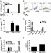
Protection by anthrax spore vaccine requires Th17 cell induction by CT. (A) Mice (10 per group) were immunized intranasally, as indicated, and monitored for survival after live-spore challenge. (B) Mice (10–12 per group) were immunized intranasally with PA-deficient irradiated spores and CT. Indicated depleting antibodies were administered intraperitonially 1 d before challenge. (C) Mice (4 per group) were immunized as indicated and then antigen challenged with irradiated spores. Three days after challenge, splenic CD4+ cells were isolated and cocultured with antigen-presenting cells pulsed with or without irradiated spores to determine spore-specific cytokine production. Data are shown as the mean of each group, and error bars represent SD. *, P < 0.05 compared with saline-treated group. (D) Mice (8 per group) immunized with irradiated spores and CT were treated with the indicated antibodies before infection. Control mice (Unimm) received saline for the immunization and depletion protocols.
Fig. 2.
Fig. 2.
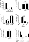
CT-treated DCs elicit Th17 cells. (A) Splenic OT-II CD4 T cells were incubated for 6 d with C57BL/6 bone marrow-derived DCs that had been treated overnight with OVA 1(0 μg/mL) in the absence or presence of CT (1 μg/mL). After anti-CD3 + anti-CD28 restimulation, supernatants were analyzed by ELISA. Data shown are representative of more than six independent experiments. (B) DCs were incubated with OVA or CT+OVA. The DCs were then fixed and resuspended in their original medium. OT-II CD4 T cells were added with or without CT. After 6 d, the T cells were restimulated and their supernatants analyzed by ELISA. Data shown are representative of two independent experiments. (C and D) DCs from C57BL/6 (B6), MyD88−/−, Nod2−/−, or Tlr4−/− mice were incubated with CT or OVA overnight before incubation with OT-II CD4+ T cells. After restimulation, supernatants were analyzed by ELISA. Data shown are representative of three independent experiments. All graphs show the mean of triplicate samples and error bars reflect standard deviation. *, P < 0.05 compared to CT-untreated group; n.d., not detected.
Fig. 3.
Fig. 3.
CT elicits Th17 cells via cAMP. (A) cAMP levels in DC lysates were measured by ELISA after 3 h incubation with media, CT, or forskolin (10 μM). (B and C) DCs were incubated overnight with media, CT, 8-Br-cAMP (500 μM), IBMX (100 μM), forskolin (10 μM), EPAC agonist (CPT-O-Me-cAMP, 500 μM), or PKA agonist (Bnz-cAMP, 500 μM) in the absence or presence of OVA. OT-II CD4 T cells were then added, restimulated, and their supernatants analyzed by ELISA. (D) DCs were treated with or without H89 (10 μM) during a 3 h incubation with media, OVA, or CT. DCs were then fixed and incubated in their original media with OT-II CD4 T cells before analysis of restimulated T cell supernatants. All data shown are representative of two independent experiments. Data are shown as the mean of triplicate samples, and error bars reflect standard deviation. *, P < 0.05 compared with media-treated group.
Fig. 4.
Fig. 4.
Secreted factors from CT-treated DCs are sufficient to induce Th17 differentiation. (A) DCs were incubated with OVA or CT+OVA and then left unfixed or fixed. OT-II CD4 T cells were then added with or without the original DC supernatant (DC supe), and IL-17 production assessed after restimulation. (B–E) Sorted naive CD4 T cells were stimulated with anti-CD3, anti-CD28, anti-IL-4, and anti-IFN-γ in the presence of media, 10% CT-CM, or IL-6+TGF-β. Cytokine production after restimulation with phorbol myristate acetate and ionomycin was determined by flow cytometry and ELISA. (F) RORγt expression in CD4 T cells, stimulated as above, was determined by quantitative RT-PCR. Fold-induction of cells treated with CT-CM or IL-6+TGF-β compared with cells incubated with media alone is shown. All data are representative of at least two independent experiments. ELISA data are shown as the mean of duplicate samples and error bars reflect SD. *, P < 0.05 compared with media-treated group; n.d., not detected.
Fig. 5.
Fig. 5.
A combination of secreted factors mediates Th17 differentiation by CT. (A and B) Cytokine production was determined after stimulation of naive CD4 T cells with 10% CT-CM and the indicated neutralizing antibodies. (C) Ten-percent CT-CM from wild-type (B6) or Cox2−/− DC, or 10% CT-CM with or without neutralizing amphiregulin antibody was used to stimulate naive CD4 T cells. (D and E) Cytokine production was determined after stimulation of naive CD4 T cells with 10% CT-CM, IL-6+TGF-β, β-CGRP inhibitor (β-CGRP8–37), or β-CGRP. (F) Cytokine production was determined after stimulation of naive CD4 T cells with the indicated factors. All data are representative of at least two independent experiments. ELISA data are shown as the mean of duplicate samples, and error bars reflect SD. *, P < 0.05 compared with CT-CM (A) or media-treated (F) group.
Fig. 6.
Fig. 6.
IL-17 is required for CT-induced antibody production after gavage. OVA-specific bronchoalveolar lavage (BAL), fecal, and serum antibody levels were determined in wild-type (B6) and IL-17-deficient mice immunized with OVA or CT+OVA intranasally (A–D, 4–6 mice per group) or by gavage (E–H, 10–12 mice per group). Intranasal experiment is representative of two independent experiments and gavage experiment is representative of three independent experiments.

Similar articles

Cited by

References

    1. Northrup RS, Fauci AS. Adjuvant effect of cholera enterotoxin on the immune response of the mouse to sheep red blood cells. J Infect Dis. 1972;125:672–673. - PubMed
    1. Elson CO, Ealding W. Generalized systemic and mucosal immunity in mice after mucosal stimulation with cholera toxin. J Immunol. 1984;132:2736–2741. - PubMed
    1. Freytag LC, Clements JD. Mucosal adjuvants. Vaccine. 2005;23:1804–1813. - PubMed
    1. Vanden Broeck D, Horvath C, De Wolf MJ. Vibrio cholerae: Cholera toxin. Int J Biochem Cell Biol. 2007;39:1771–1775. - PubMed
    1. Xu-Amano J, et al. Helper T cell subsets for immunoglobulin A responses: Oral immunization with tetanus toxoid and cholera toxin as adjuvant selectively induces Th2 cells in mucosa associated tissues. J Exp Med. 1993;178:1309–1320. - PMC - PubMed

Publication types