. 2010 Jun 1;107(22):10220-5.
doi: 10.1073/pnas.1001673107. Epub 2010 May 17.

A viral assembly factor promotes AAV2 capsid formation in the nucleolus

Affiliations

A viral assembly factor promotes AAV2 capsid formation in the nucleolus

Florian Sonntag et al. Proc Natl Acad Sci U S A. .

Abstract

The volume available in icosahedral virus capsids limits the size of viral genomes. To overcome this limitation, viruses have evolved strategies to increase their coding capacity by using more than one ORF while keeping the genome length constant. The assembly of virus capsids requires the coordinated interaction of a large number of subunits to generate a highly ordered structure in which the viral genome can be enclosed. To understand this process, it is essential to know which viral and nonviral components are involved in the assembly reaction. Here, we show that the adeno-associated virus (AAV) encodes a protein required for capsid formation by means of a nested, alternative ORF of the cap gene. Translation is initiated at a nonconventional translation start site, resulting in the expression of a protein with a calculated molecular weight of 23 kDa. This protein, designated assembly-activating protein (AAP), is localized in the host cell nucleolus, where AAV capsid morphogenesis occurs. AAP targets newly synthesized capsid proteins to this organelle and in addition fulfils a function in the assembly reaction itself. Sequence analysis suggests that also all other species of the genus Dependovirus encode a homologous protein in their cap gene. The arrangement of different ORFs that encode capsid proteins and an assembly factor within the same mRNA facilitates a timely coordinated expression of the components involved in the assembly process.


Conflict of interest statement

The authors declare no conflict of interest.

Figures
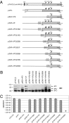
Fig. 1.
Fig. 1.
Comparison of capsid assembly using different cap gene constructs. (A) Schematic of constructs comprising the entire cap gene or 5′-deleted variants thereof used for transfection of 293T cells. Small numbers indicate nucleotide positions relative to the AAV2 genome. Arrows represent the translation start sites of the VP proteins. Mutated translation start sites are labeled with a cross. CMV: cytomegalovirus promoter; ITR: inverted terminal repeat; rep: rep gene. (B) Western blot analysis of VP protein expression was performed using mAb B1, which detects all three capsid proteins. In extracts of pKEX-VP3–, pCMV-VP3/2765–, and pCMV-VP3/2809–transfected cells, mAb B1 reacted with two polypeptide bands migrating slower than VP3. These polypeptides were a consequence of higher VP3 levels, which were not completely denatured and cannot result from VP1 or VP2 expression because the respective coding sequences were deleted. (C) Capsid formation was quantified by ELISA based on mAb A20. Detection limit: 5 × 10⁷ capsids/mL Means ± SDs of at least three independent experiments are shown; asterisks indicate constructs for which no capsids could be detected.
Fig. 2.
Fig. 2.
Trans-complementation of VP3 capsid assembly. A panel of constructs (Left) was used for complementation of VP3 expression plasmid pCMV-VP3/2809 by cotransfection into 293T cells. Samples were analyzed by Western blot (Center) using mAb B1 to detect VP3, mAb A69 to detect VP2N-gfp, and a truncated VP2 (VP2tru), mAb anti-AU1 to detect AAP-AU1, or polyclonal anti-AAP serum to detect AAP-AU1 or C-terminally truncated AAP (AAPtru). Capsid formation was quantified by ELISA based on mAb A20 (Right). Means ± SDs of at least three independent experiments are shown; asterisks indicate samples for which no capsids could be detected. (A) Construct pVP2N-gfp, containing the EcoNI-BsiWI fragment derived from the AAV2 genome and a gfp-cassette, was cotransfected with pCMV-VP3/2809 in decreasing amounts, starting with equimolar ratios. For comparison, empty vector pBS or plasmid pCMV-VP3/2696 was transfected. (B) Constructs pVP2N/ORF1cm and pVP2N/ORF2cm are derivatives of pVP2N-gfp with codon-modified ORF1 or ORF2 of the cap gene fragment, respectively (shaded boxes). See Fig. S6 for codon-modified sequences. (C) Constructs pORF2/CTG-AU1, pORF2/ATG-AU1, and pORF2/TTG-AU1 comprise the entire ORF2 of the cap gene (AAV2 nt 2717–3340) fused to sequences coding for an AU1-tag. The predicted AAP translation initation codon (CTG) was additionally mutated to ATG and TTG.
Fig. 3.
Fig. 3.
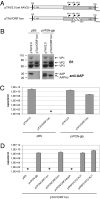
Trans-complementation of the full-length AAV2 genome containing an ORF1 codon-modified cap gene fragment. (A) Schematic of plasmid pTAV2.0, harboring the wild-type AAV2 genome, and of plasmid pTAV/ORF1cm, containing the ORF1 codon-modified EcoNI-BsiWI fragment of the cap gene (shaded box). Plasmids were cotransfected with the indicated constructs into 293T cells. (B) Western blot analysis of VP protein expression was performed using mAb B1. AAP and AAPtru were detected with polyclonal anti-AAP serum. (C and D) Capsid formation was quantified by ELISA based on mAb A20. Means ± SDs of at least three independent experiments are shown; asterisks indicate samples for which no capsids could be detected.
Fig. 4.
Fig. 4.
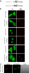
Intracellular localization and capsid assembly of VP3, NLS-VP3, and NoLS-VP3. (A) Schematic of constructs used for expression of VP3 fused to the nuclear localization signal of the SV40 large T-antigen (NLS-VP3) and of VP3 fused to the nucleolar localization signal of HIV Rev (NoLS-VP3). Indirect double immunofluorescence of HeLa cells transfected with plasmids indicated at the left. (B) Localization of total expressed capsid proteins using polyclonal VP anti-serum (VPs, green) and of assembled capsids using mAb A20 (capsid, red). (C) Detection of AAP-AU1 using a mAb against the AU1-tag (anti-AU1, red) and of nucleoli using polyclonal fibrillarin antibody (anti-fibrillarin, green).
Fig. 5.
Fig. 5.
Organization of genetic information in the AAV2 cap gene. Expression of the AAV2 cap gene occurs under the control of promoter p40. After transcription, two mRNAs are generated by alternative splicing. The translation initiation codons used for expression of the four proteins involved in AAV2 capsid formation (VP1, VP2, VP3, and AAP) are indicated. Only the minor splice product contains the translation initiation codon for VP1. The unique assembly-activating protein (AAP) is encoded by a nested, alternative ORF comprising a nonconventional CUG translation initiation codon.

References

    1. Trempe JP, Carter BJ. Alternate mRNA splicing is required for synthesis of adeno-associated virus VP1 capsid protein. J Virol. 1988;62:3356–3363. - PMC - PubMed
    1. Cassinotti P, Weitz M, Tratschin JD. Organization of the adeno-associated virus (AAV) capsid gene: Mapping of a minor spliced mRNA coding for virus capsid protein 1. Virology. 1988;167:176–184. - PubMed
    1. Becerra SP, Koczot F, Fabisch P, Rose JA. Synthesis of adeno-associated virus structural proteins requires both alternative mRNA splicing and alternative initiations from a single transcript. J Virol. 1988;62:2745–2754. - PMC - PubMed
    1. Myers MW, Carter BJ. Assembly of adeno-associated virus. Virology. 1980;102:71–82. - PubMed
    1. Wistuba A, Kern A, Weger S, Grimm D, Kleinschmidt JA. Subcellular compartmentalization of adeno-associated virus type 2 assembly. J Virol. 1997;71:1341–1352. - PMC - PubMed

Publication types

MeSH terms