. 2010 Jun 1;107(22):10202-7.
doi: 10.1073/pnas.0913897107. Epub 2010 May 17.

Extracellular sulfatases support cartilage homeostasis by regulating BMP and FGF signaling pathways

Affiliations

Extracellular sulfatases support cartilage homeostasis by regulating BMP and FGF signaling pathways

Shuhei Otsuki et al. Proc Natl Acad Sci U S A. .

Abstract

The balance between anabolic and catabolic signaling pathways is critical in maintaining cartilage homeostasis and its disturbance contributes to joint diseases such as osteoarthritis (OA). A unique mechanism that modulates the activity of cell signaling pathways is controlled by extracellular heparan endosulfatases Sulf-1 and Sulf-2 (Sulfs) that are overexpressed in OA cartilage. This study addressed the role of Sulfs in cartilage homeostasis and in regulating bone morphogenetic protein (BMP)/Smad and fibroblast growth factor (FGF)/Erk signaling in articular cartilage. Spontaneous cartilage degeneration and surgically induced OA were significantly more severe in Sulf-1(-/-) and Sulf-2(-/-) mice compared with wild-type mice. MMP-13, ADAMTS-5, and the BMP antagonist noggin were elevated whereas col2a1 and aggrecan were reduced in cartilage and chondrocytes from Sulf(-/-) mice. Articular cartilage and cultured chondrocytes from Sulf(-/-) mice showed reduced Smad1 protein expression and Smad1/5 phosphorylation, whereas Erk1/2 phosphorylation was increased. In human chondrocytes, Sulfs siRNA reduced Smad phosphorylation but enhanced FGF-2-induced Erk1/2 signaling. These findings suggest that Sulfs simultaneously enhance BMP but inhibit FGF signaling in chondrocytes and maintain cartilage homeostasis. Approaches to correct abnormal Sulf expression have the potential to protect against cartilage degradation and promote cartilage repair in OA.


Conflict of interest statement

The authors declare no conflict of interest.

Figures

Fig. 1.
Fig. 1.
Spontaneous cartilage degeneration in Sulf−/− mice. (A) Knee joints were harvested from WT and Sulf−/− mice at 1.5, 3, and 6 months of age to determine spontaneous development of changes in joint tissues. Sections were stained with safranin O and cartilage degeneration was quantified by using a modified Mankin score and a summed score (n = 6–8 animals per time point). (B) At 6 months, Sulf−/− mouse knee joints showed significantly more severe cartilage degeneration than WT. , P < 0.01. (Scale bars: low magnification, 200 μm; high magnification, 50 μm.)
Fig. 2.
Fig. 2.
Surgically induced OA is more severe in Sulf−/− mice. (A) Surgical OA was induced in WT and Sulf−/− mice at the age of 8 weeks (n = 8–10 mice per group) by resecting the medial meniscotibial ligament and harvesting the knee joints 4 weeks after surgery for safranin O staining. Arrow indicates roughening of the articular surface. (B) Sulf−/− mice showed significantly increased Mankin and summed scores compared with WT after surgery. , P < 0.01. (Scale bars: low magnification, 200 μm; high magnification, 50 μm.)
Fig. 3.
Fig. 3.
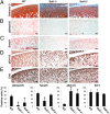
Abnormal ECM, MMP-13, ADAMTS-5, and noggin expression patterns in Sulf−/− mice. (A) Knee joint sections from mice at the age of 4 weeks were stained with safranin O and for MMP-13 (n = 6). (Scale bars: low magnification for safranin O, 200 μm; high magnification for MMP-13, 50 μm.) (B) Articular cartilage from Sulf−/− mice contained more MMP-13-positive cells especially in Sulf-1−/− compared with WT mice. , P < 0.01. (C) mRNA expression in articular chondrocytes from WT and Sulf−/− mice. col2a1, mmp-13, and noggin expression was analyzed by Taqman PCR in chondrocytes from WT, Sulf-1−/−, and Sulf-2−/− mice (n = 6–8). (D) mRNA expression of adamts-5 was examined in chondrocytes from WT, Sulf-1−/−, and Sulf-2−/− mice (n = 6). Results are shown as mRNA levels of the indicated genes relative to gapdh (*, P ≤ 0.05; , P < 0.01). (E) ADAMTS-5 expression was examined in 3-month-old WT, Sulf-1−/−, and Sulf-2−/− mice by immunohistochemistry. (Scale bars: 50 μm.)
Fig. 4.
Fig. 4.
Smad and Erk expression and phosphorylation in murine knee joints. (A–E) Knee joint sections from 2-week-old mice were stained with safranin O and immunostained for total and phosphorylated forms of Smad1 and Erk1/2. For quantification total cell numbers and positive cells were counted in cartilage from WT and Sulf−/− mice (n = 6; , P < 0.01). Results are expressed as percentage of positive cells. (Scale bars: 20 μm.)
Fig. 5.
Fig. 5.
Smad1/5 and Erk1/2 expression and phosphorylation in chondrocytes from WT and Sulf−/− mice. (A) Western blot analysis of pSmad1/5, Smad1, pErk1/2, and Erk1/2. Protein expression and phosphorylation of Smad1/5 were reduced, but Erk1/2 was increased in Sulf−/− mice. (B) Densitometry of the autoradiographs was performed with NIH Image J software (n = 4; , P < 0.01).
Fig. 6.
Fig. 6.
Sulfs regulate BMP-7-induced Smad1/5 and FGF-2-induced Erk1/2 phosphorylation in human chondrocytes. (A and B) Human articular chondrocytes were transfected with siRNA for Sulf-1 or Sulf-2. RNA was isolated for qPCR and proteins were isolated for Western blotting after 48 h. Sulfs were knocked down specifically by 50–70% (n = 5; , P < 0.01). (C) Western blotting shows the expression of Smad1/5 and Erk1/2 phosphorylation in response to BMP-7 and FGF-2 stimulation of human chondrocytes from 0 to 120 min. (D) Maximal Smad1/5 and Erk1/2 phosphorylation is observed at 60 min. Chondrocytes were transfected with Sulf siRNA to determine changes in Smad1/5 and Erk1/2 phosphorylation in response to BMP-7 and FGF-2 (each 100 ng/mL) after 60 min by Western blotting. Sulf-1 and Sulf-2 siRNA reduced Smad1/5, but increased Erk1/2 phosphorylation, and densitometry of the autoradiographs was performed with NIH Image J software (E). These representative data are from chondrocytes obtained from a 61-year-old male donor with normal articular cartilage.

Similar articles

Cited by

References

    1. Goldring MB. The role of the chondrocyte in osteoarthritis. Arthritis Rheum. 2000;43:1916–1926. - PubMed
    1. Lyons KM, Pelton RW, Hogan BL. Patterns of expression of murine Vgr-1 and BMP-2a RNA suggest that transforming growth factor-beta-like genes coordinately regulate aspects of embryonic development. Genes Dev. 1989;3:1657–1668. - PubMed
    1. Lyons KM, Pelton RW, Hogan BL. Organogenesis and pattern formation in the mouse: RNA distribution patterns suggest a role for bone morphogenetic protein-2A (BMP-2A) Development. 1990;109:833–844. - PubMed
    1. Yi SE, Daluiski A, Pederson R, Rosen V, Lyons KM. The type I BMP receptor BMPRIB is required for chondrogenesis in the mouse limb. Development. 2000;127:621–630. - PubMed
    1. Miljkovic ND, Cooper GM, Marra KG. Chondrogenesis, bone morphogenetic protein-4 and mesenchymal stem cells. Osteoarthritis Cartilage. 2008;16:1121–1130. - PubMed

Publication types

MeSH terms

Substances