Comparative Study
. 2010 Jun 20;28(18):3061-8.
doi: 10.1200/JCO.2009.26.7252. Epub 2010 May 17.

Integrated molecular genetic profiling of pediatric high-grade gliomas reveals key differences with the adult disease

Affiliations
Comparative Study

Integrated molecular genetic profiling of pediatric high-grade gliomas reveals key differences with the adult disease

Barbara S Paugh et al. J Clin Oncol. .

Abstract

Purpose: To define copy number alterations and gene expression signatures underlying pediatric high-grade glioma (HGG).

Patients and methods: We conducted a high-resolution analysis of genomic imbalances in 78 de novo pediatric HGGs, including seven diffuse intrinsic pontine gliomas, and 10 HGGs arising in children who received cranial irradiation for a previous cancer using single nucleotide polymorphism microarray analysis. Gene expression was analyzed with gene expression microarrays for 53 tumors. Results were compared with publicly available data from adult tumors.

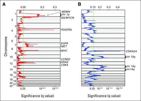
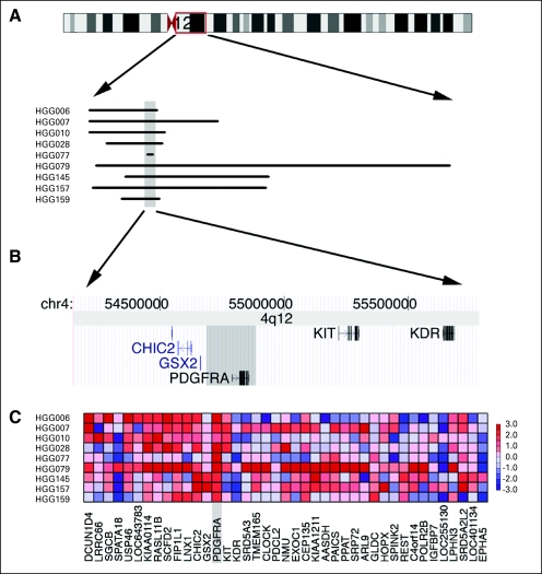
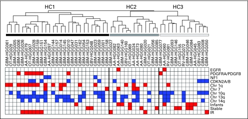
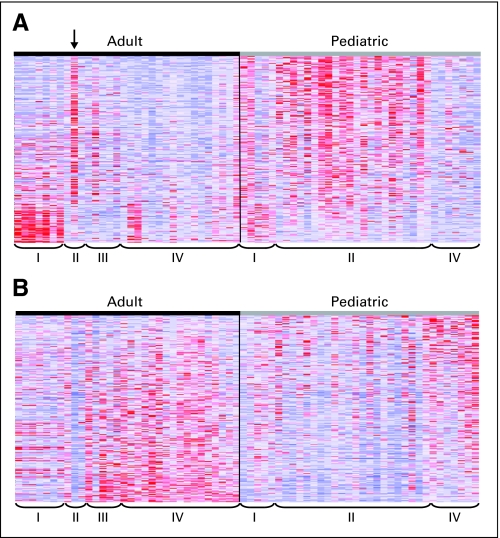
Results: Significant differences in copy number alterations distinguish childhood and adult glioblastoma. PDGFRA was the predominant target of focal amplification in childhood HGG, including diffuse intrinsic pontine gliomas, and gene expression analyses supported an important role for deregulated PDGFRalpha signaling in pediatric HGG. No IDH1 hotspot mutations were found in pediatric tumors, highlighting molecular differences with adult secondary glioblastoma. Pediatric and adult glioblastomas were clearly distinguished by frequent gain of chromosome 1q (30% v 9%, respectively) and lower frequency of chromosome 7 gain (13% v 74%, respectively) and 10q loss (35% v 80%, respectively). PDGFRA amplification and 1q gain occurred at significantly higher frequency in irradiation-induced tumors, suggesting that these are initiating events in childhood gliomagenesis. A subset of pediatric HGGs showed minimal copy number changes.

Conclusion: Integrated molecular profiling showed substantial differences in the molecular features underlying pediatric and adult HGG, indicating that findings in adult tumors cannot be simply extrapolated to younger patients. PDGFRalpha may be a useful target for pediatric HGG, including diffuse pontine gliomas.


Conflict of interest statement

Authors' disclosures of potential conflicts of interest and author contributions are found at the end of this article.

Figures

Fig 1.
Fig 1.
Most significant genomic alterations in pediatric high-grade glioma (HGG) identified by Genomic Identification of Significant Targets in Cancer (GISTIC). Significance of copy number (A) gains and (B) losses identified in 68 de novo pediatric HGGs by GISTIC is shown. Chromosome positions are displayed along the y-axis, calculated G-score is above the x-axis, and false discovery rate q values are along the lower x-axis. The green line indicates a q value threshold of 0.25.
Fig 2.
Fig 2.
PDGFRA is the target of common amplifications. (A) The extent of amplification for each tumor is shown. (B) Genes within the minimal common region and 500 kilobases upstream and 900 kilobases downstream. (C) Expression of all probes within the maximal region of amplification. The minimal common region of amplification is shown with a gray bar in all panels. Only tumors with matched expression data are shown.
Fig 3.
Fig 3.
Integrated gene expression subgroups and copy number imbalances. Distribution of significant copy number features and age information among expression subgroups that were identified by unsupervised hierarchical clustering, with dendrogram and tumor identification numbers shown above. Copy number features include amplification of EGFR, PDGFRA, and PDGFB (red); homozygous deletion of CDKN2A; focal deletion or inherited mutation of NF1 (blue); and broad regions of gain (red, chr1q and chr7) and loss (blue, chr10q, chr13q, and chr14q). “Infants” are patients younger than age 3 years at diagnosis. “Stable” indicates tumors that did not have large-scale genomic changes. IR, irradiation induced.
Fig 4.
Fig 4.
Principal component analysis (PCA) shows differences and similarities between pediatric and adult glioblastoma. PCA was generated using the 1,000 most variable probes and glioblastomas from our pediatric cohort and from published data from adult glioblastoma., Pediatric and adult tumors are shown in orange and green, respectively. Tumor subgroups are indicated by shape as follows: proneural (diamonds), proliferative (Prolif; spheres), mesenchymal (Mes; cubes), and Prolif/Mes (cones). Adult tumor subgroups are based on Lee et al. Asterisks indicate irradiation-induced tumors. Arrow indicates tumor from 53-year-old patient. Black boxes indicate tumors from infants.
Fig 5.
Fig 5.
Differential expression of PDGFRA and EGFR gene signatures in pediatric versus adult glioblastoma. Gene sets that are either upregulated in PDGFRA-amplified tumors (The Cancer Genome Atlas [TCGA] PDGFRA set) or upregulated in EGFR-amplified tumors (TCGA EGFR set) were identified using TCGA data from adult glioblastomas and used for gene set enrichment analysis (GSEA) to compare our pediatric glioblastoma data to published adult glioblastoma data., Expression heat maps of (A) the TCGA PDGFRA set genes and (B) the TCGA EGFR set genes in adult and pediatric glioblastomas are shown. Tumor subgroups are indicated by proneural (PN; I), proliferative (Prolif; II), Prolif/mesenchymal (Prolif/Mes; III), and Mes (IV). Adult tumor subgroups are based on Lee et al, and pediatric tumor subgroups are based on unsupervised hierarchical clustering. Arrow indicates tumor from a 53-year-old patient. GSEA showed significantly increased expression of the TCGA PDGFRA set (nominal P = .116, false discovery rate [FDR] = 0.121), whereas the TCGA EGFR set is downregulated in pediatric high-grade gliomas (nominal P = .229, FDR = 0.238) as well as the adult PN subclass. Overexpression of the TCGA PDGFRA set and underexpression of the TCGA EGFR set shows even greater enrichment in pediatric glioblastoma if pediatric tumors in the Mes subgroup are removed from the analysis (upregulation of PDGFRA gene set; nominal P = .048, FDR = 0.108 and downregulation of EGFR gene set, nominal P = .108, FDR = 0.108).

References

    1. Broniscer A, Gajjar A. Supratentorial high-grade astrocytoma and diffuse brainstem glioma: Two challenges for the pediatric oncologist. Oncologist. 2004;9:197–206. - PubMed
    1. Broniscer A, Baker SJ, West AN, et al. Clinical and molecular characteristics of malignant transformation of low-grade glioma in children. J Clin Oncol. 2007;25:682–689. - PubMed
    1. Furnari FB, Fenton T, Bachoo RM, et al. Malignant astrocytic glioma: Genetics, biology, and paths to treatment. Genes Dev. 2007;21:2683–2710. - PubMed
    1. Bredel M, Bredel C, Juric D, et al. High-resolution genome-wide mapping of genetic alterations in human glial brain tumors. Cancer Res. 2005;65:4088–4096. - PubMed
    1. Kotliarov Y, Steed ME, Christopher N, et al. High-resolution global genomic survey of 178 gliomas reveals novel regions of copy number alteration and allelic imbalances. Cancer Res. 2006;66:9428–9436. - PMC - PubMed

Publication types

Substances