. 2010 May 17;189(4):641-9.
doi: 10.1083/jcb.200912021.

The Dam1 complex confers microtubule plus end-tracking activity to the Ndc80 kinetochore complex

Affiliations

The Dam1 complex confers microtubule plus end-tracking activity to the Ndc80 kinetochore complex

Fabienne Lampert et al. J Cell Biol. .

Abstract

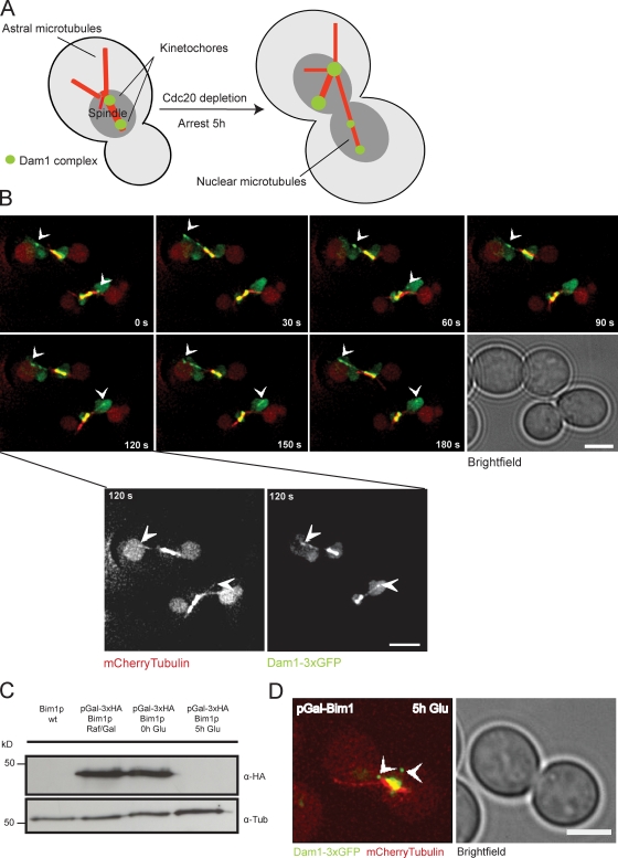
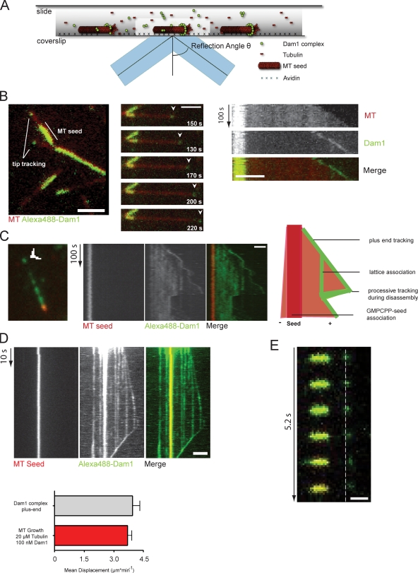
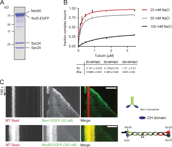
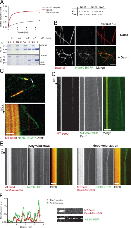
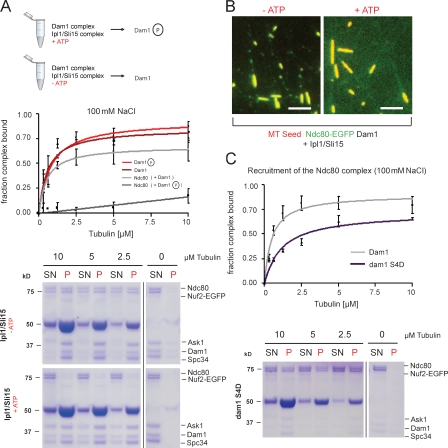
Kinetochores must remain associated with microtubule ends, as they undergo rapid transitions between growth and shrinkage. The molecular basis for this essential activity that ensures correct chromosome segregation is unclear. In this study, we have used reconstitution of dynamic microtubules and total internal reflection fluorescence microscopy to define the functional relationship between two important budding yeast kinetochore complexes. We find that the Dam1 complex is an autonomous plus end-tracking complex. The Ndc80 complex, despite being structurally related to the general tip tracker EB1, fails to recognize growing ends efficiently. Dam1 oligomers are necessary and sufficient to recruit Ndc80 to dynamic microtubule ends, where both complexes remain continuously associated. The interaction occurs specifically in the presence of microtubules and is subject to regulation by Ipl1 phosphorylation. These findings can explain how the force harvested by Dam1 is transmitted to the rest of the kinetochore via the Ndc80 complex.


Figures

Figure 1.
Figure 1.
The Dam1 complex tracks plus ends independently of Bim1p in vivo. (A) Schematic illustration of the live cell imaging strategy (Tanaka et al., 2005). (B) Time-lapse live cell microscopy showing plus end tracking of the Dam1 complex (arrowheads) during phases of microtubule growth and shrinkage (Videos 1 and 2). Insets show higher magnification views of the indicated panel (120 s). (C) Western blot analysis showing depletion of Bim1p-3xHA in a Dam1-3xGFP, mCherry-tubulin background probed with an anti-HA antibody. (D) Still image from a video depicting microtubule plus end localization of the Dam1 complex (arrowheads) in the absence of the +TIP Bim1p (Video 3). Bars, 2 µm.
Figure 2.
Figure 2.
Reconstitution of Dam1 plus end tracking in vitro using TIRF microscopy. (A) Schematic illustration of the in vitro imaging setup. (B, left) Still image of the Alexa Fluor 488–labeled Dam1 complex (100 nM) on dynamic rhodamine-labeled microtubules (MT). The complex shows preferred association with the plus end and the GMPCPP seed. (middle) A time sequence of Dam1 plus end tracking. (right) Kymographs (time/space plot) of Dam1 plus end tracking are shown (Video 4). (C) Still image and kymograph of a time-lapse video showing Dam1 decoration on unlabeled tubulin extensions. Overlay and individual channels are shown. The Dam1 complex tracks plus ends during phases of growth and shrinkage. The microtubule lattice is decorated by additional Dam1 complexes, which are collected during microtubule disassembly (Videos 5 and 6). (B and C) Arrowheads indicate Dam1 localization at the tip of the microtubule. (D) Kymograph from a streaming video recording continous Dam1 tip tracking. Error bars indicate averaged Dam1 outward displacement at the tip and lattice compared with the mean growth speed of microtubules (n ≥ 20). (E) Still series of a spike experiment depicting an individual Dam1 dot moving away from the stable seed. The dashed line indicates the position of the Dam1 signal at time 0. Bars: (B–D) 2 µm; (E) 1 µm.
Figure 3.
Figure 3.
Ndc80 is not an autonomous end-binding complex. (A) Coomassie-stained SDS-PAGE showing the reconstituted S. cerevisiae Ndc80 complex. The Nuf2 subunit is fused to EGFP. (B) The Ndc80 complex was tested for its ability to cosediment with taxol-stabilized microtubules in the presence of increasing amounts of salt. The binding affinity curves plot averaged data from three independent experiments. Error bars represent standard error. (C) Kymographs demonstrating 50 nM Bim1-EGFP tip tracking during microtubule (MT) assembly (top) and faint association of 300 nM Ndc80–EGFP with the microtubule lattice (bottom). CH, calponin homology. Bars, 2 µm.
Figure 4.
Figure 4.
The Dam1 complex recruits the Ndc80 complex to dynamic microtubule plus ends. (A) Microtubule-binding activity of Ndc80 was tested in the absence and presence of equimolar amounts of Dam1 complex and 100 mM NaCl. (B, top) TIRF micrographs showing little microtubule (MT) decoration of the Ndc80 complex under physiological ionic strength. (bottom) The Dam1 complex efficiently recruits Ndc80 to taxol-stabilized microtubules. SN, supernatant; P, pellet. Bar, 4 µm. (C) Still image and kymograph of a time-lapse video demonstrate that in the presence of 100 nM unlabeled Dam1 complex, Ndc80–EGFP tracks microtubule plus ends (Video 7). Arrowhead indicates Ndc80–EGPF at the tip of the microtubule. (D) Streaming video showing continuous tip tracking of Ndc80–EGFP in the presence of unlabeled Dam1 (Video 8). (E) The Dam1–Alexa Fluor 594 complex and Ndc80–EGFP colocalize at microtubule plus ends during microtubule polymerization and depolymerization (Video 9). (F) Intensity scan demonstrating that Dam1 signals on the microtubule lattice correlate with the signal intensity profile of the Ndc80 complex. Bars, 2 µm.
Figure 5.
Figure 5.
Phosphorylation of Dam1 by Ipl1–Sli15 prevents Ndc80 recruitment to microtubules. (A) The Ndc80 complex was tested for microtubule binding in the presence of phosphorylated and unphosphorylated Dam1 complex. (top) The phosphorylation experiments are outlined. The binding affinity curves plot averaged data from two independent experiments. Error bars represent SEM. (B) TIRF micrographs showing that the Dam1 complex phosphorylated by Ipl1–Sli15 fails to recruit the Ndc80 complex to dynamic microtubules (right). Bars, 2 µm. (C) The ability of the dam1 S4D (S20D, S257D, S265D, and S292) complex to recruit the Ndc80 complex to microtubules was tested in the presence of 100 mM NaCl. The dam1 S4D complex has a decreased ability to recruit Ndc80. The binding affinity curves plot averaged data from two independent experiments. SN, supernatant; P, pellet. Error bars represent SEM.

References

    1. Akhmanova A., Steinmetz M.O. 2008. Tracking the ends: a dynamic protein network controls the fate of microtubule tips. Nat. Rev. Mol. Cell Biol. 9:309–322 10.1038/nrm2369 - DOI - PubMed
    1. Asbury C.L., Gestaut D.R., Powers A.F., Franck A.D., Davis T.N. 2006. The Dam1 kinetochore complex harnesses microtubule dynamics to produce force and movement. Proc. Natl. Acad. Sci. USA. 103:9873–9878 10.1073/pnas.0602249103 - DOI - PMC - PubMed
    1. Bieling P., Laan L., Schek H., Munteanu E.L., Sandblad L., Dogterom M., Brunner D., Surrey T. 2007. Reconstitution of a microtubule plus-end tracking system in vitro. Nature. 450:1100–1105 10.1038/nature06386 - DOI - PubMed
    1. Brouhard G.J., Stear J.H., Noetzel T.L., Al-Bassam J., Kinoshita K., Harrison S.C., Howard J., Hyman A.A. 2008. XMAP215 is a processive microtubule polymerase. Cell. 132:79–88 10.1016/j.cell.2007.11.043 - DOI - PMC - PubMed
    1. Cheeseman I.M., Desai A. 2008. Molecular architecture of the kinetochore-microtubule interface. Nat. Rev. Mol. Cell Biol. 9:33–46 10.1038/nrm2310 - DOI - PubMed

Publication types

MeSH terms

Substances