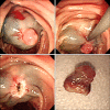
Pyogenic granuloma of the duodenum treated successfully by endoscopic mucosal resection
- PMID: 20479901
- PMCID: PMC2871565
- DOI: 10.5009/gnl.2009.3.1.48
Pyogenic granuloma of the duodenum treated successfully by endoscopic mucosal resection
Abstract
Pyogenic granuloma is a lobular capillary hemangioma that occurs mostly on the skin and the mucosal surfaces of the oral cavity and tongue. Only a few cases in other parts of the digestive tract have been reported. Gastrointestinal pyogenic granuloma is a rare cause of hemorrhage in the digestive tract, but should be considered in the differential diagnosis of patients with gastrointestinal bleeding. We report the case of a 62-year-old anemic woman found to have a pyogenic granuloma of the duodenum, which was treated adequately by endoscopic mucosal resection.
Keywords: Duodenum; Granuloma; Pyogenic.
Figures


Similar articles
-
Endoscopic Mucosal Resection of a Proximal Esophageal Pyogenic Granuloma.Case Rep Gastrointest Med. 2019 Sep 29;2019:9869274. doi: 10.1155/2019/9869274. eCollection 2019. Case Rep Gastrointest Med. 2019. PMID: 31662914 Free PMC article.
-
Pyogenic granuloma: an unrecognized cause of gastrointestinal bleeding.Virchows Arch. 2004 Jun;444(6):590-3. doi: 10.1007/s00428-004-1013-5. Epub 2004 Apr 15. Virchows Arch. 2004. PMID: 15221476
-
Pyogenic granuloma of the duodenum as an unusual cause of gastrointestinal bleeding.Rev Esp Enferm Dig. 2019 May;111(5):410-411. doi: 10.17235/reed.2019.5977/2018. Rev Esp Enferm Dig. 2019. PMID: 31021165
-
Pyogenic granuloma, an unusual presentation of peripubertal vaginal bleeding. Case report and review of the literature.J Pediatr Endocrinol Metab. 2015 Mar;28(3-4):443-7. doi: 10.1515/jpem-2014-0029. J Pediatr Endocrinol Metab. 2015. PMID: 25324441 Review.
-
Pyogenic granuloma of the sigmoid colon.Ann Diagn Pathol. 2005 Apr;9(2):106-9. doi: 10.1016/j.anndiagpath.2004.12.009. Ann Diagn Pathol. 2005. PMID: 15806519 Review.
Cited by
-
Esophageal pyogenic granuloma: endosonographic findings and endoscopic treatments.Clin Endosc. 2013 Jan;46(1):81-4. doi: 10.5946/ce.2013.46.1.81. Epub 2013 Jan 31. Clin Endosc. 2013. PMID: 23423701 Free PMC article.
-
Endoscopic Mucosal Resection of a Proximal Esophageal Pyogenic Granuloma.Case Rep Gastrointest Med. 2019 Sep 29;2019:9869274. doi: 10.1155/2019/9869274. eCollection 2019. Case Rep Gastrointest Med. 2019. PMID: 31662914 Free PMC article.
-
Pyogenic granuloma of the ampulla of Vater: unexpected cause of gastrointestinal bleeding.Clin J Gastroenterol. 2019 Feb;12(1):34-37. doi: 10.1007/s12328-018-0891-z. Epub 2018 Aug 9. Clin J Gastroenterol. 2019. PMID: 30094594
-
Gastrointestinal Pyogenic Granuloma (Lobular Capillary Hemangioma): An Underrecognized Entity Causing Iron Deficiency Anemia.Case Rep Gastrointest Med. 2016;2016:4398401. doi: 10.1155/2016/4398401. Epub 2016 Jun 15. Case Rep Gastrointest Med. 2016. PMID: 27403353 Free PMC article.
References
-
- Enzinger FM, Weiss SW. Soft tissue tumors. 4th ed. St. Louis: Mosby; 2001. Benign tumors and tumor-like lesions of blood vessels; pp. 864–865.
-
- Hartzell MB. Granuloma pyogenicus. J Cutan Dis. 1904;22:520–523.
-
- Noh KA, Moom JS, Kim KS, Kim CD, Ryu HS, Hyun JH. A case of pyogenic granuloma of esophagus. Korean J Gastroenterol. 1989;21:653–657.
-
- Okumura T, Tanoue S, Chiba K, Tanaka S. Lobular capillary hemangioma of the esophagus. A case report and review of the literature. Acta Pathol Jpn. 1983;33:1303–1308. - PubMed
-
- Craig RM, Carlson S, Nordbrock HA, Yokoo H. Pyogenic granuloma in Barrett\'s esophagus mimicking esophageal carcinoma. Gastroenterology. 1995;108:1894–1896. - PubMed
LinkOut - more resources
Full Text Sources

