

Membrane proteome analysis of cerulein-stimulated pancreatic acinar cells: implication for early event of acute pancreatitis
- PMID: 20479917
- PMCID: PMC2871617
- DOI: 10.5009/gnl.2010.4.1.84
Membrane proteome analysis of cerulein-stimulated pancreatic acinar cells: implication for early event of acute pancreatitis
Abstract
Background/aims: Cerulein pancreatitis is similar to human edematous pancreatitis with dysregulation of the production and secretion of digestive enzymes, edema formation, cytoplasmic vacuolization and the death of acinar cells. We hypothesized that membrane proteins may be altered as the early event during the induction of acute pancreatitis. Present study aims to determine the differentially expressed proteins in the membranes of cerulein-treated pancreatic acinar cells.
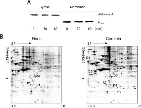
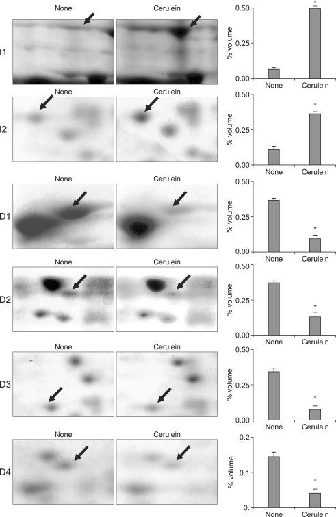
Methods: Pancreatic acinar AR42J cells were treated with 10(-8) M cerulein for 1 hour. Membrane proteins were isolated from the cells and separated by two-dimensional electrophoresis using pH gradients of 5-8. Membrane proteins were identified by matrix-assisted laser desorption/ionization-time of flight mass spectrometry (MALDI-TOF MS) analysis of the peptide digests. The differentially expressed proteins, whose expression levels were more or less than three-fold in cerulein-treated cells, were analyzed.
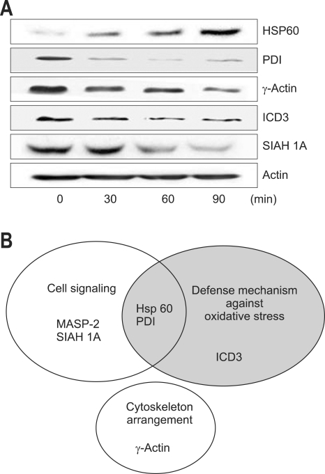
Results: Two differentially expressed proteins (mannan-binding lectin-associated serine protease-2, heat shock protein 60) were up-regulated while four proteins (protein disulfide isomerase, gamma-actin, isocitrate dehydrogenase 3, seven in absentia homolog 1A) were down-regulated by cerulein treatment in pancreatic acinar cells. These proteins are related to cell signaling, oxidative stress, and cytoskeleton arrangement.
Conclusions: Oxidative stress may induce cerulein-induced cell injury and disturbances in defense mechanism in pancreatic acinar cells.
Keywords: Cerulein; Membrane proteome; Pancreatic acinar cells; Pancreatitis.
Figures
Similar articles
-
Differentially expressed proteins in cerulein-stimulated pancreatic acinar cells: implication for acute pancreatitis.Int J Biochem Cell Biol. 2008;40(3):503-16. doi: 10.1016/j.biocel.2007.09.001. Epub 2007 Sep 16. Int J Biochem Cell Biol. 2008. PMID: 18024178
-
Proteome analysis of rat pancreatic acinar cells: implication for cerulein-induced acute pancreatitis.Proteomics. 2003 Dec;3(12):2446-53. doi: 10.1002/pmic.200300545. Proteomics. 2003. PMID: 14673795
-
Altered gene expression in cerulein-stimulated pancreatic acinar cells: pathologic mechanism of acute pancreatitis.Korean J Physiol Pharmacol. 2009 Dec;13(6):409-16. doi: 10.4196/kjpp.2009.13.6.409. Epub 2009 Dec 31. Korean J Physiol Pharmacol. 2009. PMID: 20054485 Free PMC article.
-
Peroxisome Proliferator-activated Receptor-gamma Inhibits the Activation of STAT3 in Cerulein-stimulated Pancreatic Acinar Cells.J Cancer Prev. 2017 Sep;22(3):189-194. doi: 10.15430/JCP.2017.22.3.189. Epub 2017 Sep 30. J Cancer Prev. 2017. PMID: 29018784 Free PMC article.
-
Suppression of IL-1beta expression by the Jak 2 inhibitor AG490 in cerulein-stimulated pancreatic acinar cells.Biochem Pharmacol. 2006 Nov 30;72(11):1555-62. doi: 10.1016/j.bcp.2006.07.008. Epub 2006 Aug 24. Biochem Pharmacol. 2006. PMID: 16934228
Cited by
-
Role of janus kinase/signal transducers and activators of transcription in the pathogenesis of pancreatitis and pancreatic cancer.Gut Liver. 2012 Oct;6(4):417-22. doi: 10.5009/gnl.2012.6.4.417. Epub 2012 Aug 7. Gut Liver. 2012. PMID: 23170143 Free PMC article.
-
Rhein Protects Against Severe Acute Pancreatitis In vitro and In vivo by Regulating the JAK2/STAT3 Pathway.Front Pharmacol. 2022 Mar 17;13:778221. doi: 10.3389/fphar.2022.778221. eCollection 2022. Front Pharmacol. 2022. PMID: 35370748 Free PMC article.
-
Hsp90 regulation affects the treatment of glucocorticoid for pancreatitis-induced lung injury.Mol Cell Biochem. 2018 Mar;440(1-2):189-197. doi: 10.1007/s11010-017-3166-y. Epub 2017 Aug 21. Mol Cell Biochem. 2018. PMID: 28828564
-
Loss of γ-cytoplasmic actin triggers myofibroblast transition of human epithelial cells.Mol Biol Cell. 2014 Oct 15;25(20):3133-46. doi: 10.1091/mbc.E14-03-0815. Epub 2014 Aug 20. Mol Biol Cell. 2014. PMID: 25143399 Free PMC article.
References
-
- Shirohara H, Otsuki M. Plasma cholecystokinin levels in acute pancreatitis. Pancreas. 1997;14:249–254. - PubMed
-
- Koide M, Okabayashi Y, Hasegawa H, et al. Plasma cholecystokinin concentration in patients with chronic pancreatitis measured by bioassay. Nippon Shokakibyo Gakkai Zasshi. 1989;86:2419–2424. - PubMed
-
- Mitchell CJ, Playforth MJ, Kelleher J, McMahon MJ. Functional recovery of the exocrine pancreas after acute pancreatitis. Scand J Gastroenterol. 1983;18:5–8. - PubMed
-
- Gukovsky I, Gukovskaya AS, Blinman TA, Zaninovic V, Pandol SJ. Early NF-kappaB activation is associated with hormone-induced pancreatitis. Am J Physiol. 1998;275:G1402–G1414. - PubMed
-
- Willemer S, Elsasser HP, Adler G. Hormone-induced pancreatitis. Eur Surg Res. 1992;24(Suppl 1):29–39. - PubMed
LinkOut - more resources
Full Text Sources
Research Materials
